
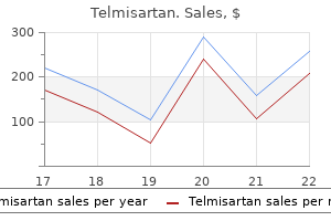
Telmisartan
Purchase telmisartan 20mg without a prescription
Quality of life scores for transplant patients are generally higher than scores for those receiving dialysis blood pressure chart for 80 year old woman generic telmisartan 80 mg overnight delivery. In most areas of care it has not been possible to estimate expenditure on social care. For example, dialysis patients incur some admitted patient costs for dialysis-related complications. If anti-hypertensive medications are prescribed in the same quantities and proportions as for the East Kent cohort, the total annual cost for England is estimated at 215 million. Costs for this proportion of activity are estimated at 53 million, as shown in Table 8. Immunosuppression costs were included for the first 12 weeks after transplantation only. No costs are counted for patients who died or experienced graft failure during the year. There are 81 episodes of live donor pre-transplantation work-up recorded in Reference Costs, nine donor screening episodes and seven post-transplant examinations. No cost estimates were available for the retrieval and transportation of kidneys from deceased donors. However, in the absence of detailed cost estimates for children, the estimates for adult care have been applied to all transplants. According to the 2010 Renal Registry report, 21, 544 people were receiving dialysis in England at the end of 2009. Implied annual per patient costs are 24, 043 for haemodialysis and 20, 078 for peritoneal dialysis, as shown in Table 11. For this reason, no further costs are counted here for drugs provided to dialysis patients by acute providers. This estimate is based on the assumption that the average patient travels to the dialysis centre three times a week, 52 weeks a year. This is equivalent to 2, 792 for each haemodialysis patient, bringing the estimated annual mean cost of haemodialysis to 26, 835 per patient. Six model specifications were tested: normal, gamma and inverse Gaussian using both identity and log links. Indeed, the risk may be higher in the undiagnosed than in the diagnosed population, owing to the lack of risk-ameliorating treatment. It is recognised, however, that the estimate of excess strokes in people receiving dialysis is subject to uncertainty as the relative risk appears to vary with ethnicity. The baseline population risk has been taken from the Oxford Record Linkage Study (data supplied to the British Heart Foundation). Given these figures, the expected number of infections in 17, 349 people (the number of people receiving haemodialysis) is 0. The estimate of total expenditure is more than twice the sum that would be produced by extrapolating from the costs in the 2002 Wanless report, Securing our Future Health: Taking a Long-term View. The total prevalence (diagnosed and undiagnosed) is also believed to be increasing. The Programme Budgeting estimates therefore include expenditure on other renal conditions. The net annual saving for this group, over a 5-year perspective, is estimated at 39 million. The implied annual per patient cost for peritoneal dialysis presented in this paper is lower than that for haemodialysis. It is not known how many renal patients are receiving conservative care in England. Annual expenditure estimates range from 3 million for the 5% scenario to 13 million for the 18% scenario, as shown in Table A1. Total expenditure estimates cover both first year costs for incident strokes and long-term care costs for those whose strokes occurred in earlier years. Chronic kidney disease: Early identification and management of chronic kidney disease in adults in primary and secondary care. Creatinine fluctuation has a greater effect than the formula to estimate glomerular filtration rate on the prevalence of chronic kidney disease. Prevalence of chronic kidney disease based on estimated glomerular filtration rate and proteinuria in Icelandic adults. Chronic kidney disease and the risks of death, cardiovascular events, and hospitalization. Systematic Review of the Effectiveness and Cost-Effectiveness of Home Versus Hospital or Satellite Unit Haemodialysis for People with End Stage Renal Failure. Chronic kidney disease as a risk factor for cardiovascular disease and all-cause mortality: a pooled analysis of community-based studies. Lowest systolic blood pressure is associated with stroke in stages 3 to 4 chronic kidney disease. Angiotensin-converting enzyme inhibitors and progression of nondiabetic renal disease. A comparative survival study of patients over 75 years with chronic kidney disease stage 5. Several epidemiologic studies reported low prevalence 6 and incidence rates of renal failure among hypertension subjects. Copyright the diagnosis of benign hypertensive nephrosclerosis is a diagnosis by exclusion. This is an open It is a clinical diagnosis based on history, physical examination, urinalysis and laboratory access article distributed under the 4 evaluation. Since kidney biopsy is rarely performed, the diagnosis is typically made in patients Creative Commons Attribution 4. Histopathologic lesions in benign distribution, and reproduction in hypertensive nephrosclerosis are characterized by vascular, glomerular and tubular changes. Prevalence, awareness, treatment and control of hypertension among United States adults 1999-2004. Prevalence of chronic kidney disease in population-based studies: Systematic review. Development of chronic kidney disease in essential hypertension during long-term therapy. Elevated blood pressure and risk of end-stage renal disease in subjects without baseline kidney disease. The deletion polymorphism of the angiotensin-converting enzyme is associated with nephroangiosclerosis. Low grade albuminuria and the risks of hypertension and blood pressure progression. Prevalence of hypertension in 1795 subjects with chronic renal disease: the modifcation of diet in renal disease study baseline cohort. Prognostic role of ambulatory blood pressure measurement in patients with nondialysis chronic kidney disease. Masked hypertension and white-coat hypertension in chronic kidney disease: A meta-analysis. Comparative epidemiology of resistant hypertension in chronic kidney disease and the general hypertensive population. Current issues in the management and monitoring of hypertension in chronic kidney disease.
Diseases
- Hyperoxaluria
- Chitayat Haj Chahine syndrome
- Heparane sulfamidase deficiency
- Alopecia universalis onychodystrophy vitiligo
- Scleroatonic myopathy
- Lysosomal alpha-D-mannosidase deficiency
- Emery Dreifuss muscular dystrophy, X-linked
Generic 20 mg telmisartan otc
The condition arises around the 4th week of gestation as the Wolffian ducts fuse with the developing cloaca hypertension nih purchase telmisartan 20mg with amex. A pair of sail-shaped valves develops adjacent to the verumontanum with appearances not unlike valves in a vein. Consequences are bilateral hydronephrosis and hydroureter, hypertrophy of the bladder detrouser and a dilated prostatic urethra. Posterior urethral valves were also seen as a secondary abnormality in two further cases. The urachus is a structure that connects the dome of the bladder to the anterior abdominal wall at the level of the umbilicus. Patent in early development but gradually obliterated so that a solid core of tissue, the median umbilical ligament, remains. This is most commonly seen in association with obstruction at the neck of the bladder or urethra. Dysplasias are less common and are characterized by generalized defective development of bone and cartilage. Ultrasonically it is not possible to give a precise pathological diagnosis of dysplasias but in general two main groups are recognized. Firstly, short limbs with severely defective ossification with or without fractures will be seen in severe osteogenesis imperfecta or hypophosphatasia. Secondly short limbs with or without spinal deformity are features of thanatophoric dysplasia, achondrogenesis, Jeune syndrome, (asphyxiating thoracic dystrophy) and Jarco-Levin syndrome amongst others. They are commonest in female infants, term deliveries, breech presentation and the left hip. Developmental dysplasia of the hip was recorded as a primary diagnosis in all but two incidences. The anomaly is not a single entity and may be considered as extrinsic, (deformation), of intrinsic (true malformation). When the finger buds have completed growth in length they are still joined by webs which break down by progressive cell death until the normal proximal web configuration is reached. Failure of cell death results in syndactyly, the commonest congenital hand abnormality. The appearance of abnormality depends on the tine of interference with the developing part. The absence of a consistent classification system may be a consequence of a lack of understanding of the pathogenesis of these abnormalities. Major limb defects are usually diagnosed at routine 2nd trimester ultrasound imaging. Limb deficiencies are characterized by either the total or partial absence of the skeletal structure of the limbs or different degrees of limb hypoplasia. Interference with the development of blood supply is the most likely mechanism in the appearance of many congenital limb deformities. Longitudinal limb deficiencies, (Q714-Q716, Q724-Q727), refer to the partial absence of a limb extending parallel to the long axis of the limb. They typically involve specific components of the limbs: preaxial (first ray: thumb or radius in the arms or first toe or tibia in the leg); postaxial (fifth ray: fifth finger or ulna in the arm, fifth toe or fibula in the leg); or central components (typically third or fourth rays in the hand or foot such as lobster-claw hand). It is variable in presentation from an isolated thumb anomaly to complete absence of the radius. In transverse limb defects, (Q710, Q712, Q713, Q720, Q722, Q723), there is complete or partial absence of distal structures of a limb in a transverse plane at the point where the deficiency begins. Often severe phocomelia, deformed carpal bones, clinodactyly, syndactyly, radial synostosis and deformed sternum. Intercalary cases, (Q711 & Q721), are where there is complete or partial absence of the proximal or mid segments of a limb but preservation of distal limb structures. There were no cases of intercalary limb deficiency recorded in the current cohort. Larsen syndrome, Freeman-Shelden syndrome and Multiple Pterigium syndrome) and non-syndromic. Sometimes these codes are used to differentiate between a presentation with primary involvement of the limbs and more complex abnormality where limb deformity is combined with other congenital anomalies. It was first identified by Pena and Shokeir in 1974 but although early descriptions resulted in the eponym it has recently been suggested that Pena-Shokeir is not a specific unitary diagnosis or syndrome, but rather a description of a clinically and genetically heterogeneous phenotype from variable aetiology, resulting from the reduction of movements in the uterus due to an intrinsic pathology regardless of the cause, and was subsequently included among the phenotypes associated with the fetal akinesia/hypokinesia deformation sequence. In this instance concerns were first raised at the 20 weeks fetal anomaly scan when bilateral talipes and persistently clenched hands were seen throughout the scan. The scan findings deteriorated with progressive scans and minimal movement was seen. Prior to termination significant polyhydramnious was becoming evident suggesting that fetal swallowing was now affected. Craniosynostosis causes distortion of the shape of the skull owing both to failure of bone growth at the prematurely closed suture site and to compensatory overgrowth at the sutures that remain open. The different types of craniosynostosis are classified by which sutures have closed prematurely. Infants born with Jarcho-Levin syndrome have short necks, limited neck motion due to abnormalities of the cervical vertebrae and short stature. In most cases, infants with Jarcho-Levin syndrome experience respiratory insufficiency and are prone to repeated respiratory infections that result in life-threatening complications. Disorders of the spine are also classified as secondary abnormalities in four further cases. Caudal Regression Sequence, (Q8980) Caudal regression sequence is a disorder that impairs the development of the lower (caudal) half of the body. Areas affected include the lower back and limbs, the genitourinary system and the gastrointestinal tract. The bones vertebrae of the lower spine, particularly the sacrum, are frequently misshapen or missing. Defects include unilateral renal agenesis, horseshoe kidney, bladder extrophy, ureteral duplication, hypospadias, cryptorchidism in the male and rectovaginal fistula in the female. Individuals with caudal regression syndrome may have malrotation of the large bowel and imperforate anus. The condition is likely to be caused by the interaction of multiple genetic and environmental factors resulting in a combination of abnormal mesoderm development and decreased blood flow to the caudal areas of the fetus. Indeed, the female infant with tetralogy of Fallot and associated sacral agenesis, (listed above), was delivered to an older mother with diabetes. There were also some minor abnormalities of the right hand listed in the post-mortem report but not coded. With the second case the booking scan had demonstrated that although the lower limbs were present they were highly flexed and held in an abnormal position underneath the baby. Post mortem examination following termination of pregnancy was to also reveal a double outlet right ventricle. Additional skeletal abnormalities can include unusually shaped clavicles and pelvic bones, and cone-shaped ends of the long bones in the arms and legs. Many infants with this condition are born with an extremely narrow, bell-shaped chest that can restrict the growth and expansion of the lungs. Life-threatening problems with breathing result, and people with asphyxiating thoracic dystrophy may live only into infancy or early childhood. Mutations in at least 11 genes have been found to cause asphyxiating thoracic dystrophy. Hypochondroplasia is a rare inherited dysplasia causing short stature not unlike a mild form of achondroplasia. Features, other than short stature, include macrocephaly, lordosis, disproportionate arms and legs with short but broad hands and feet, limitation of elbow movement but hypermobility of other joints.

Discount telmisartan 40mg
The use of unopposed (without progestogen) estrogen-hormone therapy and obesity blood pressure on apple watch order telmisartan 40mg online, which increases endogenous concentrations of estrogen, increases the risk of endometrial cancer. Additional information available to the committees responsible for subsequent updates through Update 2014 has not changed that conclusion. In comparison with non-deployed female Vietnam-era veterans, those who served in Vietnam had no excess cervical cancer mortality. A further analysis restricted to female nurses, again using the non-deployed cohort as the referent, yielded virtu ally the same nonstatistically signifcant risk of mortality from cervical cancer. Similarly, there were also very few observed uterine cancer deaths of women who served in Vietnam, served near Vietnam, or were non-deployed, with 9, 4, and 12 deaths, respectively, and no excess risk of uterine cancer mortality was found in any of the three cohorts when compared with the general population. In the inter nal comparison to non-deployed Vietnam-era veterans, uterine cancer mortality was not associated with service in Vietnam or near Vietnam. There were more deaths from ovarian cancer in the entire cohort, but no differences in the risk of ovarian cancer mortality were found among those who served in Vietnam, served near Vietnam, or were non-deployed in comparison with the general population of U. In the internal comparison with the non-deployed veterans, ovarian cancer mortal ity was increased among Vietnam veterans and among women who served near Vietnam, but neither was statistically signifcant. An analysis restricted to nurses revealed similar patterns of increased (albeit not statistically signifcant) ovarian cancer mortality, both for veterans who served in Vietnam and for veterans who served near Vietnam, when compared with non-deployed nurses. Update of the Epidem iologic Literature Relevant studies on cancers of the female reproductive system include the cervix, uterus, ovary, and vagina. No studies of female reproductive cancers among Vietnam veterans have been published since Update 2014. The mechanism of action might be related to endocrine disruption and chronic infammation. The most relevant evidence came from a follow-up study on mortality among female U. For both cervical and uterine cancers there was no evidence of increased mortality risk; however, the small observed number of deaths for these outcomes in all three cohorts limited the statistical power of the associations. However, because the rate of ovarian cancer mortality was similar between veterans who served in Vietnam (with potential exposure to herbicides) and those who served near Vietnam (who presumably were not so exposed), this evidence is equivocal. Most fndings from occupational cohorts and environmental studies where exposure was well-characterized have not found increased risks for cervical, uterine, or ovarian cancers. No new studies with suffcient exposure specifcity were identifed for the current update. The results of mechanistic studies provide more plausibility for a reduced risk of female reproductive cancers than for an increased risk. That makes prostate cancer the second most common cancer in men (after non-melanoma skin cancers); it is expected to account for about 9. The incidence of and mortality from prostate cancer varies widely with age and race. The inci dence rate of prostate cancer for men aged 75 and older decreases slightly, but remains high (432. As a group, African American men have the highest recorded incidence of prostate cancer in the world (Jemal et al. Other than race and age, the risk factors include a family history of the disease both in frst and second degree relatives (Bruner et al. There is some evidence that some elements of the W estern diet, including a high con sumption of red meat and saturated fats, may be a risk factor for prostate cancer, but these have not been conclusively identifed. Of note, selenium and vitamin E supplementation did not reduce, but rather slightly increased, prostate cancer incidence in a large clinical trial (Klein et al. The 5reductase inhibiting drugs fnasteride and dutasteride, which are widely used to treat benign enlargement of the prostate, were found to decrease the prevalence of prostate cancer by about 25% in two major randomized trials (Andriole et al. Finasteride acts by decreas ing the formation of the potent androgen metabolite 5dihydrotestosterone in the prostate. Study of the incidence of and mortality from prostate cancer is complicated by various approaches to screening for the disease in different countries and populations. In addition, fndings that show an association between an exposure and prostate cancer mortality should be examined closely to determine whether the exposed group had poorer access to screening or treatment that would have decreased the likelihood of survival. Strati fying tumors by grade and characteristics led to a stronger association between herbicide exposure and intermediate to high-grade prostate cancer and an even stronger association with more aggressive prostate cancer. In a follow-up study of 2, 783 male New Zealand veterans who had served in Vietnam and were still alive as of 1988, M cBride et al. Among Korean veterans who served in Vietnam, a total of 125 incident cases and 53 deaths from prostate cancer were identifed during the follow-up period in the cohort studied by Yi and colleagues (Yi, 2013; Yi and Ohrr, 2014; Yi et al. When compared with the general Korean population, there was a 22% statistically signifcant excess prostate cancer risk in the entire cohort (Yi, 2013), which was mostly due to a signifcant 2. Yi and Ohrr (2014) did not stratify incident prostate cancer cases according to tumor characteristics (low versus high-grade tumors) as is usually done in studies of prostate cancer incidence. Cox proportional hazards regression modeling was used to assess the relationship between exposure to Agent Orange and biochemical recurrence, secondary treatment, metastases, and prostate cancer-specifc mortality. Although Agent Orange expo sure included an additional level of service location verifcation to self-report, this measure is still only a proxy for actual initial and subsequent exposure levels. Several pesticide exposure metrics were constructed for each pesticide based on the duration and frequency of pesticide exposure. The results suggest that a genetic variation may decrease the risk of prostate cancer with exposure to dicamba. Environm ental Studies In a well-designed and conducted nested case-control study, Koutros et al. The study sample was identifed from the Janus Serum Bank cohort, a population-based research biobank consisting of almost 317, 000 individuals with an average age at enrollment of 41 years. The Janus cohort was linked with to the Cancer Registry of Norway to identify new cases of prostate cancer. Eligible cases consisted of incident metastatic prostate cancer cases with no history of cancer (except non-melanoma skin cancer) who were diagnosed from enrollment through December 31, 1999, and were diagnosed at least 2 years after serum collection. Cases (n = 150) and controls (n = 314) were matched on date of blood draw (1-year strata), age at blood draw (2-year strata), and region. The power to detect more modest associations was limited in the higher exposure level categories. After excluding women and men with missing data, the subcohort consisted of 831 subjects from which 256 controls and 110 incident cases of prostate cancer (identifed through the National Cancer Registry, a nationwide hospital cancer registry covering 99% of all cases diagnosed in South Korea) were selected. A total of 240 incident cases were identifed, and 268 controls with other diseases (except cancer) were recruited and matched to cases on ethnicity and age. Given that this is a small study that did not report information on case and control response rates, that control diagnoses were not known, and that it is not clear whether there was adjustment for potential confounders, this study is of limited utility. Because male Vietnam veterans were exposed to herbicides after adolescence, toxicologic fndings concerning early-life exposure are not particularly relevant to this population, although their exposure to herbicides could potentially infuence risk of the prostate cancer later in life. Ranch Hands and Australian Vietnam veterans that used better exposure assessment support an association between exposure to the herbicides used in Vietnam and prostate cancer. Several positive associations between exposure to specifc herbicides or their contaminants and prostate cancer have been reported from previously reviewed occupational studies. However, there is no substantial understanding of the importance of these mechanisms or how they could affect prostate cancer risk. The modeled incidence rate of testicular cancer in 2014 for all races combined for men ages 65 years and over (which would include most Vietnam veterans) is 1.

Discount telmisartan 20mg online
The hair follicle containing a comedone is surrounded by lymphocytic infltrate in papular acne blood pressure reading chart telmisartan 40mg sale, and neutrophilic infltrate in pustular acne. The lesions appear as vesico pustules which may rupture and are followed by characteristic yellowish crusts. M/E the characteristic feature is the subcorneal pustule which is a collection of neutrophils under the stratum corneum. Often, a few acantholytic cells and gram-positive bacteria are found within the pustule. The upper dermis contains severe infammatory reaction composed of neutrophils and lymphoid cells. Depending upon the clinical appearance and location, they are classifed into different types described below. Epidermodysplasia verruciformis is of special clinical signifcance as it may undergo malignant change. M/E Prototype of verruca is common viral wart having following features: i) Papillomatosis (papillary folds). Clinically, the lesions are often multiple, discrete, waxy, papules, about 5 mm in diameter and are seen more frequently on the face and trunk. M/E Typical lesion consists of sharply circumscribed cup-like epidermal lesion growing down into the dermis. The proliferating epidermal cells contain the pathognomonic intracytoplasmic eosinophilic inclusion bodies called molluscum bodies. Vaccinia (cowpox) is primarily a disease of the 523 teats and udders of cows but humans are infected by milking the infected animals. Varicella (chickenpox) and herpes zoster (shingles) are both caused by a common virus, varicella-zoster virus. M/E the characteristic feature of viral exanthemata is the formation of intra epidermal vesicles or bullae due to cytopathic effects of viruses. This is followed by intracellular oedema and ballooning degene ration that progresses on to rupture of the cells with eventual formation of vesicles or bullae. These include some of the common dermatophytes such as Trichophyton rubrum and Pityrosporum. Clinically, these fungal infections are labelled according to the region involved. These are as follows: i) Tinea capitis occurring on the scalp, especially in children. M/E Fungal hyphae (or mycelia) and arthrospores of dermatophytes are present in the stratum corneum of skin, nails or hair. Tubercle bacilli are present in very small numbers that are hard to demonstrate by acid-fast staining. Cutaneous manifestations appear as presenting feature in about a quarter of patients and include erythema nodosum, or brown-red jelly-like papules or plaques with central clearing. Fibrinoid necrosis and presence of intracellular inclusions such as asteroid bodies are some other features which may be seen. M/E the centre of the lesion shows a well-demarcated focus of complete collagen degeneration. These foci are surrounded by an infltrate composed 524 largely of histiocytes and some mononuclear infammatory cells forming a palisade arrangement and are therefore also referred to as palisading granu lomas. The discoid variety is more common which is generally benign, while systemic form may be fatal, usually from renal involvement. Morphea consists of lesions limited to the skin and subcutaneous tissue, while progressive systemic sclerosis consists of extensive involvement of the skin and the subcutaneous tissue and has visceral lesions too. M/E There is thickening of the dermal collagen extending into the subcutaneous tissue. The epidermis is often thin, devoid of rete ridges and adnexal structures, and there is hyalinised thickening of the walls of dermal arterioles and capillaries. M/E the bullae are suprabasal in location so that the basal layer remains attached to dermis like a row of tombstones. Intraepidermal abscesses composed almost entirely of eosinophils are diagnostic of pemphigus vegetans. M/E Superfcial subcorneal bullae are found which contain acantholytic epidermal cells. The distribution of clinical lesions is similar to lupus erythematosus involving face. M/E the characteristic distinguishing feature from pemphigus is the subepidermal location of the non-acantholytic bullae. With passage of time, there is some epidermal regeneration from the periphery at the foor of the bulla. The bullous cavity contains fbrin network and many mononuclear infammatory cells and many eosinophils. Dermal changes seen in infammatory bullae consist of infltrate of mononuclear cells, a few eosinophils and neutrophils. The disease has an association with gluten-sensitive enteropathy (coeliac disease). Both dermatitis herpetiformis and gluten-sensitive enteropathy respond to a gluten-free diet. M/E the early lesions of dermatitis herpetiformis consist of neutrophilic micro-abscesses at the tips of papillae, producing separation or blister between the papillary dermis and the epidermis. The older blisters contain fair number of eosinophils causing confusion with bullous pemphigoid. Direct immunofuorescence shows granular deposits of IgA at the papillary tips in dermatitis herpetiformis. The condition occurs due to hypersensitivity to certain infections and drugs, and in many cases, it is idiopathic. As the name suggests, the lesions are multiform such as macular, papular, vesicular and bullous. Stevens Johnson syndrome is a severe, at times fatal, form of involvement of skin and mucous membranes of the mouth, conjunctivae, genital and perianal area. As the scales are removed by gentle scrapping, fne bleeding points appear termed Auspitz sign. Commonly involved sites are the scalp, upper back, sacral region and extensor surfaces of the extremities, especially the knees and elbows. M/E Fully-developed lesions show: i) Acanthosis with regular downgrowth of rete ridges to almost the same dermal level with thickening of their lower portion. The lesions are distributed symmetrically with sites of predilection being fexor surfaces of the wrists, forearms, legs and external genitalia. A special manifestation of idiopathic calcinosis cutis is tumoral calcinosis in which there are large subcutaneous calcifed masses, often accompanied by foreign body giant cell reaction. Idiopathic calcinosis of the scrotum consists of multiple asymptomatic nodules of the scrotal skin. Tumours and tumour-like lesions may arise from different components of the skin such as surface epidermis, epidermal appendages and dermal tissues. Each of these tissues may give rise to benign and malignant tumours as well as tumour-like lesions. Another 527 group of tumours have their origin from elsewhere in the body but are cellular migrants to the skin. Though considered by many authors to include common viral warts (verrucae) and condyloma acuminata, true squamous papillo mas differ from these viral lesions. M/E Squamous papillomas are characterised by hyperkeratosis, acanthosis with elongation of rete ridges and papillomatosis.
Mountain Flax. Telmisartan.
- Are there safety concerns?
- How does Mountain Flax work?
- Dosing considerations for Mountain Flax.
- Vomiting, purging stool from the bowel, and other conditions.
- What is Mountain Flax?
Source: http://www.rxlist.com/script/main/art.asp?articlekey=96347
Telmisartan 20mg lowest price
If there is no recurrence without medication in four years blood pressure medication bruising purchase genuine telmisartan line, the risk may then become acceptable for medical certification. Consideration should not be given until a four-year seizure-free and medication-free observation period has been achieved. With normal studies and no risk factors, recurrence risk after four years approximates that of the normal population. Embolic stroke (artery to artery or cardio-embolic source) must also be considered. In the young, additional factors must be considered such as hypercoaguable states, patent foramen ovale, and arteriopathies. Clearly the existence of any persistent neurological deficit must be addressed in terms of functional compromise. Beyond the first year, recurrence risk is about four per cent per year, with some variability depending on stroke subtype. Medical certification is appropriate when cause and risk factors have been identified and addressed and a recurrence risk has been assessed. Recurrent stroke may cause sudden incapacitation, and a recurrence risk exceeding one per cent per year is not acceptable. A recurrence-free observation period is appropriate prior to medical certification following ischaemic stroke, and this will vary dependent upon mechanism and risk factors. If an individual with arterial dissection has no recurrence in one year, risk recurrence thereafter is less than one per cent per year. Vascular malformations including cavernous angiomas may also lead to intracerebral bleeding, sometimes with complete recovery. Though surgical cure of a vascular malformation might preclude re-bleeding, the risk of residual seizures may still bar certification. There are exceptions in which tissue destruction is minimal and recovery is complete or near complete. A one to two-year observation period is appropriate following haemorrhagic stroke. A full neurological evaluation indicating satisfactory recovery and freedom from relevant risk factors may allow medical certification at that time. Aneurysms ordinarily arise from major arteries at the base of the brain (Circle of Willis) and are thought to develop from congenital changes in the muscular wall of the artery and degenerative changes in the internal elastic lamina. Death occurs in 23 per cent, and half of the survivors have significant disability. Sequelae may include focal neurological deficit, seizures, and cognitive impairment. Absent these conditions and with a period of symptom-free observation, medical certification may be possible. If there is no recurrence within one year, statistics reveal an acceptably low risk of recurrence thereafter. In another specific condition, called peri-mesencephalic or pre-pontine subarachnoid haemorrhage, recurrence risk is low. Partial obliteration of an aneurysm with residual lumen may present an unacceptable risk. For subarachnoid haemorrhage of unknown cause, a one-year observation period is also warranted. The presence of a vascular malformation (cavernous angioma, arteriovenous malformation) requires individual evaluation. Residual malformation, haemosiderin deposition and other factors will affect risk for recurrent haemorrhage or seizure, and medical certification may not be possible. Most head injuries, including some with a linear skull fracture, do not involve brain injury. Liberal use of modern imaging techniques may indicate parenchymal injury (localized haemorrhage) in individuals with no clinical signs or symptoms of injury. It is important to determine the nature and severity of injury as part of the evaluation. Symptomatic medications are often employed, precluding medical certification until the condition subsides. Medical records and current neurological functioning will provide information regarding persistent deficit. When indicated, detailed neuropsychological testing by a qualified examiner may document the presence or absence of any cognitive residual sequelae. With penetrating injuries involving violation of the cranial vault, the risk is high and may approach 40 per cent. In more commonly occurring closed head injuries, risk is a much lower five per cent. Cerebral contusion, parenchymal haematoma, post-traumatic amnesia beyond one day, depressed skull fracture and subdural haematoma confer increased risk. With penetrating injuries, 97 per cent of the risk will have been achieved in three years, though some elevated risk still persists ten years after the injury. Depending upon severity, focal neurological deficit may warrant a six months to two years period of observation for maximal neurological recovery. In individuals with neuropsychological residual changes, usually indicating significant traumatic brain injury, a one to five-year observation period is warranted depending upon severity of cognitive impairment. Careful cognitive evaluation for permanent impairment should then precede medical certification. The presence of blood (hence iron) in the brain parenchyma is thought to play an aetiological role in the development of post-traumatic epilepsy. Simple uncomplicated epidural haematoma without parenchymal blood might allow medical certification following a one to two-year observation period. Subdural haematoma is often associated with underlying cortical contusion, increasing risk of post-traumatic epilepsy. Significant risk is present in the first two years post injury, though it declines with time. With intraparenchymal haematoma, a two-year period of observation is warranted due to the presence of parenchymal blood. Seizure risk also exists with diffuse axonal injury, and a period of one to two years of observation is appropriate. In such cases, however, an observation period up to five years may be appropriate. Neurological symptoms may include headaches and vomiting related to increased intracranial pressure, seizures, focal neurological deficit related to mass effect or infiltration, cognitive changes, and visual field defects related to pituitary neoplasms. Benign parenchymal growths include ependymoma, choroid plexus papilloma, and colloid cyst (considered a cyst rather than a neoplasm). Though craniopharyngiomas are benign, they may invade adjacent neural tissue and are subject to recurrence. At times there may be residual neoplastic tissue, since complete excision carries the risk of creating a neurological deficit. In such instances, medical certification may be possible, conditional upon satisfactory follow-up with serial imaging studies and current status reports. Posterior fossa neoplasms, which characteristically do not lead to seizures, are an exception. Ordinarily limitations have to be imposed, with certification being conditional on periodic evaluation for tumour recurrence. Eventual recurrence is the rule, though with low grade glial neoplasms this may occur indolently over many years. Seizures are a risk, and subtle neurological impairment depending upon location is an additional concern. These features ordinarily preclude medical certification, though some cases of cure appear in the literature. There may be very rare exceptions following a long recurrence-free and symptom-free interval. Others follow a slowly progressive temporal profile, lending themselves to monitoring measures that can identify the point of compromise to flight safety. Over 90 per cent of affected individuals experience hand tremor, 33 per cent head tremor, 16 per cent voice tremor, and 12 per cent leg tremor. Handwriting, fine movements such as using a screwdriver or threading a needle, and drinking soup from a spoon, may be affected. Treatment with aeromedically acceptable beta-adrenergic blocking medicines is often highly effective.
Quality telmisartan 40mg
Suppression is a positive inhibitory reflex developed to allow the visual cortex to ignore the visual information coming from a deviating eye so as to avoid diplopia prehypertension range chart buy 20mg telmisartan with mastercard. In alternating strabismus the suppression changes from one eye to the other depending on which eye is being used. The size, shape and density or depth of the suppression scotoma is different in different types of strabismus. In most squinting persons with suppression, the whole area of the visual field of the deviating eye that overlaps the fixing eye is suppressed. Thus, the deviating eye always contributes to the overall binocular field of vision in a strabismic patient in two ways. Neither the area corresponding to the blind spot of the fixing eye nor the peripheral temporal crescent area in the deviating eye is suppressed. The binocular field is smaller (narrower) in esotropic patients and larger (wider) in exotropic patients. The retinal midline divides the temporal retina and one side of the brain from the nasal retina and the other side of the brain. The image of the fixation object always falls on the same side of the retina of the deviating eye and is suppressed. If, however, the deviation is changed from esotropia to exotropia or vice versa, this is a new situation and diplopia is triggered. It is the change in position of the retinal image from one half of the retina to the other half that triggers the change from suppression to diplopia and vice versa whenever the visual fields overlap. Thus the risk to get outside the suppression area and become diplopic is the risk to change from esotropia to exotropia or vice versa. The monofixation syndrome is characterized by a minor heterotropia with paracentral fixation and good peripheral fusion. The risk of diplopia is minimal and depends on the peripheral fusional amplitude, which maintains ocular alignment. To make a patient aware of the images perceived by the deviated eye, one must reduce the retinal illuminance in the fixating eye until the patient sees double. This is best done 16 with a series of red filters of increasing density in the form of a ladder (Sbisa bar). The patient fixates a small light source, and the filters are placed in front of the fixating eye. Some patients see double with a light density filter; others require a heavier-density filter before they recognize their diplopia. The lighter the density of the filter needed to produce diplopia the more superficial is the suppression indicating an increased risk of diplopia. In individuals with normal fusion, placing graduated neutral-density filters in front of either eye will, at a certain density level, prevent fusion and induce two lights either together (orthophoria) or apart from each other (diplopia with heterophoria). Symptoms of asthenopia include redness, dryness, discomfort, a feeling of heaviness in the eyes and inability to use the eyes for more than a short period of time. The symptoms may indicate decreased accommodation, ametropia or heterophoria, sometimes with reduced fusional 16 Sbisa bar: a Bagolini filter bar, manufactured by Sbisa Industriale SpA, Italy. Other conditions such as conjunctivitis and anterior uveitis may cause similar symptoms. Patients with asthenopia require full ocular examination including refraction, measurement of accommodation and evaluation of ocular alignment and binocular status. Double vision (diplopia) means that a single object is seen in two different locations. Single vision in gaze straight ahead, down and to the sides is required for safety. Some individuals who have diplopia only in the extremes of up-gaze to the sides may be acceptable for flying duty. Persons with alternating strabismus may note a shift in the apparent position of objects when they alternate fixation and be disturbed by this. Alternating strabismic patients who always fixate with the same eye for distance and the other eye for near will not experience shift in location and may be fit for flight. Changes in refraction may result in an unstable fixation pattern incompatible with safe flying. The evaluation of binocular vision can be considered under screening tests and detailed assessment. The applicant who is asymptomatic and has no past history of strabismus treatment with patching, orthoptics or surgery should be evaluated with regard to visual acuity, refraction, ocular motility and general health of the eyes. Ocular alignment should be tested with cover testing using the appropriate spectacle correction or contact lens correction. Sensory testing with the Worth four-dot test, measurement of stereopsis and measurement of fusional amplitudes are useful in evaluation of the binocular status. Applicants who do not normally pass the screening tests mentioned ought to be examined by an eye specialist. Based on a full sensory and motor evaluation of the applicant, the specialist may be able to estimate the risk of diplopia or shift in location. Symptoms of diplopia or location shift or a high risk of these would disqualify the applicant for class 1 and 2 certificates. The medical examiner should be familiar with the demands likely to be imposed upon hearing, equilibrium and speech during flight and other aviation duties. It contains methods for comprehensive assessment of applicants in whom there is a suspicion or overt manifestation of ear, nose and throat pathology. It further serves as a guide in the assessment of normal, presumably healthy, applicants for aviation personnel licences. The examiner must make certain that the functions of hearing, equilibrium and speech required for the safe performance of aviation duties can be reliably carried out by the applicant. When the examiner is unable to visualize the tympanic membrane and where the hearing is markedly impaired due to obstruction, an applicant should obtain proper treatment and present himself later for completion of the examination. The tympanic membrane is slightly cone-shaped, like the diaphragm of a loudspeaker. Both the concavity of the tympanic membrane and its position relative to the auditory canal normally vary somewhat and may be greatly altered in disease. The short process stands out like a tiny knob at the upper end of the long process (or handle). The malleus is the key structure in dividing the tympanic membrane into its four quadrants. A line drawn perpendicular to the malleus at the level of the umbo (lower end of the malleus) gives four quadrants: anterior superior, anterior inferior, posterior superior and posterior inferior. Any retraction of the tympanic membrane will displace the cone of light inferiorly. The evidence of injury may vary from slight hyperaemia to a ragged perforation of the tympanic membrane. Perforations usually heal but the healed area is thinner, more transparent and also more flaccid when alternating positive and negative pressures are produced, as with a pneumatic otoscope. Any perforations should be described as small or large, marginal or central, and their location given by quadrant or as numbers on the clock. Atrophic parts of the tympanic membrane are of special concern as they may rupture when exposed to even a small increase in differential pressure. A sudden perforation during descent may cause alternobaric vertigo and lead to acute incapacitation. Because of their fragility, atrophic areas should be treated aeromedically as if they were true perforations. Grey white masses of debris may be a sign of cholesteatoma which also can lead to acute incapacitation with vertigo and/or hearing loss. Granulation tissue in the general area of the tympanic membrane usually indicates protrusion of the tissue from the middle ear through a small perforation in the tympanic membrane. An applicant should not be declared fit until all of these conditions have been fully examined and evaluated. The otoscopic findings 2 of the aerotitis media can be classified into 5 or 6 levels according to Teed. In the 6-level Teed classification, grade 0 is a condition with subjective symptoms but no otological signs, grade 1 diffuse redness and retraction of the tympanic membrane, grade 2 slight haemorrhage and retraction of the tympanic membrane, grade 3 gross haemorrhage and retraction of the membrane, grade 4 free blood or fluid in the middle ear, and grade 5 perforation of the tympanic membrane.

Buy generic telmisartan from india
There is considerable variation in symptoms from family to family and among members of the same family blood pressure 50 year old male cheap telmisartan 20mg overnight delivery. For example, if your parent had kidney cancer, it does not mean you will develop a kidney tumor. So far, researchers have not been able to find any patterns that would allow one to predict which symptoms a person will develop based on the particular genotype they have. All people have two copies of the fumarate hydratase gene, one from their mother and one from their father. The other is altered, meaning that it has a change in Page 12 it and does not work very well. When a person has only one working copy of the gene, their cells make less fumarase than normal, but enough to be healthy. However, fibroids are very common in the general population and are rarely diagnostically useful on their own. However, the incidence of kidney tumors in the European group is reported as much lower. Some laboratories that can test for fumarase activity find difficulties because of problems in calculating and interpreting the results. Examples are benign adrenal tumors and Leydig testicular cancer which develops in the Leydig cells the cells in the testes that release the male hormone, testosterone. It gives family members who do not have symptoms the ability to discover whether they have the gene alteration by a simple blood test. As we learn more, the exact alteration in your gene will become increasingly important to your own health maintenance. Things to keep in mind regarding genetic testing: It may complicate your ability to obtain life or health insurance. The results may be difficult to interpret, and it is best to undertake genetic testing through a genetic professional (geneticist or genetic counselor) who can help you understand the results and their implications for yourself and your family. If you have your children tested before their age of consent and they are found positive there are implications for future life or health insurance, and mortgage applications as well as the start of a life-long screening process. This is a difficult decision to make between health safety and financial implications. The actual risk figure depends on the closeness of the relationship starting as high as 50% with a first degree relative (parent, child or sibling). The alteration cannot however skip generations so the more genetic testing a family has Page 16 the more precise the risk figure will be for each individual ranging from 0% to 50%. In other words, if one of your parents is at risk, but tests negative, then you will not be at risk. It is important to keep in mind that your family members may be overwhelmed when they receive this information. It can sometimes take several months to obtain genetic testing results, but once one genetic alteration is identified within the family, testing of additional family members is faster and less costly. Periodic screening with scans will catch a tumor early, so that it can be treated. Life and Health Insurance A useful link describing the general implications for insurance (applies to all genetic conditions not just to Birt-Hogg-Dube) is. Many states have enacted state laws to protect their citizens from genetic discrimination by health insurers. Health insurance protection: Group and individual health insurers may not use your genetic information to set eligibility, premium or contribution amounts; Health insurers may not request or require that you take a genetic test. Employment protection: Employers may not use your genetic information to make decisions involving hiring, firing, job assignments and promotions; Employers may not request, require or purchase genetic information about you or one of your family members. Employers with fewer than 15 employees and the military are not required to abide by the employment protections. Page 18 Having Children Deciding whether to have children when there is a 50% chance of inheriting a problem is a difficult decision to make. Before making any decision you may wish to speak with a geneticist or genetic counselor about possible testing options and their implications. Just as uterine leiomyomas grow from smooth muscle of the uterus, cutaneous leiomyomas are rare benign tumors that grow from smooth muscles in the skin. The arrectores pilorum (singular arrector pili, also called piloerectus muscles) are small smooth muscles that are attached to hair follicles. They tend to grow anywhere on the body and limbs, but rarely on the face, hands or feet. These bumps can be very small, but sometimes large, can range in color from skin-colored to light brown to red, and tend to grow in mosaic clusters, but can also be solitary. Some people have a single leiomyoma, but many people develop small clusters of leiomyomas. Once a leiomyoma appears, it does not normally go away and others may appear over the following years. A skin biopsy involves minor surgical removal of some of the skin bump, after which the tissue is sent to a pathology lab. An anesthetic agent is injected under the skin around the leiomyoma and once the area is numb, a small sample of tissue is taken. Slides are prepared from the tissue and examined under a microscope by a pathologist to determine whether the diagnosis is piloleiomyoma or some other type of growth. The exact cause of pain has not been understood, but there is a thought that the leiomyoma has trapped nerve cells. The variation is not just from patient to patient, but also within one patient and can increase over time. Some patients find a cold sensitivity to such an extent that they even consider moving to a warmer country. Some find that if a pain develops in one leiomyoma it acts as a trigger to all the others to become also painful for hours or days at a time. Sometimes a leiomyoma that grows initially without having any pain symptom can start to become irritable and painful. When pain does occur most patients describe it as excruciating, like having a knifepoint stab. There is a reported case of extensive multiple piloleiomyomas being successfully removed by surgery and reconstructed with a flap technique. Sometimes the leiomyomas will grow back after removal, possibly because some tissue was left behind, or there were new ones growing in a cluster. You should talk with a dermatologist about what is best for your type of skin growth. If you have painful cutaneous leiomyomas, you may want to get a contact name from info@hlrccinfo. As with any treatment Page 24 you should first discuss and agree its suitability with your physician and or dermatologist. The links below have a lot of information about Lyrica including descriptions of warnings and side effects which seem important to study before deciding to take it. Significant pain relief has also been reported with pulsed hysocine butyl bromide see. This article mentions many calcium channel blockers like nifedipine, phenoxybenzamine, doxazocine, gabapentin and topical 9% hyoscine hydrobromide.

Purchase telmisartan in united states online
Immune system: Reduced IgG response to antigens peripheral neuropathy purchase telmisartan 80mg on line, frequent and more severe infections. In fatty liver due to chronic alcoholism, the following mechanisms are involved except: A. Enzymatic digestion is the predominant event in the following type of necrosis: A. Mechanism of mammalian apoptosis involves the most important role of the following protein: A. Specifc or adaptive immunity is specifc and is characterised by antigenic specifcity. It too has 2 main components: a) Humoral: consisting of antibodies formed by B cells. Just as other haematopoietic cells, all three subtypes of lymphocytes are formed from lymphoid precursor cells in the bone marrow. Mast cells and basophils are involved in mediating infammation in allergic reactions and have a role in wound healing. Their main role is in molecular interaction between various cells of the immune system and are critical in innate as well as in adaptive immune responses. Allografts are those in which the donor is of the same species but of a different genotype. Presently, surgical skills exist for skin grafts and for organ transplants such as kidney, heart, lungs, liver, pancreas, cornea and bone marrow. The development of immunosuppressive drugs has made the survival of allografts in recipients possible. Immunodefciency disorders are characterised by defcient or absent cellular and/or humoral immune functions. Hypersensitivity reactions are characterised by hyperfunction or inappropriate response of the immune system and cover the various mecha nisms of immunologic tissue injury. These are: sodium hypochlorite (liquid chlorine bleach) (1-10% depending upon amount of contamination with organic material such as blood, mucus), formaldehyde (5%), ethanol (70%), glutaraldehyde (2%), b-propionolactone. Latent period and immune attack In an inactive infected T cell, the infection may remain in latent phase for a long time, accounting for the long incubation period. Generally, in an immunocompetent host, the biologic course passes through following 3 phases: 1. Clinical category B Includes symptomatic cases and includes conditions secondary to impaired cell-mediated immunity. Disease progression occurs in all untreated patients, even if the disease is apparently latent. These include antiretroviral treatment, aggressive treatment of opportunistic infections and tumours. Tests for defects in immunity these tests are used for diagnosis as well as for monitoring treatment of cases. Depending upon the rapidity, duration and type of the immune response, these 4 types of hypersensitivity reactions are grouped into either immediate or delayed type: 1. Immediate type in which on administration of antigen, the reaction occurs immediately (within seconds to minutes). Immune response in this type is mediated largely by humoral antibodies (B cell mediated). Delayed type in which the reaction is slower in onset and develops within 24-48 hours and the effect is prolonged. Type I reaction includes participation by B lymphocytes and plasma cells, mast cells and basophils, neutrophils and eosinophils. It may manifest as a local irritant (skin, nose, throat, lungs etc), or sometimes may be severe and life threatening anaphylaxis. Local anaphylaxis: i) Hay fever (seasonal allergic rhinitis) due to pollen sensitisation of conjunctiva and nasal passages. The reaction occurs about 24 hours after exposure to antigen and the effect is prolonged which may last up to 14 days. Generation and maintenance of tolerance or anergy by T and B lymphocytes in the body. The mechanisms by which the immune tolerance of the body is broken causes autoimmunity. Immunological factors Failure of immunological mechanisms of tolerance initiates autoimmunity as follows: i) Polyclonal activation of B cells ii) Generation of self-reacting B cell clones iii) Decreased T suppressor and increased T helper cell activity. Depending upon the type of autoantibody formation, the autoimmune diseases are broadly classifed into 2 groups: 1. Organ specifc (Localised) diseases In these, the autoantibodies formed react specifcally against an organ or target tissue component and cause its chronic infammatory destruction. Organ non-specifc (Systemic) diseases these are diseases in which a number of autoantibodies are formed which react with antigens in many tissues and thus cause systemic lesions. Systemic or disseminated form is characterised by acute and chronic infammatory lesions widely scattered in the body and there is presence of various nuclear and cytoplasmic autoantibodies in the plasma. Discoid form is characterised by chronic and localised skin lesions involving the bridge of nose and adjacent cheeks without any systemic manifestations. Diffuse scleroderma in which the skin shows widespread involvement and may progress to involve visceral structures. All the three forms of infammatory myositis appear to have an autoimmune etiology. It emerges that on the basis of morphology and physical characteristics, all forms of amyloid are similar in appearance, but they are chemically heterogeneous. Non-fbrillar components which include P-component predominantly; and there are several other different proteins which together constitute the remaining 5% of amyloid. These consist of delicate, randomly dispersed, non-branching amyloid fbres having 4-6 fbrils, each measuring 7. Chemical analysis of fbril proteins of amyloid reveals heterogeneous nature of amyloid. Ab2-microglobulin (Ab2M) this form of amyloid is seen in cases of long-term haemodialysis (for 10-12 years). It is structurally related to C-reactive protein, an acute phase reactant, but is not similar to it. By electron microscopy, it has a penta gonal profle (P-component) or doughnut-shape with an external diameter of 9 nm and internal diameter of 4 nm. Apolipoprotein-E (apoE) It is a regulator of lipoprotein metabolism and is found in all types of amyloid. Pool of amyloidogenic precursor protein is present in circulation in 41 different clinical settings and in response to stimuli. A nidus for fbrillogenesis, meaning thereby an alteration in microenvironment, to stimulate deposition of amyloid protein is formed. Partial degradation or proteolysis occurs prior to deposition of fbrillar protein which may occur in macrophages or reticuloendothelial cells. This takes place by monoclonal proliferation of plasma cells, B lymphocytes, or their precursors. Primary amyloidosis is often severe in the heart, kidney, bowel, skin, peripheral nerves, respiratory tract, skeletal muscle and tongue (macroglossia). Secondary or reactive amyloidosis occurs typically as a complication of chronic infectious.
Generic 80 mg telmisartan overnight delivery
Anomaloscopes give both qualitative and quantitative assessment of the colour vision deficiency blood pressure chart readings for ages purchase genuine telmisartan on-line. These instruments are difficult to use, expensive, and not generally available but may be found in major clinics and research centres. The red-green types are inherited as a sex-linked recessive trait which is typically manifest in men and transmitted by women. There is less information available about tritanopia which may be polygenetic and inherited as an irregular dominant trait. The more important causes include: a) Tapeto-retinal degenerations and pigmentary retinopathies; b) Chorioretinitis from any cause including macular lesions; c) Optic neuropathy from any cause including advanced glaucoma; d) Drug toxicity affecting the macula or the optic nerve. Sildenafil (Viagra) is a drug which is widely used in the treatment of erectile dysfunction in males that has been shown to cause light sensitivity and bluish colour tinge of viewed objects in 3 to 11 per cent of users. These effects may last up to five hours or longer and could be dangerous in situations where correct colour identification of blue or green light is required. Some Contracting States test all flight crew and air traffic controllers on a regular basis and test each eye separately using a method which screens for yellow-blue defects in addition to the more common red-green defects. This allows detection of the uncommon but important acquired colour vision defects. Studies of colour perception in the aviation environment have so far been limited. Further research in this area is required to determine precisely the importance of colour perception and what defects can be allowed without affecting safety. In many cases the problems will be treatable, allowing the applicant to reapply after successful therapy. Cornea a) History of recurrent keratitis, corneal ulcers, corneal scars or vascularization which interferes with vision. Uveal tract a) History of anterior uveitis except on a single occasion and without sequelae. Any history of posterior uveitis (choroiditis) or signs of chorioretinal scars except minor scars not affecting central or peripheral vision when tested by ordinary clinical methods. Retina and optic nerve a) Any of the tapeto-retinal degenerations of the retina including pigmentary retinopathies. Lens a) Lens opacities (cataract) affecting visual acuity, visual field or causing glare. Above normal intraocular pressure not accompanied by demonstrable optic nerve damage does occur (ocular hypertension). The most accurate method is by applanation or flattening of the cornea utilizing a contact tonometer mounted on a slit-lamp. Such instruments are expensive and not usually available to non-specialist physicians. Hand-held instruments such as the Perkins tonometer are satisfactory, less expensive and may be practical in situations where fairly large numbers of screening examinations are done. Indentation instruments such as the Schiotz tonometer are widely available and reasonably accurate if they are properly maintained and correctly used. After ten to fifteen seconds to allow the anaesthetic to work, the examiner uses thumb and forefinger or middle finger to hold the eyelids open without pressing on the eye. The applicant is instructed to look straight upwards (looking at his own finger held up in front of the eyes is helpful) while the tonometer is lowered gently onto the centre of the cornea, care being taken to keep the instrument vertical. Gentle fluctuation of the tonometer needle is a good indication that the instrument is correctly positioned and is transmitting the normal ocular pulsations. Standard tables (Friedenwald tables) are used to determine the intraocular pressure. For a given scale reading the ocular pressure will depend on which tonometer weight was used. If the glaucoma is secondary to some underlying disease such as anterior uveitis, the treatment will be that of the underlying disease. It can be treated with laser or conventional surgery but in most parts of the world topical drug therapy is the initial treatment of choice. Laser therapy or filtering surgery is used for patients whose glaucoma cannot be satisfactorily controlled with medications. The main groups of pharmaca used for treating primary open angle glaucoma are the following: a) Epinephrine derivatives. They are useful in flight crew because they produce no significant blurring of vision but can cause local irritation of the eyes and also systemic effects such as cardiac arrhythmia. They induce miosis and accommodative blurring of vision, especially in young individuals and for this reason are generally not allowed in flight crew. They are potent, but may have numerous systemic side effects including bradycardia, central nervous system effects, and aggravation of asthma. Examples of topical carbonic anhydrase inhibitors include dorzolamide and brinzolamide. These drugs work by reducing aqueous humour production and by increasing uveoscleral outflow. These are useful because they simplify the treatment regimen and lead to better patient compliance. Such mixtures have the side effects of their components, and those containing pilocarpine will not be suitable for most flight crew. Examples of available combinations are dipivefrin/levobunolol, pilocarpine/timolol, and dorzolamide/ timolol. Fitness for flying will depend on what medications are required to control the disease and what side effects, if any, these produce. New generations of aircraft and navigation systems together with improved instrumentation and new ways to manage increasingly crowded airspace bring with them challenges to flight crew, ground support staff, air traffic controllers and those charged with supporting the health of aviation workers and improving the comfort and safety of their workplace. Improved surgical techniques and better medical management of many disorders enable individuals who might have had to stop working in the aviation environment to continue safely and effectively. This is most likely to occur in the sections dealing with refractive surgery and with glaucoma medications. Updating will be required in a few years to keep pace with further developments in medical science and to make new adjustments to the changing occupational demands of flight crew and air traffic controllers, the paramount concern remaining the safety of aviation. A significant defect of binocular vision implies either the presence of or increased risk of visual symptoms incompatible with safe flying. In a traditional ophthalmological meaning of the terms, an applicant may show anomalous or absent binocular vision without demonstrating symptoms significant for safe flying. On the other hand, an applicant may demonstrate apparently normal binocular vision, which in some situation may decompensate, resulting in symptoms incompatible with safe flying. Evaluating binocular vision in relation to aviation medicine thus implies establishment of how the two eyes cooperate and an assessment of the stability of this cooperation. After this, cerebral integration of the two images (sensory fusion) occurs so that the observer sees the object as single, at a given distance and in a particular direction. Traditionally, the normal binocular vision is considered to have three elements: simultaneous perception, fusion and stereopsis. The presence and maintenance of normal binocular vision requires precise coordination of the movements of the two eyes to ensure that the object of regard is imaged on corresponding retinal points. Fusion is the blending of the visual information from the two eyes into a single, unified perception and, as mentioned, has both sensory and motor components. The motor component can be measured by determining the ability to overcome prismatic displacement of the retinal image in a given direction. Such measurements of the fusional reserve are called fusional amplitudes and normally are greater at near than at distance and much greater horizontally than vertically.