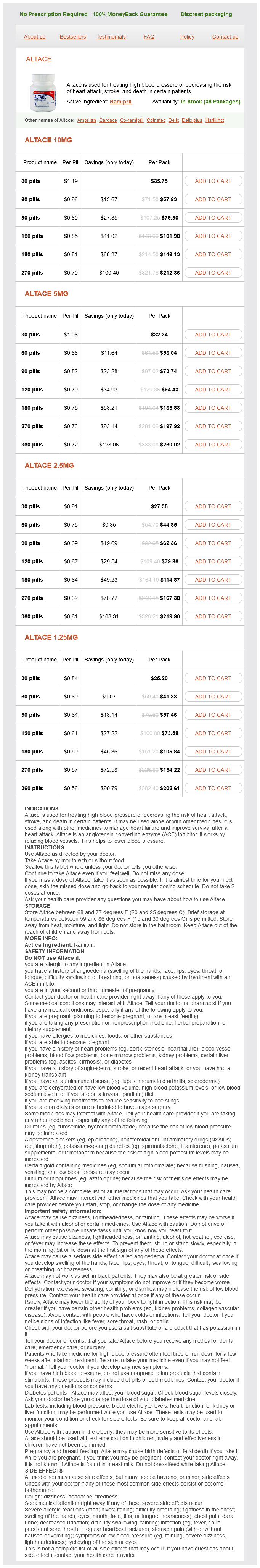
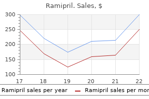
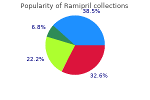
Ramipril
Discount ramipril 10 mg with amex
Several genes have also been identified that play a role in causing holoprosencephaly blood pressure control chart order ramipril. Individuals who have Down syndrome may also have heart defects, digestive problems such as gastroesophageal reflux or celiac disease, hearing loss, and cancer of blood-forming tissue (leukemia). Down syndrome also appears to be is associated with an increased risk of Alzheimer disease Down syndrome is usually caused by the presence of an extra chromosome number 21, called trisomy 21, which means each cell in the body has three copies of chromosome 21 instead of the usual two copies. Most cases of Down syndrome are not inherited, but occur as random events during the formation of egg or sperm. Many males with fragile X syndrome have characteristic physical features that become more apparent with age such as a long and narrow face, large ears, prominent jaw and forehead, unusually flexible fingers, and enlarged testicles after puberty. Women with Turner syndrome are often shorter than average and are usually unable to conceive children because they lack ovarian function. Other features of Turner syndrome can include extra skin on the neck, puffiness or swelling of the hands and feet, skeletal abnormalities, heart defects, and kidney problems. Developmental delays, learning disabilities, and behavioral problems are may also be present, although these characteristics vary among affected females. Most males who have Klinefelter syndrome have one extra copy of the X chromosome in each cell. The presence of an extra X chromosome interferes with male sexual development causing their testicles to develop abnormally, and leading to low levels of the hormone testosterone beginning during puberty. A lack of testosterone can lead to breast development, reduced facial and body hair, and an inability to father children. Boys who have Klinefelter syndrome may have learning disabilities and difficulty with speech and language development. Klinefelter syndrome is not inherited, but usually occurs as a random event during the formation of egg or sperm. Individuals who have Bloom syndrome are usually much smaller than average, and often have a high-pitched voice and characteristic facial features including a long, narrow face; small lower jaw; and prominent nose and ears. They tend to develop pigmentation changes that often appear as a butterfly-shaped patch of reddened skin on the face. Other features of the Bloom syndrome may include learning disabilities, mental retardation, chronic lung problems, diabetes, and immune deficiency that leads to recurrent pneumonia and ear infections. Men with Bloom syndrome are usually not able to father children because they do not produce sperm. Chromosome instability in Bloom syndrome also results in a high risk of cancer in affected individuals. Bloom syndrome is inherited in families in an autosomal recessive pattern, which means both copies of the gene in each cell have mutations. The signs and symptoms of Canavan disease usually begin in early infancy; however, the course of the disorder can be quite variable. Infants with Canavan disease usually appear normal for the first few months of life. By age 3 to 5 months, these infants begin to have developmental delays in motor skills, weak muscle tone, large head size, and mental retardation. They may also develop feeding and swallowing difficulties, seizures, and sleep disturbances. Canavan disease is inherited in an autosomal recessive pattern, which means both copies of the gene in each cell have mutations. Characteristic features of Fabry disease include episodes of pain, particularly in the hands and feet; clusters of small, dark red spots on the skin; a decreased ability to sweat; cloudiness of the front part of the eye; and hearing loss. Individuals with Fabry disease are also at risk for potentially life-threatening complications such as progressive kidney damage, heart attack, and stroke. Fabry disease is inherited in an X-linked pattern; however, unlike other X-linked disorders, Fabry disease causes significant medical problems in many females who have one altered copy of the mutated gene. The disorder causes disturbances in autonomic nerve cells, which control involuntary actions such as digestion, breathing, production of tears, and the regulation of blood pressure and body temperature. It also affects activities related to the senses, such as taste and the perception of pain, heat, and cold. Problems related to this disorder first appear during infancy and include poor muscle tone, feeding difficulties, poor growth, lack of tears, frequent lung infections, and difficulty maintaining body temperature. Developmental delays in walking and speech, are usually present, although some affected individuals do not show signs of developmental delay. Familial dysautinomia is inherited in an autosomal recessive pattern, which means both copies of the gene in each cell have mutations. For some affected individuals, however, the initial episode occurs much later in life. The episodes usually last 12 to 72 hours and may vary in severity and length of time between attacks. A buildup of protein deposits occurs in some cases of familial Mediterranean fever and this can lead to kidney failure if left untreated. This condition is inherited in an autosomal recessive pattern, which means both copies of the gene in each cell have the mutations. Rarely, familial Mediterranean fever may be inherited in an autosomal dominant pattern, which means one copy of an altered gene is sufficient to cause the disorder. Fanconi anemia causes the bone marrow to stop making enough new blood cells for the body to function normally. Fanconi anemia can also cause the bone marrow to make many abnormal blood cells, which can lead to serious health problems such as cancer. This condition is inherited in an autosomal recessive pattern, which means both copies of the gene in each cell have the mutations. In affected individuals the abnormal lipid metabolism causes harmful amounts of lipids to accumulate in the spleen, liver, lungs, bone marrow, and brain. Type A presents during infancy and is characterized by an enlarged liver and spleen, failure to thrive, and progressive deterioration of the nervous system. Children born with Niemann-Pick, Type A generally do not survive past early childhood. They usually attain a maximum developmental age of 15 months in language and motor function, although their receptive abilities are more advanced. The welfare of the donor remains paramount, and vigilance in donor care and management is essential to ensure that appropriate safeguards are in place to protect individuals and to inspire public confidence. These guidelines are intended to act as a resource for the transplant community, and to underpin best practice in living donor kidney transplantation. These include the ethical and medico-legal aspects of donor selection, medical and pre-operative donor evaluation, identification of high risk donors, the management of complications, and expected outcome. Scenarios that present an increased level of risk to the potential recipient, such as antibody incompatible transplantation, recurrent disease and transplantation in the context of other co-morbidities, are also included. Guidance is provided on the most appropriate investigations to be considered to assist clinical decision-making, and the best surgical approaches when faced with different clinical scenarios. Initially published in 2000 (2) and revised in 2005 (3) and 2011 (4), the guidelines have achieved international repute. This fourth edition has used the framework of previous editions but has been significantly updated in the light of new data and changing practice. In updating these guidelines, areas of interest were identified with input from clinicians and patient representatives. A systematic review of the relevant literature and synthesis of the available evidence was undertaken by selected relevant clinical experts. Draft proposals were amended by the editorial committee and the appropriate levels of evidence added to recommendations. With a handful of exceptions, conference presentations have not been included and the publication cut-off date for evidence was July 2017. All practitioners need to undertake clinical care on an individualised basis and keep up to date with changes in the practice of clinical medicine. These guidelines represent the collective opinions of a number of experts in the field and do not have the force of law. They contain information/guidance for use by practitioners as a best practice tool. The opinions presented are subject to change and should not be used in isolation to define the management for any individual patient. The guidelines are not designed to be prescriptive, nor to define a standard of care. United Kingdom Guidelines for Living Donor Kidney Transplantation, Second Edition, 2005.
Generic ramipril 10 mg with mastercard
However hypertension complications ramipril 5mg online, net marks are not always evident on dolphins known to have been entangled and there should be some caution in the interpretation of this finding (Duignan et al. Acute pulmonary lesions indicative of asphyxiation were present in both Hector?s dolphins and in the bottlenose and common dolphins known to have died as a result of capture in fishing gear. These animals also appeared to have acute subendocardial cardiomyopathy (hyper-contraction and fibre fragmentation) of the thickest part of the left ventricular wall consistent with coagulative myocytolysis or coagulative necrosis. Both lesions are morphologically similar particularly in the peracute to acute stage of lesion development. Generally cardiac lesions take hours to develop to a stage where necrosis is unequivocal. In humans with myocardial infarction, necrosis is not seen for up to twelve hours post infarction (Kumar et al. However ultrastructural changes as determined by electron microscopy can be seen after two hours. Thus too little time may elapse between the onset of a lesion in the dolphin myocardium and the death of the animal. This problem can only be addressed by conducting necropsies on fresh unfrozen dolphins as soon as possible after death by entanglement. Of the nine Hector?s dolphins that were beachcast, two (22%) have a high probability, based on observed lesions, of having died as a result of entanglement in fishing gear. One juvenile animal appears to have died suddenly from blunt trauma, but the origin of that trauma could not be determined. Two others had severe parasitic pneumonia and that may have played a role in their death. The Maui?s dolphin died as a result of severe pulmonary infection by the opportunistic terrestrial fungus, Aspergillus fumigatus. In this case the fungus invaded the pulmonary artery from the lung and caused intra thoracic haemorrhage that caused death. Aspergillosis is extremely rare in dolphins worldwide, but has been reported in striped dolphins, Stenella coeruleoalba, and bottlenose dolphins debilitated by morbillivirus infection (Domingo et al. A previous case was reported for a juvenile male Hector?s dolphin with fulminating pulmonary and cerebral aspergillosis (Duignan et al. In neither case was morbillivirus implicated based on virus isolation and immunohistochemical staining of tissues for morbillivirus antigen (Duignan et al. However, the underlying cause for immunosuppression in Hector?s and Maui?s dolphins remains unresolved. Assistance with necropsies, sorting and identifying stomach was provided on occasion by A. Knight for virology, Dr Christina Lockyer, Age 20 Duignan & Jones?Autopsy of cetaceans, 2002/03 Dynamic, Denmark, for consultation on age determination. We acknowledge the goodwill of the Department of Pathobiology and the Institute of Veterinary Animal and Biomedical Sciences in provision of facilities, storage space, use of the chiller, space for freezers. The status of Hector?s dolphin, Cephalorhynchus hectori (van Beneden), in New Zealand waters. Geographical variation in Hector?s dolphin: recognition of new subspecies of Cephalorhynchus hectori. Preliminary study of the male reproductive cycle in common dolphins, Delphinus delphis, in the eastern North Atlantic. Conservation of Hector?s dolphins: a review of studies that led to the establishment of the Bank?s Peninsula marine mammal Sanctuary. Pathologic and immunocytochemical studies of morbillivirus infection in striped dolphins (Stenella coeruleoalba). Autopsy of cetaceans incidentally caught in fishing operations 1997/98, 1999/2000, and 2000/01. Autopsy of cetaceans incidentally caught in commercial fisheries, and all beachcast specimens of Hector?s dolphins, 2001/02. Distribution and abundance of Maui?s dolphins (Cephalorhynchus hectori maui) along the North Island west cost, New Zealand. National Oceanic and Atmospheric Administration Technical Memorandum, National Marine Fisheries Service?Southwest Fisheries Science Centre 198. Mass mortality of common dolphins (Delphinus delphis) in south west England due to incidental capture in fishing gear. Morbilliviral disease in Atlantic dolphins (Tursiops truncatus) from the 1987?1988 epizootic. Age and body length characteristics of Cephalorhynchus commersoni from incidentally caught specimens off Tierra del Fuego. Assessment of reproductive status of female fin and sei whales taken off Iceland, from a histological examination of the uterine mucosa. Changes in ovaries of the short-finned pilot whale Globicephala macrorhynchus, with age and reproductive activity. Notes on Hector?s dolphins Cephalorhynchus hectori (van Beneden) from New Zealand. Quantifying abundance of Hector?s dolphins between Farewell Spit and Milford Sound. Until 1840, poisoning was often thought to be a way to ?get away with murder?, and it worked because there were no visible signs of foul play. In that year, a French woman named Marie Lafarge became the first person to be convicted of murder by poisoning thanks to new methods for arsenic testing, and the field of forensic toxicology was born. Forensic toxicology takes it a step further, including a number of related disciplines to assist in the detection and interpretation of drugs and poisons in medicolegal death investigations, human performance issues;. Establish if substances are present and whether or not they represent legitimate use or exposure, such as prescribed medications or workplace exposures Forensic toxicology can also be used to determine drugs and dosing for hospital patients, for example in therapeutic drug monitoring and emergency clinical toxicology; identify crimes where toxicants are used to poison or sedate; resolve cases of driving under the influence; and establish whether drugs have been used to improve human performance, as in sport ?doping?. For the purposes of this module, we will be primarily discussing post-mortem forensic toxicology. Principles of Post- mortem (Death Investigation) Forensic Toxicology Sixteenth century scientist Paracelsus gave us the adage ?the dose makes the poison?. Basically, he surmised that a certain amount of every substance, even water and air, can be toxic and those amounts can differ somewhat from person to person and substance to substance. Therein lies one of the most basic challenges of toxicology: is it the quantity of the toxin or the make-up of the person? Forensic toxicology applies analytical toxicology to the purposes of the law, and includes the analysis of a variety of fluids and tissue samples to determine the absence or presence of drugs and poisons. Once the analytical component is complete, the toxicologist has the equally challenging aspects of interpreting the findings. In post-mortem investigations, suspected drug overdoses are clear situations where toxicology is required to establish if an excessive intake of the drug occurred and, if so, whether this contributed to death. Conversely, toxicology can eliminate the possibility of a drug overdose if concentrations are not capable of causing death, given all other factors. This means that toxicology testing can produce a positive result even in cases where drug use is not mentioned in the police circumstances. This is not surprising given the wide availability of potentially toxic substances, both legal and illegal. In addition, concentrations of substances change after death making any interpretation difficult, no matter the concentration. In many cases, poisons may be detected by the toxicology laboratory but are not necessarily a cause of death, rather their presence may be relevant in the circumstances of death. For example, alcohol and impairing drugs are found in about half of all drivers killed in motor vehicle crashes in Australia, nearly one-third (31%) of all traffic-related deaths in the United States and in a significant proportion of other accidental deaths. Alcohol and/or drugs are also found in a significant number of other deaths reported to the coroner; for example, in suicides that include non-drug related intentional deaths. In death cases where natural disease is partially to blame, drugs that indicate an underlying disease are often detected, such as drugs that have been used to treat a condition or pain. Regardless how the individual died, toxicology testing can determine whether levels of toxic substances may have contributed to this death. How It?s Done How the evidence is collected Specimens sent for toxicology testing are usually collected by the forensic pathologist (who may also be an appointed ?medical examiner? or ?coroner? in some jurisdictions) or mortuary technician during an autopsy. Specimens must be properly identified, labelled and sealed as soon as practicable after collection. All specimens pertaining to a case must be collected and bagged separately in tamper-proof containers. Like any other evidence, the chain of custody must be preserved at all times, from the mortuary through the laboratory testing, reporting and storage, for court purposes. If the continuity of evidence is compromised, it can result in the case being dismissed in court.
Best ramipril 10mg
Thus any conditions that affects levels of thyroid binding proteins will affect the total (but not the free) T4 hormone levels prehypertension epidemiology consequences and treatment purchase 1.25 mg ramipril with visa. For example, estrogens and acute liver disease will increase thyroid binding, while androgens, steroids, chronic liver disease and severe illness can decrease it. For example, a substantial proportion of seriously ill patients will have abnormal thyroid function in the absence of true thyroid disease, due to "sick euthyroid syndrome. Total and Free Triiodothyronine (T3) the total T3 test measures the concentration of triiodothyronine in the serum. The T3 is increased in almost all cases of hyperthyroidism and usually goes up before the T4 does. The T3 is decreased during acute illness and starvation, and is affected by several medications including Inderal, steroids and amiodarone. Resin Thyroid Uptake (T-uptake) these assays have been variously referred to as T3-uptake, T4-uptake and thyroid-uptake tests, depending on the assay design. All are used in exactly the same manner and for the same purpose, not as stand-alone assays, but in combination with total T4 or total T3 assays. The resin T3/T4 uptake is used to assess the binding capacity of the serum for thyroid hormone. This is used to help determine if the total T4 is reflecting the free T4, or if abnormalities in binding capacity are responsible for changes in T4 values and thus this test is only useful in conjunction with Total T4 or Total T3. If there is an increase in binding capacity, more labeled hormone will be bound to the binding proteins and thus less will be left free in the serum. The free labeled hormone in the serum is measured and usually reported as a percent of the total labeled hormone added. Another way of putting this is that if the Total T4 or Total T3 deviates from normal in one direction and the Resin T3 uptake deviates in the opposite direction, then the abnormality is due to changes in binding capacity, otherwise it is secondary to a true change in thyroid function. For example, if the binding capacity is increased because of high estrogens, the free labeled hormone will be decreased and the Resin T3 uptake will be decreased. The thyroid peroxidase enzyme (responsible for iodinating tyrosine residues in the thyroglobulin molecule) was subsequently identified as the major microsomal component recognized by these autoantibodies. New, improved assays, designed in the wake of this insight, have been rapidly replacing the older antimicrosomal antibody assays. This assay is also used to monitor response to immunotherapy, to identify at-risk individuals (with family history of thyroid disease), and as a predictor of postpartum thyroiditis. Total hormone levels also adjust accordingly, to maintain free thyroid hormone levels within the euthyroid range. In certain situations, the knowledge that grossly abnormal thyroid hormone levels are not the consequence of a thyroid disorder may be very reassuring. Thus tests for remaining thyroid tissue are particularly important for monitoring thyroid cancer patients for residual, metastasized, and recurring thyroid tissue after the thyroid has been completely removed. Historically, the only procedure available for this purpose has been the total body scan. It is not used for initial documentation of hyperthyroidism, but as a secondary test to differentiate between "true" and "other" forms of hyperthyroidism. After this work has been published by InTech, authors have the right to republish it, in whole or part, in any publication of which they are the author, and to make other personal use of the work. Any republication, referencing or personal use of the work must explicitly identify the original source. As for readers, this license allows users to download, copy and build upon published chapters even for commercial purposes, as long as the author and publisher are properly credited, which ensures maximum dissemination and a wider impact of our publications. Notice Statements and opinions expressed in the chapters are these of the individual contributors and not necessarily those of the editors or publisher. No responsibility is accepted for the accuracy of information contained in the published chapters. The publisher assumes no responsibility for any damage or injury to persons or property arising out of the use of any materials, instructions, methods or ideas contained in the book. Publishing Process Manager Igor Babic Technical Editor Teodora Smiljanic Cover Designer InTech Design Team First published February, 2012 Printed in Croatia A free online edition of this book is available at Chamindrani Mendis-Handagama Chapter 13 Congenital Hypothyroidism due to Thyroid Dysgenesis: From Epidemiology to Molecular Mechanisms 229 Johnny Deladoey Chapter 14 Consideration of Congenital Hypothyroidism as the Possible Cause of Autism 243 Xiaobin Xu, Hirohiko Kanai, Masanori Ookubo, Satoru Suzuki, Nobumasa Kato and Miyuki Sadamatsu Preface this book provides both the basic and the most up-to-date information on the clinical aspect of hypothyroidism. This first part offers general and elaborated view on the basic diagnoses in overt and subclinical hypothyroidism, autoimmune thyroid diseases and congenital hypothyroidism. Researchers and clinicians experts provide results of their long time experience and results of their own scientific work. This information may be helpful for all of physician not only endocrine specialization. This first part contains many important specifications and innovations for endocrine practice. I would like to thank all of authors who had helped in the preparation of this book. Drahomira Springer Institute of Clinical Biochemistry and Laboratory Diagnostics, General University Hospital, Prague, Czech Republic Part 1 Introduction 1 Hypothyroidism Osama M. Ahmed2,3 1Physiology Division, Zoology Department, Faculty of Science, Beni-Suef University, 2Lab of Comparative Endocrinology, Catholic University, Leuven, 3Zoology Department, Faculty of Science, Beni-Suef University, 1,3Egypt 2Belgium 1. Introduction Hypothyroidism is caused by insufficient secretion of thyroid hormones by the thyroid gland or by the complete loss of its function. The share of hypothyroidism among other endocrine diseases is gradually increasing. The idiopathic form of hypothyroidism occurs mainly in females older than 40 years. Treatment, however, is nearly always completely successful and allows a patient to live a fully normal life (Potemkin, 1889; Thomas, 2004; Roberts and Ladenson, 2004). History Hypothyroidism was first diagnosed in the late nineteenth century when doctors observed that surgical removal of the thyroid resulted in the swelling of the hands, face, feet, and tissues around the eyes. The term myxoedema (mucous swelling; myx is the Greek word for mucin and oedema means swelling) was introduced in 1974 by Gull and in 1878 by Ord. On the autopsy of two patients, Ord discovered mucous swelling of the skin and subcutaneous fat and linked these changes with the hypofunction or atrophy of the thyroid gland. The disorder arising from surgical removal of the thyroid gland (cachexia strumipriva) was described in 1882 by Reverdin of Geneva and in 1883 by Kocher of Berne. Causes and incidence Many permanent or temporary conditions can reduce thyroid hormone secretion and cause hypothyroidism. About 95% of hypothyroidism cases occur from problems that start in the thyroid gland. Secondary and tertiary hypothyroidism is caused by disorders of the pituitary gland and hypothalamus respectively (Lania et al. Only 5% of 4 A New Look at Hypothyroidism hypothyroid cases suffer from secondary and tertiary hypothyroidism (Potemkin, 1889). Primary hypothyroidism may also occur as a result of insufficient introduction of iodine into body (endemic goiter). In iodine-replete communities, the prevalence of spontaneous hypothyroidism is between 1 % and 2 %, and it is more common in older women and ten times more common in women than in men (Vanderpump, 2005 and 2009). Primary hypothyroidism may also occur as a result of hereditary defects in the biosynthesis of thyroid hormones (due to defect in the accumulation of iodine by the thyroid gland or defect in the transformation of monoiodotyrosine and diiodotyrosines into triiodothyronine and thyroxine) or may be caused by hypoplasia and plasia of the thyroid gland as a result of its embryonic developmental defect, degenerative changes, total or subtotal thyroidectomy (Potemkin, 1889). The most prevalent cause of central hypothyroidism, including secondary and tertiary subtypes, is a defective development of the pituitary gland or hypothalamus leading to multiple pituitary hormone deficiencies, while defects of pituitary and hypothalamic peptides and their receptors only rarely have been identified as the cause of central congenital hypothyroidism (Grueters et al. Pituitary Although not every case of secondary hypothyroidism has Secondary gland a clear-cut cause, it is usually caused by damage to the pituitary gland, as by a tumor, radiation, or surgery. Secondary hypothyroidism accounts for less than 5% or 10% of hypothyroidism cases. Classification of hypothyroidism according to the origin of cause (Simon, 2006; Aminoff, 2007; Elizabeth and Agabegi, 2008). Grades of hypothyroidism Hypothyroidism ranges from very mild states in which biochemical abnormalities are present but the individual hardly notices symptoms and signs of thyroid hormone deficiency, to very severe conditions in which the danger exists to slide down into a life- threatening myxoedema coma. Hypothyroidism is thus a graded phenomenon, in which the first stage of subclinical hypothyroidism may progress via mild hypothyroidism towards overt hypothyroidism (Table 2) ( Reverdin, 1882).
Order generic ramipril line
In contrast hypertension classification purchase ramipril paypal, the minute free hormone fractions readily enter cells by specific membrane transport mechanisms to exert their biological effects. Unfortunately, the physical techniques used for separating the minute free 108 National academy of clinical biochemistry laboratory support for the diagnosis Carayon P. Routine clinical laboratories typically use a variety of free hormone tests that estimate the free hormone concentration in the presence of protein-bound hormone. Accordingly, a nomenclature of free T4 and free T3 estimate methods has been set up:. The free hormone methods used by most clinical laboratories (indexes and immunoassays) do not employ physical separation of bound from free hormone and do not measure free hormone concentrations directly! There is currently no consensus as to the best criteria to use for evaluating these free T4 estimate methods. Studies available from manufacturers vary widely in the number of compounds studied and in the concentrations tested. There is closer agreement between the reference intervals of the various ligand assays used by clinical laboratories than there is between the various methods that employ physical separation. Problems are expected and effectively occur from time to time with ?new methods? put on the market. Methods are often based on non-isotopic immunometric assay principles and are available on a variety of automated immunoassay analyzer platforms. Most of the current methods are capable of achieving a functional sensitivity of 0. However, given the high prevalence of mild (subclinical) hypothyroidism in the general population, it is likely that the current upper limit of the population reference range is skewed by the inclusion of persons with occult thyroid dysfunction. Cellular damage occurs when sensitized T- lymphocytes and/or autoantibodies bind to thyroid cell membranes causing cell lysis and inflammatory reactions. Alterations in thyroid gland function result from the action of stimulating or blocking autoantibodies on cell membrane receptors. In iodide sufficient areas, TgAb is primarily determined as an adjunct test to serum Tg measurement, because the presence of TgAb can interfere with the methods that quantitate Tg. In iodide deficient areas, serum TgAb measurements may be useful for detecting autoimmune thyroid disease in patients with a nodular goiter and for monitoring iodide therapy for endemic goiter. Laboratory tests that determine the cell-mediated aspects of the autoimmune process are not currently available. Although autoantibody tests have inherent clinical utility in a number of clinical situations, these tests should be selectively employed. The prevalence of thyroid autoantibodies is increased when patients have non-thyroid autoimmune diseases such as type 1 diabetes and pernicious anemia (254). The clinical significance of low levels of thyroid autoantibodies in euthyroid subjects is still unknown (256). These include amiodarone therapy for heart disease, interferon-alpha therapy for chronic hepatitis C and lithium therapy for psychiatric disorders (75,259-262). However, changes in autoantibody concentrations often reflect a change in disease activity. These terms correspond to the molecular entities (immunoglobulins) which react with the specified autoantigens recognized by the laboratory test. Note: Pregnant women who are euthyroid after receiving prior antithyroid drug treatment for Graves? disease have a negligible risk for fetal or neonatal hyperthyroidism. An elevated serum Tg concentration is a non-specific indicator of thyroid dysfunction. Approximately two thirds of these patients have an elevated pre-operative serum Tg level that confirms the tumor?s ability to secrete Tg, and validates the use of serum Tg measurements as a post-operative tumor marker (307). In contrast, when the pre-operative serum Tg concentration is not elevated above normal, there is no evidence that the tumor is capable of Tg secretion, and the value of an undetectable post-operative serum Tg value is less reassuring. In such patients a detectable post-operative serum Tg could represent a large amount of tumor. The bias between different Tg methods may result from differences between the Tg-free matrix used to dilute standards and patient serum, or differences in the epitope recognition by the different Tg antibodies used by individual manufacturers. Ideally, the diluent used for standards should be Tg-free/TgAb-free human serum or alternatively, a non-serum matrix that has been selected to produce a signal (radioactive counts, relative light units etc) that is identical to Tg-free/TgAb-free human serum. These method-to-method differences are greater than the goal for maximum imprecision required for monitoring individual patients and precludes the interchangeable use of different Tg methods for long- term follow-up of thyroid cancer patients. Before changing the Tg method the laboratory should consult with physician users and compare results between the old and proposed new method using specimens from both TgAb- negative and TgAb-positive patients. Undetectable serum Tg results cannot be used to indicate the absence of tumor in a TgAb-positive patient. A detectable Tg level indicates that Tg is present, but concentrations may be underestimated. Detectable serum Tg results should not be used as the sole factor for determining the presence of residual thyroid tissue or tumor. A low serum Tg concentration can be a useful parameter for confirming the diagnosis of thyrotoxicosis factitia and/or investigating the etiology of congenital hypothyroidism 14. These are autosomal dominant inherited multiglandular syndromes with age-related penetrance and variable expression. The gene responsible for these diseases is known to be located on the chromosome sub-band 10q11. In countries like the United States where genetic testing is readily available, surgery for gene carriers is based on genetic testing alone and provocative tests are rarely used. In some countries Pg has become difficult to obtain and the majority of surgeries are now performed based on genetic testing alone. Increased serum calcitonin release occurs with autoimmune thyroid diseases (Hashimoto?s thyroiditis or Graves? disease). This now allows physicians to screen for the condition before the first biological signs appear. Currently in many developed countries, genetic studies are the first line approach for this diagnosis. For accurate disease prediction however, it is necessary that positive genetic screening results be followed with an exhaustive survey of both the healthy and affected members of the family 14. It follows therefore that the measurement of iodine intake from foodstuffs or medications has clinical relevance. In the clinical laboratory, iodine measurements are used primarily for epidemiological studies or for research. To date, the major application of iodine analysis is to assess the dietary iodine intake of a given population. Iodine measurement in thyroid or breast tissue has been performed as part of research studies. An active laboratory-physician interface ensures that high quality, cost-effective assays are used in a logical sequence, to assess abnormal thyroid disease presentations and to investigate discordant thyroid test results. The National Academy of Clinical Biochemistry, Laboratory Medicine Practice Guidelines, Laboratory Support for the Diagnosis and Monitoring of Thyroid Disease;. The authors are all consultant cellular pathologists, reporting thyroid histology and/or cytology, some of whom hold or have held office with various stakeholder organisations. They have between them contributed to national guidance in this area, and to published papers, research and other professional organisations with an interest in the area of thyroid pathology. Date active January 2016 Date for review January 2021 Comments this document replaces the 1st edition of Guidance on the reporting of thyroid cytology specimens, published in 2009. In accordance with the College?s pre-publications policy, this document was on College website for consultation from 27 October to 24 November 2015. Twenty-three items of feedback were received and the document was amended accordingly. You may download, display, print and reproduce this document for your personal, non-commercial use. Apart from any use as permitted under the Copyright Act 1968 or as set out above, all other rights are reserved. Requests and inquiries concerning reproduction and rights should be addressed to the Royal College of Pathologists at the above address. This ensures that accurate diagnostic and prognostic information is available to clinicians for optimal patient care and ensures appropriate management for specific clinical circumstances. It may rarely be necessary or even desirable to depart from the guidelines in the interests of specific patients and special circumstances. The clinical risk of departing from the guidelines should be carefully considered by the reporting pathologist; just as adherence to the guidelines may not constitute defence against a claim of negligence, so a decision to deviate from them should not be deemed negligent or a failure of duty of care. The guidelines themselves constitute the tools for implementation and dissemination of good practice.
Cheap ramipril 5mg visa
Acknowledgements this work was partially supported by Grant for Scientific research project 3/2009 of the Medical Faculty prehypertension diet order ramipril pills in toronto, Trakia University, Stara Zagora from Ministry of Education, Science and Sports Bulgaria. Hashimoto?s Disease - Involvement of Cytokine Network and Role of Oxidative Stress in the Severity of Hashimoto?s Thyroiditis 115 Akita K. Hashimoto?s Disease - Involvement of Cytokine Network and Role of Oxidative Stress in the Severity of Hashimoto?s Thyroiditis 117 De Zwart L. Stress hormones, Th1/Th2 patterns, pro/anti- inflammatory cytokines and susceptibility to disease. Selenium supplementation in patients with autoimmune thyroiditis decrease thyroid peroxidase antibodies concentrations. Hashimoto?s Disease - Involvement of Cytokine Network and Role of Oxidative Stress in the Severity of Hashimoto?s Thyroiditis 119 Karanikas G. Interferon-gamma-inducing factor enhances T helper 1 cytokine production by stimulated human T cells: synergism with interleukin-12 for interferon-gamma production. Hashimoto?s Disease - Involvement of Cytokine Network and Role of Oxidative Stress in the Severity of Hashimoto?s Thyroiditis 121 Noel R. A novel costimulatory factor for gamma interferon induction found in the livers of mice causes endotoxic shock. Th1 and Th2 serum cytokine profile characterize patients with Hashimoto?s thyroiditis (Th1) and Graves? disease (Th2). Interleukin-18 enhances lipopolysaccharide-induced interferon-gamma production in human whole blood cultures. Hashimoto?s Disease - Involvement of Cytokine Network and Role of Oxidative Stress in the Severity of Hashimoto?s Thyroiditis 123 Spilianakis C. Introduction the importance of the thyroid gland for the human body is largely due to its production of hormones necessary for appropriate energy levels and an active life. They also have critical effects on energy metabolism and on the metabolism of nutrients and inorganic ions. To such an extent is every tissue impacted in one way or another by thyroid hormones that a given degree of thyroid dysfunction is highly likely to result in multiorgan failure, this often mimicking different diseases (Saranac et al. Its incidence has increased dramatically over the past few decades afflicting up to 2% of the general population. The clinical manifestations of acquired hypothyroidism in childhood differ from those in adults. The classic manifestations also occur in children, but are not so prominent (Table 1). Instead, the most important sign of acquired hypothyroidism in childhood is growth failure (Table 2). Weight tends to increase and in most instances weight for age is greater than height for age. The retardation of bone age in hypothyroidism usually equals or exceeds the retardation in linear growth (Fisher, 1999; Hall, 1989). Clinical signs of overt hypothyroidism Growth retardation Bone age retardation Muscle hypertrophy pseudohypertrophy Sexual disorders Delayed puberty Precocious puberty Table 2. Clinical signs of acquired hypothyroidism unique to childhood (Fisher, 1990) Segmental vitiligo, hypopigmented rings surrounding dark naevi ("halo naevi"), leucotrichia, premature greying of the hair, and alopecia areata are all, like typical vitiligo, associated with autoimmune disorders and are asigned as clinical markers of autoimmunity (Hall, 1989). Examples from clinical praxis of overt, late diagnosed hypothyroidism in children 3. Muscular pseudohypertrophy of solar muscles was present and this phenomenon has been referred to as the Kocher-Debre- Semelaigne syndrome. The sella turcica was enlarged and the rare radiologic signs of neglected hypothyroidism were present: osteosclerosis of the skull base and "motocyclist?s sunglasses sign" (Fig. The severe impairment of linear growth led to dwarfism, characterized by limbs that are disproportionately short compared with the trunk. Besides the growth retardation, the child appeared younger than his age because of sexual infantilism (Fig. The treatment with Na-l-thyroxine accelerated growth and resulted in normal final height and allowed for pubertal progression. This catch-up growth was adequate to compensate the preexistant growth retardation (Fig. Different Faces of Chronic Autoimmune Thyroiditis in Childhood and Adolescence 127 Fig. Bone age retardation of hypothyroid patient (a) corresponds to 8 years versus chronological age of 14 years and 7 months. Disproportionately short stature with pubertal delay and growth curve of the Case 1. At admittion she was 13 years old, inert, apathetic, bradicardic and with dry skin. Weight gain during past 2 years was reported and she was put on unrewarding diets. The most prominent laboratory findings were elevated liver enzyms (oxalacetic and piruvic transaminase), lactat dehydrogenase and creatinin phosphokinase as well as triglycerids (Table 3). Ultrasound image showed small gland with inhomogene structure and hypoechogenic zones (Fig. Both cases are illustration of thyroid hormone effects in almost all tissues in the body. Because the long-standing hypothyroidism the dose of Na-l-thyroxine was increased gradually to prevent cardiac failure. When clinical features, such as loss of body hair, raise the possibility of pituitary hypothyroidism, it is dangerous to treat the patient with thyroid hormone without checking the plasma cortisol and if necessary correcting adrenocortical deficiency (Hall, 1989). Unlike insulin and cortisol levels, which fluctuate widely in response to food ingestion and stress, thyroid hormones are typically maintained at a constant level that keeps the metabolic machinery running in a proper metabolic rate. Thyroid hormones are crucial for survival both in rodents and humans (Zimmerman-Belsing et al. After 3 to 4 years of age thyroid hormone deficiency is not associated with mental retardation, but delayed somatic and linear bone growth. Bone maturation, measured as bone age, also is delayed, diaphyseal bone growth is reduced, and epiphyseal growth and mineralisation largely cease. The effects of thyroid hormones on somatic and skeletal growth are mediated 130 A New Look at Hypothyroidism by stimulation of growth hormone and growth factors synthesis and action. Growth hormone synthesis by pituitary cells is known to be thyroid hormone dependent. Catch-up growth is defined as a linear growth rate greater than expected for age after a period of growth inhibition. Growth inhibiting conditions conserve the limited proliferative capacity of growth plate chondrocytes, thus showing the normal process of growth plate senescence. When the growth-inhibiting condition resolves, the growth plates are less senescent and therefore growth more rapidly than normal for age (Marino et al. If the hypothyroid state is prolonged prior to treatment, catch-up growth may be incomplete. The reasons for diagnostic pitfalls, beacause of clinical ambiguity are chalanging for pediatricians and endocrinologists. Tunbridge recorded in adults clinical features that included cold intolerance, a dry skin, lack of energy, puffiness around the eyes, acroparaesthesiae and weight gain, and the signs elicited were those of periorbital swelling, scaling of the skin and a slow pulse rate (minor degrees of hypothyroidism) (Hall, 1989). In children even subclinical form of hypothyroidism has impact on growth, weight regulation, bone maturation and pubertal development. While the mild clinical picture of hypothyroidism is expected in children, the appearance of opposite, hyperfunction signs in subclinicaly hypothyroid subjects, is intriguing. Transient shift from blocking to stimulating antibodies may provoke hyperthyroid signs in hypothyroid subject (Song et al. We believe that it is particularly important to draw attention to this problem in pediatric patients. Attenuation of spontaneous nocturnal growth hormone secretion in children with hypothyroidism and its correlation with plasma insulin-like growth factor I concentrations. Increased risk of autoimmune thyroid disease in hepatitis C vs hepatitis B before, during, and after discontinuing interferon therapy. Thyrotropin-releasing hormone: distribution, role and importance (article in Serbian).
Purchase ramipril 1.25 mg with mastercard
If symptoms persist for longer than 5 days or recur heart attack jarren benton lyrics discount ramipril 2.5 mg on line, administer corticosteroids, prednisone 1?2 mg/kg/day or equivalents, followed by a taper for Grade 2 diarrhea or colitis. Initiate hormone replacement therapy or medical management of hyperthyroidism as clinically indicated. Adrenal Insufficiency: Monitor patients for clinical signs and symptoms of adrenal insufficiency. For Grade 2 or higher adrenal insufficiency, initiate prednisone 1to 2 mg/kg/day or equivalents, followed by a taper and hormone replacement as clinically indicated. There was insufficient information to adequately characterize the median duration of adrenal insufficiency. Type 1 Diabetes Mellitus: Monitor patients for hyperglycemia or other signs and symptoms of diabetes. Hypophysitis: For Grade 2 or higher hypophysitis, initiate prednisone 1-2 mg/kg/day or equivalents, followed by a taper and hormone replacement therapy as clinically indicated. For suspected Grade 2 immune-mediated adverse reactions, exclude other causes and initiate corticosteroids as clinically indicated. For severe (Grades 3 or 4) adverse reactions, administer corticosteroids, prednisone 1 to 2 mg/kg/day or equivalents, followed by a taper. If uveitis occurs in combination with other immune-mediated adverse reactions, evaluate for Vogt-Koyanagi-Harada syndrome, which has been observed with other products in this class and may require treatment with systemic steroids to reduce the risk of permanent vision loss. Gastrointestinal: pancreatitis, including increases in serum amylase or lipase levels General: systemic inflammatory response syndrome, histiocytic necrotizing lymphadenitis Hematological: autoimmune hemolytic anemia, immune thrombocytopenic purpura. Neurological: Guillain-Barre syndrome, myasthenia syndrome/myasthenia gravis, demyelination, immune-related meningoencephalitis, aseptic meningitis, encephalitis, facial and abducens nerve paresis, polymyalgia rheumatica, autoimmune neuropathy, and Vogt-Koyanagi- Harada syndrome. In patients with urothelial carcinoma, the most common Grade 3 or higher infection was urinary tract infections, occurring in 6. For Grade 1 or 2 infusion-related reactions, consider using pre-medications with subsequent doses. Adverse reactions leading to interruption occurred in 35% of patients; the most common (? The most frequent serious adverse reactions (> 2%) were urinary tract infection, hematuria, acute kidney injury, intestinal obstruction, pyrexia, venous thromboembolism, urinary obstruction, pneumonia, dyspnea, abdominal pain, sepsis, and confusional state. Adverse reactions leading to interruption occurred in 27% of patients; the most common (> 1%) were liver enzyme increase, urinary tract infection, diarrhea, fatigue, confusional state, urinary obstruction, pyrexia, dyspnea, venous thromboembolism, and pneumonitis. The most frequent serious adverse reactions (>2%) were febrile neutropenia, pneumonia, diarrhea, and hemoptysis. The study2 excluded patients with active or prior autoimmune disease or with medical conditions that required systemic corticosteroids. The most frequent serious adverse reactions (>1%) were pneumonia, sepsis, dyspnea, pleural effusion, pulmonary embolism, pyrexia and respiratory tract infection. The most frequent serious adverse reactions were pneumonia (2%), urinary tract infection (1%), dyspnea (1%), and pyrexia (1%). These included pneumonia, respiratory failure, neutropenia, and death (1 patient each). The most frequent adverse reaction requiring permanent discontinuation in >2% of patients was infusion-related reactions (2. The detection of antibody formation is highly dependent on the sensitivity and specificity of the assay. Additionally, the observed incidence of antibody (including neutralizing antibody) positivity in an assay may be influenced by several factors including assay methodology, sample handling, timing of sample collection, concomitant medications, and underlying disease. For these reasons, comparison of the incidence of antibodies to atezolizumab in the studies described above with the incidence of antibodies in other studies or to other products may be misleading. Based on its mechanism of action, fetal exposure to atezolizumab may increase the risk of developing immune-mediated disorders or altering the normal immune response. As human IgG is excreted in human milk, the potential for absorption and harm to the infant is unknown. No overall differences in safety or effectiveness were observed between patients aged 65 years or older and younger patients. Atezolizumab is an Fc-engineered, humanized, non-glycosylated IgG1 kappa immunoglobulin that has a calculated molecular mass of 145 kDa. Each 20 mL vial contains 1200 mg of atezolizumab and is formulated in glacial acetic acid (16. Each 14 mL vial contains 840 mg of atezolizumab and is formulated in glacial acetic acid (11. The systemic accumulation ratio for every 2 weeks administration and every 3 weeks administration was 3. The effect of severe renal impairment or moderate or severe hepatic impairment on the pharmacokinetics of atezolizumab is unknown. Drug Interaction Studies the drug interaction potential of atezolizumab is unknown. Animal fertility studies have not been conducted with atezolizumab; however, an assessment of the male and female reproductive organs was included in a 26-week, repeat-dose toxicity study in cynomolgus monkeys. Weekly administration of atezolizumab to female monkeys at the highest dose tested caused an irregular menstrual cycle pattern and a lack of newly formed corpora lutea in the ovaries. This study excluded patients who had: a history of autoimmune disease; active or corticosteroid-dependent brain metastases; administration of a live, attenuated vaccine within 28 days prior to enrollment; or administration of systemic immunostimulatory agents within 6 weeks or systemic immunosuppressive medications within 2 weeks prior to enrollment. Tumor response assessments were conducted every 9 weeks for the first 54 weeks and every 12 weeks thereafter. Thirty-five percent of patients had non-bladder urothelial carcinoma and 66% had visceral metastases. Twenty percent of patients had disease progression following prior platinum-containing neoadjuvant or adjuvant chemotherapy. Twenty-eight percent of patients had non-bladder urothelial carcinoma and 56% had visceral metastases. Thirty-one percent of patients had disease progression following prior platinum-containing neoadjuvant or adjuvant chemotherapy. Both cisplatin-eligible and cisplatin-ineligible patients are included in the study. This study excluded patients who had: a history of autoimmune disease, active or corticosteroid-dependent brain metastases, administration of a live, attenuated vaccine within 28 days prior to enrollment, or administration of systemic immunostimulatory agents within 6 weeks or systemic immunosuppressive medications within 2 weeks prior to enrollment. Tumor response assessments were conducted every 9 weeks for the first 54 weeks and every 12 weeks thereafter. In this study, the median age was 66 years, 78% were male, 91% of patients were White. Twenty-six percent had non-bladder urothelial carcinoma and 78% of patients had visceral metastases. Nineteen percent of patients had disease progression following prior platinum-containing neoadjuvant or adjuvant chemotherapy. Forty-one percent of patients had received 2 or more prior systemic regimens in the metastatic setting. Seventy-three percent of patients received prior cisplatin, 26% had prior carboplatin, and 1% were treated with other platinum-based regimens. The demographic information is limited to the 800 patients enrolled in Arms B and C where efficacy has been demonstrated. The majority of patients were White (82%), 13% of patients were Asian, 10% were Hispanic, and 2% of patients were Black. Clinical sites in Asia (enrolling 13% of the study population) received paclitaxel at a dose of 175 mg/m while the remaining 87% received paclitaxel at a dose of 200 mg/m. Tumor assessments were conducted every 6 weeks for the first 48 weeks, then every 9 weeks thereafter. The majority of patients were white (90%), 2% of patients were Asian, 5% were Hispanic, and 4% were Black. Patients with a history of autoimmune disease, symptomatic or corticosteroid-dependent brain metastases, or requiring systemic immunosuppression within 2 weeks prior to enrollment were ineligible. Tumor assessments were conducted every 6 weeks for the first 36 weeks and every 9 weeks thereafter. The trial excluded patients with a history of autoimmune disease, administration of a live attenuated vaccine within 4 weeks prior to randomization, administration of systemic immunostimulatory agents within 4 weeks or systemic immunosuppressive medications within 2 weeks prior to randomization; or untreated or corticosteroid-dependent brain metastases. Tumor assessments were performed every 8 weeks (? 1 week) for the first 12 months after Cycle 1, day 1 and every 12 weeks (? 1 week) thereafter. The demographic and baseline disease characteristics of the study population were well balanced between the treatment arms.
Order cheap ramipril on line
Hemoglobin An intracellular erythrocyte protein that is responsible for the transport of oxygen and carbon dioxide between the lungs and body tissues blood pressure zones purchase ramipril with paypal. Hemoglobin distribution A measure of the distribution of hemoglobin width within an erythrocyte population. It is derived from the hemoglobin histogram generated by the Bayer/Technicon instruments. Hemoglobin Method of identifying hemoglobins based on electrophoresis differences in their electrical charges. Hemoglobinopathy Disease that results from an inherited abnormality of the structure or synthesis of the globin portion of the hemoglobin molecule. In hemolytic anemia this term refers to the premature destruction of erythrocytes. Hemolytic anemia A disorder characterized by a decreased erythrocyte concentration due to premature destruction of the erythrocyte. Hemolytic transfusion Interaction of foreign (nonself) erythrocyte reaction antigens and plasma antibodies due to the transfusion of blood. There are two types of transfusion reactions: immediate (within 24 hours) or delayed (occurring 2 to 14 days after transfusion). Hemosiderin A water insoluble, heterogeneous iron?protein complex found primarily in the cytoplasm of cells (normoblasts and histocytes in the bone marrow, liver, and spleen); the major long-term storage form of iron. Readily visible microscopically in unstained tissue specimens as irregular aggregates of golden yellow to brown granules. Hemosiderinuria Presence of iron (hemosiderin) in the urine; result of intravascular hemolysis and disintegration of renal tubular cells. Hemostasis the localized, controlled process that results in arrest of bleeding after an injury. Heparin A polysaccharide that inhibits coagulation of blood by preventing thrombin from cleaving fibrinogen to form fibrin. Commercially available in the form of a sodium salt for therapeutic use as an anticoagulant. The abnormal shape is due to a horizontal interaction defect with abnormal spectrin, deficiency or defect in band 4. Hereditary spherocytosisA chronic hemolytic anemia caused by an inherited erythrocyte membrane disorder. The vertical interaction defect is most commonly due to a combined spectrin and ankyrin deficiency. Secondary to membrane loss, the cells become spherocytes and are prematurely destroyed in the spleen. Hereditary A rare hemolytic anemia inherited in an stomatocytosis autosomal dominant fashion. The erythrocyte becomes dehydrated and appears as either target or spiculated cells. Hexose-monophosphate A metabolic pathway that converts glucose-6- shunt phosphate to pentose phosphate. Histogram A graphical representation of the number of cells within a defined parameter such as size. Hodgkin lymphoma Malignancy that most often arises in lymph (disease) nodes and is characterized by the presence of Reed-Sternberg cells and variants with a background of varying numbers of benign lymphocytes, plasma cells, histiocytes, and eosinophils. Homologous Consists of two morphologically identical chromosomes that have identical gene loci, but may have different gene alleles as one member of a homologous pair is of maternal origin and the other is of paternal origin. On Romanowsky stained blood smears, it appears as a dark purple spherical granule usually near the periphery of the cell. Hydrops fetalis A genetically determined hemolytic disease (thalassemia) resulting in production of an abnormal hemoglobin (hemoglobin Bart?s,? Hypercoagulable state A condition associated with an imbalance between clot promoting and clot inhibiting factors. Hypereosinophilic A term used to describe a persistent blood syndrome eosinophilia over 1. This can be brought about by an increase in the number of cells replicating, by an increase in the rate of replication, or by prolonged survival of cells. The stimulus for the proliferation may be acute injury, chronic irritation, or prolonged, increased hormonal stimulation; in hematology, a hyperplastic bone marrow is one in which the proportion of hematopoietic cells to fat cells is increased. Hypocellularity Decreased cellularity of hematopoietic precursors in the bone marrow. Hypochromic A lack of color; used to describe erythrocytes with an enlarged area of pallor due to a decrease in the cell?s hemoglobin content. Hypofibrinogenemia A condition in which there is an abnormally low fibrinogen level in the peripheral blood. It may be caused by a mutation in the gene controlling the production of fibrinogen or by an acquired condition in which fibrinogen is pathologically converted to fibrin. Hypogammaglobulinemi A condition associated with a decrease in a resistance to infection as a result of decreased? Hypoplasia A condition of underdeveloped tissue or organ usually caused by a decrease in the number of cells. A hypoplastic bone marrow is one in which the proportion of hematopoietic cells to fat cells is decreased. Idiopathic Pertains to disorders or diseases in which the pathogenesis is unknown. The irf may be helpful in evaluating bone marrow erythropoietic response to anemia, monitoring anemia, and evaluating response to therapy. Immune hemolytic An anemia that is caused by premature, immune anemia mediated, destruction of erythrocytes. Diagnosis is confirmed by the demonstration of immunoglobulin (antibodies) and/or complement on the erythrocytes. Immune response Body?s defense mechanism, which includes producing antibodies to foreign antigens. Immunoblast A T or B lymphocyte that is mitotically active as a result of stimulation by an antigen. The cell is morphologically characterized by a large nucleus with prominent nucleoli, a fine chromatin pattern, and abundant, deeply basophilic cytoplasm. Immunoglobulin Molecule produced by B lymphocytes and plasma cells that reacts with antigen. Consists of two pairs of polypeptide chains: two heavy and two light chains linked together by disulfide bonds. Immunohistochemical Application of stains using immunologic stains principles and techniques to study cells and tissues; usually a labeled antibody is used to detect antigens (markers) on a cell. Ineffective erythropoiesisPremature death of erythrocytes in the bone marrow preventing release into circulation. Infectious lymphocytosisAn infectious, contagious disease of young children that may occur in epidemic form. The most striking hematologic finding is a leukocytosis of 40?50 X 109/L with 60?97% small, normal-appearing lymphocytes. The leukocyte count is usually increased, which is related to an absolute lymphocytosis. Serologic tests to detect the presence of heterophil antibodies are helpful in differentiating this disease from more serious diseases. Internal quality control Program designed to verify the validity of program laboratory test results that is followed as part of the daily laboratory operations. The term intrinsic is used because all intrinsic factors are contained within the blood. Intrinsic factor A glycoprotein secreted by the parietal cells of the stomach that is necessary for binding and absorption of dietary vitamin B12. Ischemia Deficiency of blood supply to a tissue, caused by constriction of the vessel or blockage of the blood flow through the vessel. Jaundice Yellowing of the skin, mucous membranes, and the whites of the eye caused by accumulation of bilirubin. Karyorrhexis Disintegration of the nucleus resulting in the irregular distribution of chromatin fragments within the cytoplasm. Killer cell Population of cytolytic lymphocytes identified by monoclonal antibodies. Involved in several activities such as resistance to viral infections, regulation of hematopoiesis, and activities against tumor cells.
Ramipril 2.5mg fast delivery
This is particularly the individual: the assessment and determination important with partially decomposed remains arteria zygomatica purchase generic ramipril from india, of age, sex, ancestry and stature. Age: this is estimated within a range, and is (c) Cleaning/sampling: If their condition not exact. The older the individual, the wider the allows, all the bones and teeth should be range. From the foetal stage to approximately washed with simple running water and 25 years of age, the human skeleton undergoes no other product. Several to capture any material that might be indicators are evaluated, including dentition dislodged by the water. In a case of severely development, length of long bones and the decomposed skeletal remains, however, appearance and fusion of epiphyses in early washing can be detrimental. A brush with soft ages; and, in later stages of development, the bristles, such as a toothbrush, may be used pubic symphysis and the morphology of the sternal to remove dirt, special care being taken with end of the fourth rib. Once development stops, worn bones, such as the epiphyses of the long degenerative changes begin to appear, especially bones and the faces of the pubic symphysis. Sex: the sexual dimorphism in the skeleton separately, to prevent their loss is seen after puberty, so before that period In cases where the remains are not completely determination of sex is not very reliable. In older reduced to bone, and soft tissue is still individuals, there are two main ways to determine attached, a non-chemical method should be the sex: used for cleaning, under strict control. Such a process shall only be undertaken once (a) Morphological traits in specifc areas of the the forensic pathologist has evaluated and pelvis and skull, and properly documented the remains and the (b) Metric assessments, which involve necessary samples have been collected. Once measurements of various dimensions of limb the remains have been washed, they should bones and articular surfaces be allowed to dry, preferably away from light and without exposing the bones to the sun. A In cases where the remains are fragmented or fan can be used to speed up the process no bones diagnostic of sex are available, a genetic analysis (amelogenin) could also be (d) Sampling: the main reason for taking applied to determine the sex. Determining the samples when analysing skeletal remains sex (amelogenin) is undertaken as standard in is to perform a genetic analysis that could the genetic analysis of bones. It is assessed by evaluating the skeleton and the number of individuals specifc traits in the skeleton, mainly in the skull, represented, the anthropologist has to decide that can be present or absent, or present to some how many samples to take. At the same time, several measurements more complicated in commingled cases, and can be taken in the skull and post-cranial skeleton. Usually, and produce an assessment of the ancestry of the two or three healthy teeth and a suffcient skeleton under analysis, when appropriate. Forensic taphonomy is a feld that studies estimated following one of two methods: the various changes to the human body after death. On some occasions this may enable (a) Measuring the height/length of some specifc post-mortem changes observed in the skeleton to bones (skull, spine, femur, tibia and talus), be understood (e. In (b) Measuring one complete long bone (such as that sense, archaeological dating methods, using the femur, tibia or humerus) or the combination objects associated with the remains, such as coins of two such bones (femur and tibia ideally), or cartridge case, may provide a better general and applying a regression formula to the result estimation. In all cases, information on must meet standards accepted by the scientifc sex and ancestry are required in order to select community. Remaining analysis and report used, they must be endorsed by publication in reputable peer-reviewed publications. After a biological profle has been established, the analysis continues with the following steps: 291. All analysis must be properly documented, with photos, drawings, notes and specifc forms. A (a) Analysis of any indicator of ante-mortem precise record of the samples taken from the trauma, pathological conditions or remains must be kept, the samples must be skeletal variations (which may or may not be correctly labelled and records of security and the symptomatic) that can provide information chain of custody kept. If the remains are to be about the cause and circumstances of the buried before formal identifcation, the process death or specifc information leading to must be properly documented. This includes identifcation recording the exact location of the disposal of the (b) Analysis of possible post-mortem remains, proper labelling of the container holding changes in the bones, due to taphonomic the remains, and appropriate notation on the processes (see Paragraph 289) that could chain-of-custody form. The fnal forensic anthropology report must include relating to peri-mortem trauma all information relating to the reception of the remains; procedures followed for the analysis; (c) Dental analysis, to contribute to age estimation samples taken and to whom they were given; and possibly even identifcation. Establishing the period of time since death report must be integrated with those produced by is diffcult, especially in cases of skeletal remains. For cases of forensic interest, covering periods from a few days to up to 30 or 40 years, there is no scientifc method, relying on an analysis of bones or teeth, to establish whether a person died one, fve, or ten years ago. Circumstantial information and other material with the skeletal remains, or in some instances even satellite imagery, may help to establish when the events (or burial) took place. Glossary Abrasion Superfcial injury involving the skin; often called a scratch, scrape or graze. Accountable Subject to a system that is designed to ensure the proper discharge of responsibility by a person or institution. Ancestry In relation to forensic anthropology, the biological heritage of the remains. Ante-mortem data Data about a named individual while alive that can be used to compare with post-mortem data collected from the body, usually for the purpose of identifying the body. In relation to a dead body, a change (for example resulting from resuscitation or post-mortem damage) imitating pathology, disease or injury occurring in life. Autopsy In this document, the examination of a dead body involving its external and internal examination and incorporating the results of special tests (including radiology). The internal examination involves, but is not limited to , examining the contents of the cranium, chest and abdomen. Biological profle A term used in forensic anthropology to refer to the evaluation of skeletonized human remains to make conclusions about the age, sex, ancestry and stature of those remains, to aid in their identifcation. Bruise An injury characterized by extravasation of blood into the surrounding tissues. Cause of death the underlying cause (the disease, condition or circumstance initiating the chain of events resulting in death), possibly proceeding through more immediate (or proximate) causes, concludes the logically linked statements that constitute the cause of death. Thus, the cause of death of a young man who was shot in the chest resulting in massive haemorrhage as the bullet passed through the heart and the lungs should be recorded as: I(a) Haemorrhage (due to) I(b) Perforation of the heart and lungs (due to) I(c) Gunshot wound to the chest. Chain of custody A process enabling the complete history of the custody of an exhibit to be tracked and (of an exhibit) recreated; that is, who has had care and control of the exhibit from the time it was frst secured to the present. Commingled remains Usually in relation to skeletal remains, the mixing of the remains of two or more individuals, for example in a mass grave. Contamination the location on a person or object of material, whether obvious or not, from another source. Such contamination can be useful in forensically linking a suspect to a crime scene; or it can be confusing and damaging to justice (e. It is ?unnatural? when its causes are external, such as intentional injury (homicide, suicide), negligence, or unintentional injury (death by accident). In the early hours and days after death, (post mortem) some of the changes can be mistaken for injuries (e. Defence injuries/ Injuries/wounds sustained by a victim resulting from attempts at self-defence during an wounds assault. Epiphysis(es) the end(s) of particularly, but not only, long bones; the process of the fusion of the epiphysis(es) with the shaft of the long bone allows conclusions to be drawn about the age of the person. Professional ethics concentrates on the behaviour and attitudes of members of a particular profession. Exhibits Physical evidence thought to be relevant to the investigation of a crime or death that are labelled, recorded as exhibits and kept securely so that they cannot be interfered with or contaminated. Fingerprints (latent) Fingerprints present on a surface requiring a technical process to make them visible. Forensic the examination of human skeletal material to answer medico-legal questions including anthropology those of identifcation. Forensic the use of the skills employed in the study of ancient remains and objects for the purposes archaeology of the law, usually in relation to the excavation, recovery and evaluation of scenes and sites. Forensic ballistics/ these two categories of forensic science are often used interchangeably; in this document, Firearms and they refer to the examinations leading to conclusions of forensic value about gunshot toolmarks wounds and the projectiles recovered from them. Forensic doctor For the purposes of this document, a certifed medical doctor who is authorized to perform forensic post-mortem examinations. Forensic entomology the study of insects in a forensic setting, most often in forensic pathology as an indicator of the minimum time since death.