
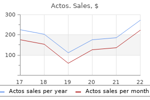
Actos
Buy actos 45 mg line
Less month scores when compared to the frst trimester scores common problems included epigastric burning early symptoms diabetes in dogs buy generic actos on-line, (p = 0. We found the rate in which women focused itching and vaginal secretion (Table 1a, b). There was a statistically signifcant change in the the postpartum fourth month quality of life scores direction of 3. The rate of sexual There was a signifcant change in the direction of function also decreased in the third trimester. Furthermore, the sexual function scores of the women increased and changed in a positive direction. In a study of 518 women from Turkey between the We observed a signifcant change in the direction ages of 18 55 years by Oksuz and Malhan in 2006[14], of 6. If it includes staining, the Body image and sexual function are integral parts instance of fecal incontinence increases to 5%. Additionally, in the study, the fecal literature, this study determined a weak, negative incontinence rate among the 61 cases was determined relationship between body image during intercourse, as 1. The study results showed that frequent urinary In the 99-case prospective study that Eason et al incontinence was most seen in the third trimester of conducted in 2004[18], urinary incontinence rates were pregnancy. In studies of the frst year after reason were most frequent in the fourth postpartum delivery, urinary incontinence was found to be at a rate month, compared to the frst and third trimester. In our study, the urinary incontinence As a result, pregnancy and delivery negatively rate was 10. This study found during intercourse, urinary incontinence and urinary incontinence occurs in one of every four encopresis. We would like to thank all the participants and staf It was determined that problems during intercourse in the Gynaecological diseases and Delivery Clinics occurred more in the pregnancy period compared to of an Education and Research Hospital of Turkey the pre-pregnancy and postpartum period (fi2 = 53. Project Number: incontinence in the fourth month of the postpartum 6242 period, as well as the third trimester, where there was a negative impact on quality of life. Confict of interest: the authors declare that they have Fecal incontinence has been observed in 1. Biopsychosocial factors psychosocial well-being in pregnancy: A comparison associated with dyspareunia in a community sample of of exercisers and non-exercisers. Efects behavior of mothers in the early postpartum period in of carrying a pregnancy and of method of delivery on Turkey. Urinary incontinence in incontinence during pregnancy and the postpartum pregnancy. Int Urogynecol J Pelvic Floor Dysfunct 2009; Urogynecological Association, Abstract Book 2002; p. Obstet Gynecol 2007; Urinary incontinence on women aged over 18 and its 109:922-928. Forty eight (98%) of the smoking Subjects: the study included male students randomly students wanted to quit smoking. The reason they were selected from 12 secondary schools from four administrative ready to quit smoking was to save their health (64. Results: the study showed that the overall smoking Conclusion: To reduce the prevalence of smoking, intensive prevalence was 12. Over 90% of these deaths important cause of preventable morbidity and are caused directly by tobacco use, whilst about premature death[1]. The diseases which frequently 10% are the result of non-smokers being exposed to occur among smokers include cardiovascular diseases, secondhand smoke[5]. If current trends continue, it is respiratory diseases and cancer[2], such as coronary projected that by 2030, tobacco will be responsible for heart disease, lung cancer and tuberculosis[3]. Globally, more than 8 million deaths each year and 80% of these it is estimated that there are about 1 billion smokers premature deaths will be among people living in low and in 2014, smokers consumed 5. Address correspondence to: Yousif Mohammed Almosaad, PhD, Faculty of Public Health and Health Informatics, Qassim University, Albukayriyah, Saudi Arabia. The study the prevalence of smoking among young people: 7% sample was calculated using the following formula in Oman, 18% in Kuwait, 23% in Iraq, 25% in Saudi and was found to be 379 students[16]. Sample selection In Sudan, it has been estimated that the prevalence of Students were chosen proportionately from each cigarette smoking in adults is 24% in 2009, according school in the study area. However, the was used to select students from the school list of prevalence rates of smoking among young people is students made by the researcher prior to distribution 13. All students in identified smoking as a behavior learnt and initiated these schools were invited to take part in the study. The earlier the individual consent of students was implied by completing and initiated smoking, the higher the likelihood of being returning the questionnaire. In addition, adolescents who smoked were more likely to be Data collection involved in other high-risk behaviors[12]. The research instrument used for this study was Previous studies of smoking among young people a pre-tested, structured questionnaire. Intrapersonal factors constructed to estimate cigarette smoking prevalence, associated with adolescent smoking include lower smoker behavior and types of smoking products used. In addition, the perceived high smoking prevalence among peers, as data for the study was collected by twelve interviewers well as perceived positive reactions of parents and who were trained to conduct face-to-face interviews. In contrast, having best friends and family members who smoked, perceived Statistical analysis less cordial family relationship, unsatisfactory In order to meet the study objectives, the data academic achievements, perceived lower socioobtained from the research instrument was analyzed. The purpose of Socio-demographic characteristics of students this study was to assess the prevalence of smoking and A total of 379 students completed the questionnaire. Relating to the class level, 150 descriptive, cross sectional study of 3,87,377 students (39. A similar attitude was reported among study In relation to the source of cigarettes, 25 (51%) of the subjects regarding male smokers. The most important reason place of smoking, the majority of the smokers reported that made them ready to quit smoking was to save that they smoked in public places (63. 3: Reasons why students are ready to quit smoking 28 Smoking Behavior among Male Students in Secondary Schools in Khartoum LocalitySudan March 2018 Three hundred and sixty-three (95. The prevalence of smoking was higher respondents supported the rule that prevented among elder students (11. Majority of the the prevalence of smoking was higher among respondents reported that the factors that lead others students in third-year secondary schools (6. The respondents suggested a list of strategies Moreover, students who had a smoking person in for smoking control. These suggestions included his family were significantly more likely to become implementing health education programmes (58. However, significant associations were observed the smokers reported that smoking has different between students family size, monthly income types of benefits. These benefits include becoming and students per diem with the smoker status (p < more social, innovative and happy 4). This is enough evidence Smoking among young people is associated with to encourage decision makers to increase efforts to a range of problems. About 13% of the students in this study smoking was due to influence of friends and family reported that they were smokers. These results are consistent with other studies that have been carried out around the world, such which have found that young people take up smoking as studies from United Kingdom and Kenya, where largely due to peer pressure. In 2011, Amin et al in Alprevalence of smoking among secondary students Hassa found that smoking of close relatives and friends were 12% and 12. A an important role in the increased risk of smoking study conducted in Malaysia, for example, found that among male secondary school students.
Order 30mg actos overnight delivery
It has a complicated gene activity controlling system which is related with cells and tissue differentiation in ontogenesis diabetes medications in pregnancy purchase 45 mg actos fast delivery. So the human genome contains 3 billons nucleotide pairs, which is enough to make more than 2 millions structural genes. To explain physiological value of genetic texts, that means to explain the relationships between genetic sequences and physiological and hereditary traits. To defend human genome from mutation preventing rise of genetic load (ecology aspect of program). The chromosome theory of inheritance state that main part of heredity information is in a nucleus, but it is also possible that part of information is in cytoplasmic organelles such as mitochondrion and chloroplast. Plants and animals ova have cytoplasm, which is rich in cell organelles, but sperm has very few organelles. So that means that traits which are encoded in cytoplasmic genes are inherited in mother line of pedigree. In yeast mitochondria it was detected the genes responsible for respiratory enzymes synthesis. In bacterial cells there are three types of plasmids: containing sex factor F, factor R and factor col colicinogenic. The plasmid with R factor may be transmitted to other cell during conjugation and play an important role in changing bacteria hereditary properties. Colicinogenic plasmids contain special genes which encode special proteins colicins. The material for cytoplasmic inheritance is genes of plasmids, mitochondria and still unknown factors. The system of cellular genetic apparatus includes nucleus genome and cyto100 plasmic genome. This apparatus is presented by chromosomes and their genes in nucleus and by plasmogenes of organelles in the cytoplasm. Accordingly, it may be distinguished two variants of heredity autosomal, and linked with Xchromosome or Y-chromosome. And also due to character of gene expression it can be distinguished dominant and recessive hereditary. The heredity linked with sexual chromosomes and linked heredity at all was described in a chapter 7. As it was said above, monogenic heredity is heredity of traits controlled by one gene. The cross in which parents are analyzed by one alternative traits pair is called monohybrid cross, by two pairs dihybrid cross and by many pairs polyhybrid cross. Having analyzed F2 generation hybrids Mendel formulated Second Law of Heredity or Law of Segregation: in crossing two heterozygous individuals analyzed by one alternative traits pair, we can predict phenotypic ratio 3:1 and genotypic ratio 1:2:1 (pic8. The outcome of such cross can be illustrated by a Punnett square, suggested by English geneticist Reginald Crundall Punnett. It can be formulated by this way: genes in gametes of hybrids are discrete (pure) and not blended. The cause why traits are not blended is that genes for these traits are in different homologues chromosomes. To analyze a genotype of individual with dominant phenotype we can use a testcross. It is because of individual with dominant phenotype may be either homozygous or heterozygous. To perform dihybrid cross Mendel took homozygous organisms having two pairs of alternative traits. It states that genes located on different chromosomes assort independently o fone another. To make cross scheme easy to write we may use so called phenotypic radical it is dominant genes oforganism, which determines it phenotype. The same probability of all kinds of gamete formation by all hybrids while monohybrids cross. The same probability of all kinds of gamete formation on a basis of independent assortment of non homologous chromosomes in meiosis while dihybrid and polyhibryd cross. That means splitting occurs in haploid gametes, on a level of genes and chromosomes, but it is analyzed in diploid organisms on a level of traits. During such period many environmental factors may act on gametes and developing organisms. Therefore, to analyze it, we need to use several statistic methods which allow to prove inherited theoretical ratio principle or to deny it. Analyzing patterns of heredity in garden pea Mendel worked with several traits pairs. Among them are achondroplasia, diabetes insipidus, albinism, pancreas fibrosis, syndactilia, glaucoma, hemophilia, and so on. Thus, if in 1958 it was known only 412 hereditary human traits, in 1978-2511, in 1981-3217. The level of trait expression depends on number of dominant polymeric genes that means on gene dose. If all genes in genotype are dominant, skin pigmentation is maximal like in native Africans (P1P1P2P2P3P3P4P4).
Discount actos 30 mg with amex
Adenocarcinomas these are the most common malignant cellular component in the effusions metabolic disease hyperparathyroidism cheap 30 mg actos otc. They mostly represent metastasis from primary adenocarcinomas such as from the stomach, lung, breast, colon, and ovary. Squamous cell carcinoma Effusion may rarely have malignant squamous cells in it and represent metastasis from carcinoma lung, oesophagus or uterine cervix. Lymphomas-leukaemias Effusions may sometimes have malignant cells of leukaemia and lymphoma in line with primary disease in the body. Samples are obtained by masturbation or coitus interruptus after observing at least 4 days of sexual abstinence and assessed on the following lines: 1. Viscosity and pH When ejaculated, semen is fairly viscid but liquefes in about 10 to 30 minutes. Motility Normally, within 2 hours of ejaculation, at least 60% of the spermatozoa are vigorously motile; in 6 to 8 hours 25 to 40% are still motile. Fructose Seminal fructose estimation (normal levels 150-600 mg/dl) complements cytological analysis. Low levels of seminal fructose indicate obstruction at the level of ejaculatory ducts. A minimum of at least three specimens collected on three successive days should be examined. The cytology specimen is collected during fbreoptic endoscopy of the part being visualised. The initial morning specimen is discarded as cells deteriorate extremely quickly in acidic urine, and the morphology of cells accumulating overnight in bladder urine is distorted to an extreme degree. As these fuids are often exudative in character, they may clot after removal from respective cavities. Methods of fxation vary depending upon the type of staining employed: fi Material for exfoliative cytodiagnosis is usually wet-fxed i. These smears are then stained with Papanicolaou (Pap) or haematoxylin and eosin (H & E) stains. Where ethanol is not available, 100% methanol, 95% denatured alcohol, or 85% isopropyl alcohol (isopropanolol) may be used. Smears prepared at the bedside as well as those pre pared in the laboratory from fuid samples are immediately placed in 95% ethanol without allowing them to dry prior to fxation. The best preservative for general use is 50% ethanol in volumes equal to that of the fuid sample. Strands of ropy mucus are also selected (exfoliated cells adhere to mucus strands). Commonly employed techniques for processing of fuids are as under: 630 Sediment smears the sample is poured into 50 ml centrifuge tubes and centrifuged at 600 g for 10 minutes. Following centrifugation, the supernatant is decanted and smears prepared from the sediment or cell button by recovering the material with a glass pipette or a platinum wire loop. Smear preparation from samples collected in a preservative require albuminised slides as cell adhesiveness is reduced by prefxation. Cytocentrifuge and membrane flter preparations these methods are most useful for small volume fuids of low cell content. The interested reader is referred to specialised texts for descriptions of these methods. Nuclear stain gives basophilic colour to the nucleus while the two cytoplasmic stains impart the orange and cyanophilic tints to cytoplasm respectively. This branch includes fne needle aspiration cytology, imprint cytology, crush smear cytology and biopsy sediment cytology. The technique has gained wide acceptance in the last four decades and is increasingly being used to sample a wide variety of body tissues. Palpable lesions commonly sampled are: breast masses, enlarged lymph nodes, enlarged thyroid and super fcial soft tissue masses. Syringe holders such as the Franzen handle permit a single-hand grip during aspiration, employing disposable syringes. On reaching the lesion, the plunger of the syringe is retracted and at least 10 ml of suction applied while moving the needle back and forth within the lesion; the direction or angle of the needle may be changed to access different areas of the lesion. Poorly-prepared smears with distorted cellular morphology will frustrate the best efforts of the most competent cytopathologist, and often result in errors of interpretation or in failure to arrive at any specifc diag nosis. Half the number of smears are immediately immersed in 95% ethanol, transported to the laboratory in the fxa tive (wet-fxed), and used for Papanicolaou or H&E staining. The remaining smears are air-dried, wrapped in tissue paper for transport to the laboratory, and stained by Romanowsky stains. Plain X-ray flms are usually adequate for lesions within bones and for some lesions within the chest. In patients with coagulopathies such as haemophilia, aspiration of joint spaces, chest and abdominal viscera is contraindicated. Liver Estimation of prothrombin time is an essential pre-requisite for aspiration of the liver. Prostate Transrectal aspiration in acute prostatitis may cause bacteraemia/septicaemia. Testis Aspiration is extremely painful in acute epididymo-orchitis and should be deferred till such time the acute infammatory process subsides. The reliability of the test, thus, depends upon the adequacy of the sample and its representative character. These smears are preferred by many workers as they allow recognition of tissue architecture to some degree, in addition to better cytological details. The method may be useful in the rapid diagnosis of bone tumours as histological sections are usually obtained after many days on account of the delay necessitated by decalcifcation. For cytomorphological recognition of cancer, the following characteristics are used to determine the presence or absence of cancer except: A. For pleural effusion, if a delay of more than 12 hours is anticipated for processing, it should be fxed in: A. The following features characterise wet-fxed smears over airdried smears except: A. These clinical cases (case 1 to 30) given at the end of most chapters are structured clinical exercises to stimulate the students of pathology to apply their knowledge and skills gained from the study of particular disease/s covered in that chapter in pathology to clinical settings as encountered in real life. He has been smoking bidis for 25 years, and gives history of having productive cough with foul smelling expectoration for 15 years, interspersed with haemoptysis off and on. His pulse rate is 90 per minute, respiratory rate 45 per minute, and blood pressure 130/90 mmHg. Her blood pressure and pulse are not recordable; while respiration rate is 40/min. He gives history of progressive fatigue and weight loss of about 20 kg during the last 6 months. She gives history of two abortions in the last 6 months and heavy and irregular menstrual bleeding for the same duration. The mother herself is also mildly anaemic which, according to her, has remained like this ever since she remembers and it did not improve with any medication. The facial appearance of the child shows prominence of forehead and cheeks and slightly protruding upper teeth. On palpation of the abdomen, the spleen is enlarged by two fngers below the costal margin and the liver is just palpable. On abdominal examination, the spleen is just palpable but the liver is not enlarged. On examination, the child is found to have mild hepatosplenomegaly but no lymphadenopathy. He gives history of intermittent pain over the sternum earlier too almost once a week which would go away when he sits down. Joint pain is feeting type involving multiple major joints, associated with redness and swelling.
Buy actos 45 mg cheap
The early typical tumor is a slightly elevated the most common intraoral locations are the papule or nodule with a translucent border and palate and upper lip diabetes symptoms uk type 2 generic actos 15 mg free shipping, and less commonly the bucsmooth, hyperkeratotic, or crusted surface. At a later a painless rubbery mass that grows slowly, is stage, tumors may appear as a large nodule with slightly mobile, and seldom may be ulcerated. Malignant Neoplasms Mucoepidermoid Carcinoma Adenoid Cystic Carcinoma Mucoepidermoid carcinoma or tumor is a maligAdenoid cystic carcinoma, or cylindroma, is a nant tumor of the salivary glands. It represents malignant neoplasm of the salivary glands with a about 2 to 3% of the tumors of major salivary characteristic histopathologic pattern. It repreglands and 6 to 9% of the minor salivary gland sents about 2 to 6% of all parotid gland tumors, tumors. The biologic behavior of the neoplasm but 15% of all submandibular gland tumors, and varies from moderate to high-grade malignancy. It equally the tumor affects almost equally men and affects men and women and is usually seen in women, most often between 30 and 50 years of patients more than 50 years of age. Clinically, an intraoral tumor appears as a Adenoid cystic carcinoma is the most common painless proliferating rubbery swelling that often malignant tumor of minor salivary glands. A common clinical finding is most frequently located on the palate, followed by the development of cysts within the tumor with the buccal mucosa, lips, and tongue. About 60% of all appears as a slightly painful, enlarging mass that intraoral tumors are found in the palate, tongue, may later ulcerate. The tumor is prone to infiltrate the perineural spaces and usually has a adenoma, mucocele, necrotizing sialometaplasia, poor prognosis. Malignant Neoplasms Malignant Pleomorphic Adenoma Clear Cell Adenocarcinoma Malignant pleomorphic adenoma or carcinoma in Clear cell adenocarcinoma is a very rare variant of pleomorphic adenoma is a rare tumor of the saliadenocarcinoma. It is slightly more frequent in vary glands with a istologic pattern showing areas women than men, particularly after 50 years of characteristic of pleomorphic adenoma mixed age. The tumor is usually located in the parotid with areas shoving evidence of malignancy. The palate is the should include other malignant neoplasms of most commonly affected site, followed by the minor salivary glands, squamous cell carcinoma, buccal mucosa, lips, and tongue. Adenocarcinoma Adenocarcinoma is a malignant salivary gland tumor with a potential for high-grade malignant behavior, which cannot be placed in any other group of carcinomas. The palate is the site usually involved, followed by the buccal mucosa, lips, tongue, and other areas. Clinically, it appears as a firm swelling that enlarges and is usually associated with ulceration and pain. The differential diagnosis includes other malignant salivary gland tumors and squamous cell carcinoma. It probably originates from primiminor salivary glands, or terminal duct carcinoma, is a form of adenocarcinoma, locally persistent, tive mesenchymal cells, such as endothelial cells. The mean age at onset is 50 years and Jewish patients and those of Mediterranean dewomen are affected more frequently than men. In scent, and it involves primarily the skin and occasionally the oral mucosa and has an indolent the great majority of cases the lesion occurs on the palate (frequently at the junction of soft and hard course. Clinically, it and it involves primarily the skin and lymph appears as a painless, firm swelling or an elevated nodes, but rarely the oral mucosa and usually has nodule that is rarely ulcerated. The clinical the differential diagnosis should include pleocourse of this form is indolent, but sometimes can morphic adenoma, other malignant minor salivary be very aggressive, involving the viscera, but gland tumors, and lymphomas. The classic form is frequently located on Fibrosarcoma the skin and seldom in other mucosae and internal Fibrosarcoma of oral soft tissues is an extremely organs. It affects men more often than women (ratio about 8: 1) 50 to 70 years of age and prorare malignant tumor of mesenchymal origin. Clinically, the skin lesions are has also been described in neonates and older characterized by multiple macules, plaques, children. It is usually located on the gingiva, bucnodules, and tumor lesions of purplish or dark mucosa, palate, tongue, and lips. The feet, hands, nose, and cal the tumor appears as an exophytic mass, soft or ears are the most common sites of involvement. Clinically, the oral the differential diagnosis should include lesions present as multiple or solitary red, brownperipheral ossifying fibroma, fibroma, malignant reddish, soft, or ulcerated elevated plaques or fibrous histiocytoma, and other malignant connectumors. Radiotherapy, interferon-A, and chemotherapy or surgical excision in small localized lesions. Malignant fibrous histiocytoma is one of the most common soft tissue sarcomas of late adult life. Approximately 60 cases have been Treatment consists of surgical removal and described so far, and the majority appear in the radiotherapy. Clinically, the tumor presents as a quickly developing exophytic painless mass, of reddish-brown color, with or without ulceration Hemangiopericytoma. Hemangiopericytoma is a rare neoplasm originatthe differential diagnosis includes postextraction ing from blood vessel wall pericytes. Benign and granuloma, peripheral giant cell granuloma, and malignant forms exist and are difficult to distinother malignant tumors of mesenchymal origin. It affects equally both sexes, usually before Laboratory test to establish the diagnosis is histhe age of 50 years, and is extremely rare in the topathologic examination. Clinically, it presents as a well-cirTreatment is surgical removal, radiotherapy, and cumscribed, firm, painless tumor of red or normal chemotherapy. Hemangioendothelioma is a rare malignant neoplasm that originates from blood vessel endotheLaboratory test. Surgical removal is the treatment of oral cavity, where the tongue, palate, gingiva, and choice. Clinically, it presents as an elevated firm tumor with characteristic deep red color. The differential diagnosis includes hemangioma, pyogenic granuloma, peripheral giant cell 31. Malignant Neoplasms Malignant Melanoma Chondrosarcoma Malignant melanoma occurs primarily in the skin Chondrosarcoma is a relatively common maligand originates from melanocytes. Primary common in men than women between 30 and 60 oral melanoma is uncommon and represents 0. However, in Japan, oral melanoma makes coma is subclassified as primary when it arises de up 7. The tumor novo and secondary when it arises from a preexistmay develop de novo or in association with a ing benign cartilage tumor. Clinimelanoma of the oral mucosa affects equally both cally, the tumor presents as a painless, hard swellsexes, usually after 40 years of age. The great ing that progressively enlarges, causing extensive majority of the lesions (about 70 to 80%) occur on bone destruction with pain and loosening of the the palate, upper gingiva, and alveolar mucosa. Occasionally, a large, erythematous, lobuthe rest appear on the lower gingiva, buccal lated, and ulcerated mass may present in the oral mucosa, tongue, floor of the mouth, and lips. Mesenchymal chondrosarcoma According to clinical and histopathologic criteria, is a rare histologically distinct variant of chonmalignant melanoma is classified in 3 forms: nodudrosarcoma that may also occur in the maxillolar melanoma, which clinically presents as an elefacial area. Osteosarcoma Metastatic Tumors Osteosarcoma is the most common primary maligMetastases in the jaws or oral mucosa represent nant neoplasm of bone. The jaws are affected in 6 to 7% of from carcinomas of the gastrointestinal tract, the cases, the mandible and maxilla equally often. The tumor usually appears about 10 years later Metastatic tumors of the oral mucosa are usualthan a primary tumor elsewhere in the skeleton. The diagnosis is made after hisLaboratory test to confirm the diagnosis is histopathologic examination. Treatment is related to the type of neoplasia and the therapy of the primary tumor. Osteosarcoma of the mandible presenting as a hard swelling at the angle of the mandible. Malignancies of the Hematopoietic and Lymphatic Tissues Leukemias hepatosplenomegaly, generalized lymphadenopathy, etc. Leukemias are a heterogeneous group of maligthe oral mucosa is affected more frequently in nant neoplastic disorder of the blood-forming tisthe acute leukemias, and up to 80% of patients sues characterized by defects in the maturation present oral manifestations during the course of and proliferation of leukocytes.
Cheap 45mg actos with amex
Antigens diabetes symptoms xanax discount actos 15mg, antibodies and immune complexes in cerebrospinal fluid of patients with cerebral gnathostomiasis. Etiology: the agent of this disease is Gongylonema pulchrum,aspiruroid nematode of the family Thelaziidae, whose main hosts are ruminants, swine, and wild boars. It is also found in horses, carnivores, monkeys, rodents, and other animals (Cappucci et al. The adult parasite lives in the esophageal mucosa and submucosa of the definitive hosts, but can also be found in the rumen and oral cavity. The eggs are eliminated to the exterior with the feces, and must be ingested by an intermediate host for the life cycle to continue. These hosts are several species of coprophilic beetles of the genera Aphodius, Blaps, Ontophagus, and others. Ruminants acquire the parasitosis upon ingesting the small beetles with grass or other infested food, and swine become infected by coprophagia. In slaughterhouses in Ukraine, the parasite was found in 32% to 94% of adult cattle, 39% to 95% of sheep, and 0% to 37% of swine. The Disease in Man: the lesions caused by the parasite are mainly irritative, due to its movement through the mucosa and submucosa; parasites have been found actively moving in the submucosa of lips, gums, hard palate, soft palate, and tonsils. Two cases described in China included bloody sialorrhea and eroded and bleeding patches on the esophageal mucosa. According to observations in Iran, there were no lesions that would indicate that the infection produced a pathologic condition. On the other hand, in the former Soviet Union, lesions, sometimes important, of the esophagi of infected bovines have been found, with hyperemia, edema, and deformations of the organ. Likewise, the infection is blamed for occlusions of the esophagus due to a reflex reaction caused by irritation of the nerve receptors. Source of Infection and Mode of Transmission: Ruminants and other animals become infected by ingesting coleopterans containing third-stage larvae. Man is an accidental host who does not play any role in the maintenance of the parasite in nature and probably is infected by the same mechanism. Salads and raw vegetables are thought to be the vehicles by means of which man ingests the small beetles. The maintenance of the parasite in nature is assured by its broad diffusion and prevalence among herbivores, swine, and other animals (definitive hosts), and the large number of susceptible species of beetles (intermediate hosts). The highest rates corresponded to several species of Aphodius and Geotrupes; the number of larvae ranged between 1 and 193. Diagnosis: Most of the human cases were diagnosed because the patient felt something moving in the submucosa of the oral cavity or observed the parasite emerging from the mouth. Specific diagnosis is done by extracting the parasite and identifying it under the microscope. The eggs are not always found by fecal examination, even when flotation or sedimentation methods are used. The parasites can be detected by postmortem examination of the esophagus (ruminants) or the tongue (swine). Control: Because of the rarity and mildness of human infection, special control measures are not justified. Individual protection can be obtained by observing the rules of personal, food, and environmental hygiene. Moreover, it would not be feasible to adopt measures aimed at protecting animals at pasture from ingestion of beetles. A comparison of parasitic helminths and arthropods from two subspecies of fox squirrels (Sciurus niger) in Florida. It has been identified in man, but parasites that seem to belong to the same species have been found in wild carnivores and in the agouti. Eggs, larvae, and adults of the ascarid are continually found in the abscesses produced by the parasite in man, suggesting ongoing reproduction in the lesion (Moraes et al. In cats, the larvae were released in the stomach and migrated through the esophagus, pharynx, trachea, otorhinopharynx, and cervical lymph nodes, to mature into adults in any of these organs 9 to 20 days after infection (Campos et al. Geographic Distribution and Occurrence: the disease occurs in Latin America and the Caribbean. It is very rare: only 19 human cases were known up to 1982 (7 in Brazil, 1 in Costa Rica, 5 in Suriname, 5 in Trinidad and Tobago, and 1 in Venezuela) (Volcan et al. Between 1982 and 2000, 7 more cases were described (1 in Bolivia, 5 in Brazil, and 1 in Mexico). The Disease in Man: the disease begins with a tumor in the neck, mastoid apophysis, tonsils, maxillae, or paranasal sinuses. Eventually, it opens to the surface of the skin, releasing pus, in which adult parasites, larvae, and eggs are intermittently found. Fistulas form, and may open in the nasopharynx, in which case purulent material and parasites are eliminated through the nose and mouth. The case of a girl in Mexico began with a hard, lobulate tumor in the neck, measuring 3 cm by 5 cm, with a purulent central pustule that contained parasites and that had been developing for six months. Neither repeated treatment with thiabenzadole nor surgical removal improved the picture (Vargas-Ocampo and Alvarado-Aleman, 1997). Three Brazilian patients had fistulous abscesses in the area of the neck and ear, and a mastoid process containing parasites; two of them had central nervous system involvement. Treatment with anthelmintics and surgical removal of the abscesses produced temporary improvement, but there were relapses in two of the cases (Veloso et al. Treatment with ivermectin, a veterinary anthelmintic, was successful in the other case (Bento et al. The Disease in Animals: Just two cases have been described, both in Brazil, of fistulated abscesses in cats (Amato and Pimentel-Neto, 1990). The parasite has also been discovered in the trachea of a bush dog (Speothos venaticus)(Volcan and Medrano, 1991). The rarity of the human infection would indicate that man is an accidental host and is unable by himself to maintain the parasite in nature. In a review of the genus Lagochilascaris, the possibility was suggested that man is infected by ingesting embryonated eggs (possibly eliminated by another animal species), and that the third-stage larva ascends to the trachea, but rather than being swallowed, as occurs with the larva of Ascaris lumbricoides, it would become established in the retropharyngeal region. Diagnosis: Specific diagnosis is made by identifying the parasite found in lesions. Control: Lack of knowledge about the transmission cycle of this parasite to man prevents determination of effective control measures. Two cases of fistulated abscesses caused by Lagochilascaris major in the domestic cat. Novos casos de infeccao humana por Lagochilascaris minor Leiper, 1909, encontrados no estado do Para, Brasil. Infeccion natural de Speothos venaticus (Carnivora: Canidae) por estadios adultos de Lagochilascaris sp. Etiology: the agents of this disease are the nematodes Mammomonogamus (Syngamus) laryngeus and M. The former is a parasite of the laryngotracheal region, and the latter is a parasite of the nasal fossae of bovines, bubalines, and occasionally sheep, goats, and deer. Since they remain in permanent union and the female has the vulva near the anterior end, they look like the letter Y. The development cycle of syngamids in mammals is not well known; it is believed to be similar to that of the fowl parasite Syngamus trachea. The eggs deposited by the parasite in the tracheal mucus are swallowed and eliminated with the feces. In the external environment, the infective larvae (third stage) can develop within or outside of the egg. Herbivores are infected by ingesting the infective larva, inside or outside of the egg, when they consume contaminated fodder or water. The infection can also probably be produced by ingestion of paratenic hosts, such as earthworms, snails, and several types of arthropods, as happens with avian S. In a slaughterhouse in the state of Sao Paulo, Brazil, 27 (45%) of 60 slaughtered cows were found to be infected (Santos and Fukuda, 1977), as were 18 (37.
Buy generic actos 15mg on line
Int J Oral Sklavounou A diabetes type 1 memes buy cheap actos 15mg on-line, Laskaris G: Frequency of desquamative ginMaxillofac Surg 17:106, 1988. Oral Surg Dupre A, Christol B, Lassere J: Geographic lip: A variant of 56:405,1983. J Oral Pathol Med 20:425, treatment with combined local triamcinolone injections and 1991. Diagnosis, prevention Fenerli A, Papanikolaou S, Papanikolaou M, Laskaris G: and treatment. Med J Malayvulgaris: Clinical, histologic and immuniostochemical sia 4:302, 1977. J Oral Surg papillomavirus type 13 and focal epithelial hyperplasia of the 38:841,1980. Odontostomatol Prog 32:68, Seifert G, Donath K, Gumberz C: Mucozelen der Speicheldrii1978. Extravasation-Mucozelen (Schleimgranulome) and ReLaskaris G, Papanicolaou S, Angelopoulos A: Focal epithelial tentions-Mucozelen (Schleim-Retentionscysten). An update of the classification and diagnostic criteria of oral Oral Surg 58:667, 1984. Oral Ficarra G: Oral lesions of iatrogenic and undefined etiology Surg 71:714, 1991. J Oral Pathol Med 22:235, croanatomy of the lateral border of the tongue with special 1993. Oral Oncol, Eur J Cancer tion: A new side-effect of azidothymidine therapy in patients 2813:39,1992. Bacterial Infections Oda D, Me Dougal L, Fritsche T, Worthington P: Oral histoAbell E, Marks R, Wilson J: Secondary syphilis: A plasmosis as a presenting disease in acquired immunodeficlinicopathological review. Zachariades N, Papanikolaou S, Koundouris J: Scrofula: A Holst E, Lund P: Cervico-facial actinomycosis. Medicine Almeida O, Jorge J, Scully C, Bozzo L: Oral manifestations of (Baltimore) 56:457, 1977. Aronson K, Soltani K: Chronic mucocutaneous candidosis: A Malden N: An interesting case of adult facial gangrene (from review. A the hard palate: First clinical sign of undiagnosed pulmonary clinicopathologic study. Oral Surg 62:262, Budtz-Jorgensen E: the significance of Candida albicans in 1986. Oral Surg 47:323, Borradori L, Saada V, Rybojad M, et al: Oral intraepidermal 1979. Friedman-Birnbaum R, Bergman R, Aizen E: Sensitivity and Sun A, Wu Y-C, Liang L-C, Kwan H-W: Circulating immune specificity of pathergy test results in Israeli patients with complexes in recurrent oral ulcers. Oral Surg prognosis for dermatomyositis, with special refference to its 16:551,1963. Arch Dermatol sialographic findings of parotid glands and histopathologic 120:941,1984. J Oral Pathol Med Aboobaker J, Bhogal B, Wojnarowska F, et al: the localiza19:81,1990. Furue M, Iwata M, Tamaki K, Ishibashi Y: Anatomical disAlbrecht M, Banoczy, Dinya E, Tamas G Jr: Oceurence of tribution and immunological characteristics of epidermolysis oral leukoplakia and lichen planus in diabetes mellitus. J Invest Dermatol 97:259, Imamura S, Yanase K, Taniguchi S, et al: Erythema mul1991. Pediatr Dermatol 8:288, zation of basement membrane components in mucous mem1991. Acta Kawasaki T, Kosaki F, Okawa S, et al: A new infantile acute Derm Venereol (Stockh) 64:70, 1984. J Am Kazmierowski J, Wuepper K: Erythema multiforme: Immune Acad Dermatol 23:1275,1990. Laskaris G, Sklavounou A: Warty dyskeratoma of the oral Prost C, Colonna De Leca A, Combemale P, et al: Diagnosis mucosa. Cicatricial pemphigoid in a 6-year-old child: Report of a case Laskaris G, Triantafyllou A, Economopoulou P: Gingival and review of the literature. Ophthalmologbetween linear IgA disease and benign mucous membrane ica1183:122, 1981. Oral Surg Kostmann R: Infantile genetic agranulocytosis: A review with 76:453,1993. J Oral Pathol Logothetis J, Economidou J, Costantoulakis M, et al: Med 21:326, 1992. Oral Surg 23:573, cleidocranial dysplasia: A rare combination of genetic ab1967. Oral Kerem B, et al: Identification of the cystic fibrosis gene: Surg 62:524, 1986. Nutritional Disorders Occurence and oral involvement in six adolescent and adult Afonsky D: Stomatitis in nutritional deficiences. Int J Oral Bovopoulou O, Sklavounou A, Laskaris G: Loss of intercelluSurg 3:256, 1974. Anatomy, pathophysiology and clinical miologic and histologic study of oral cancer and leukoplakia description. Diagnostic procedure and comprehenmicroscopic study of epithelial surface patterns. Silverman S Jr, Gorsky M, Lozada F: Oral Leukoplakia and malignant transformation: A follow-up study of 257 patients. Chierci G, Silverman S Jr, Forsythe B: A tumor registry study Surgery 23:670,1948. Acta Derm Venereol [Suppl] (Stockh) low-grade adenocarcinoma of minor salivary glands: A 85:77,1979. Proc Hirshberg A, Leibovich P, Buchner A: Metastases to the oral Finn Dent Soc 71:58, 1975. Oral of mucous membranes: A clinicopathologic study of 13 cases Surg 71:708, 1991. Oral Surg 58:413, Triantafyllou A, Laskaris G: Clear cell adenocarcinoma of the 1984. Am J nant fibrous histiocytoma, myxoid variant metastatic to the Patho132:83, 1956. Laskaris G, Papavasiliou S, Bovopoulou O, Nicolis G: AssociAm J Roentgenol Radium Ther Nucl Med 123:471, 1975. Laskaris G, Triantafyllou A, Bazopoulou E: Solitary plasmacytoma of oral soft tissues: Report of a case and review of literature. Oral Surg topathologic features of a series of 464 oral squamous cell 41:441, 1976. Tirelli U, Carbone A, Monfardini S, et al: Malignant tumors in Oral Surg 45:246,1978. Papanicolaou S, Pierrakou E, Patsakas A: Intraoral blue Lesions with and without naevus sebaceous and basal cell nevus. Am J Surg Ide F, Umemura S: A microscopic focus of traumatic neuroma Pathol 15:233, 1991. Kakarantza-Angelopouuou E, Nicolatou O, Anagnostopoulou Rapidis A, Triantafyllou A: Myxoma of the oral soft tissue. S: Verruciform xanthoma of the palate: Case report with J Oral Maxillofac Surg 41:188,1983. Mat Med Seifert G, Miehlke A, Haubrich J, Chilla R: Diseases of the Greca 8:226, 1980. Odontostomatol Progr osteoma of the jaw: Report of case and review of the 24:195,1970. Georg Thieme, Triantafyllou A, Laskaris G: Papillary syringadenoma of the Stuttgart, 1959. Pathology-diagnosis-treatment-facial Triantafyllou A, Sklavounou A, Laskaris G: Benign fibrous surgery. Tumorlike Lesions oral salivary glands: A demographic and histologic study of 426 cases. Clinicopathologic study of 224 new cases relationship of its pathogenesis to its clinical characteristics.
Buy actos 30mg fast delivery
As a treatment for abnormal electrical activity in the heart diabetic eating cheap 45mg actos with mastercard, more invasive treatments can be performed, such as the placement of a pacemaker, implantable defibrillator, or other devices to regulate the rhythm of the heart. These invasive treatments are surgical procedures, and the codes are located in the Surgery section, Cardiology subsection, Pacemaker or Implantable Defibrillator (3320233273). There are also Surgery codes for operative procedures to correct electrophysiologic problems of the heart (33250-33266), when the electrical problems are corrected surgically by incision, excision, or ablation. For example, code 33250 reports an operative ablation performed for patients with conduction disorders such as WolffParkinson-White syndrome, in which there is a short circuit between the atria and ventricles. This is a congenital defect that results in rapid heartbeats due to a muscle fiber that remains after the heart developed. This fiber would usually not be present in the normally developed heart and when it is present it interrupts normal conduction. The surgeon ablates the fibers by means of a small wire that destroys the fibers and restores normal heart rhythm. You will be learning more about the electrical conduction system of the heart later in this chapter. Nuclear cardiology Nuclear Cardiology is a diagnostic specialty that plays a very important role in modern cardiology. A physician who specializes in nuclear cardiology uses radioactive radiologic procedures to aid in the diagnosis of cardiologic conditions. Cardiovascular coding in the surgery section the Cardiovascular System subsection (33010-37799) of the Surgery section contains diagnostic and therapeutic procedure codes that are divided on the basis of whether the procedure was performed on the heart/pericardium or on arteries/veins. The Heart and Pericardium subheading (33010-33999) contains codes for procedures that involve the repair of the heart and coronary vessels, such as placement of pacemakers, repair of valve disorders, and graft/bypass procedures. In the Arteries and Veins subheading (34001-37799) are the same types of procedures but for noncoronary (nonheart) vessels. For example, a thromboendarterectomy is the removal of a thrombus (stationary obstruction) and a portion of the lining of an artery. When a thromboendarterectomy is performed on a coronary artery, you would assign a code from the Heart and Pericardium subheading; but if the procedure was performed on a noncoronary artery, you would assign a code from the Arteries and Veins subheading. Heart and pericardium the Surgery section, Cardiovascular System subsection, Heart and Pericardium subheading (33010-33999) contains procedures that are performed both percutaneously and through open surgical sites. There are always many revisions and additions in this subheading each year to reflect the many advances in this important specialty. Numerous notes are located throughout the subheading, and they must be read prior to coding. Codes in the Heart and Pericardium subheading are for services provided to repair the heart. Pericardiocentesis codes 33010 and 33011 are divided based on initial or subsequent service. Pericardium Pericardiocentesis (33010, 33011) is a procedure in which the surgeon withdraws fluid from the pericardial space by means of a needle inserted percutaneously into the space as illustrated in. Watch for these directional features throughout the Cardiovascular System subsection. The fluid withdrawn during a pericardiocentesis is then examined for microbial agents (such as tuberculosis), neoplasia, or autoimmune diseases (such as lupus or rheumatoid arthritis). The pericardiocentesis codes are divided on the basis of whether the service was initial or subsequent. A tube pericardiostomy (33015) uses the same procedure described above, but a catheter is left in the pericardial sac/space leading to the outside of the body to allow for continued drainage. The remaining procedures in the Pericardium category (3302033050) are open surgical procedures for the removal of clots, foreign bodies, tumors, cysts, or a portion of the pericardium to create a window to allow pericardial fluid to drain into the pleural space. Cardiac tumor A procedure performed to remove a tumor of the pericardium is reported using a code from the Pericardium category (33020-33050), but if a tumor is removed from the heart, you would select a code from the category Cardiac Tumor (33120, 33130). There are only two tumor-removal codes in the Cardiac Tumor category, one for a tumor that is removed from inside the heart (intracardiac) and one for a tumor that is removed from outside the heart (external). Both procedures are open surgical procedures that involve opening the chest, spreading the ribs, and excising the tumor. Transmyocardial revascularization Laser transmyocardial revascularization describes a procedure in which areas of cardiac ischemia (reversible muscle damage) are exposed to a laser beam to create holes in the surface of the heart. This procedure encourages new capillary growth, thereby revitalizing the damaged area by increasing the blood flow in the area. This procedure can be performed alone, as the only surgical procedure performed (33140), or at the time of another open cardiac procedure (add-on code 33141). Pacemaker or implantable defibrillator A pacemaker and implantable defibrillator (33202-33273) are devices that are inserted into the body to electrically shock the heart into regular rhythm (as illustrated in. When a pacemaker is inserted, a pocket is made and a generator and lead(s) are placed inside the chest. Sometimes, only components of the pacemaker are reinserted, repaired, or replaced. You need to know three things about the service provided to correctly code the pacemaker: 1. Where the electrode (lead) is placed: atrium, ventricle, or both ventricle and atrium 2. Whether the procedure involves initial placement, replacement, upgrade or repair of all components or separate components of the pacemaker 3. The approach used to place the pacemaker (epicardial or transvenous) C O D I N G S H O T A single pacemaker has one lead (atrium or ventricle), a dual pacemaker has two leads (one lead in the right atrium and one in the right ventricle), and a biventricular pacemaker has three leads (one in the right atrium, one in the right ventricle, and one on the left ventricle via the coronary sinus vein). The two approaches that are used when inserting a pacemaker are epicardial (on the heart) and transvenous (through a vein), and the codes are divided according to the surgical approach. The epicardial approach involves opening the chest cavity and placing a lead on the epicardial sac of the heart. A pocket is formed in either the upper abdomen or under the clavicle, and the pacemaker generator is placed into the pocket. The wires are then connected to the pacemaker generator and the chest area is closed. The transvenous approach involves accessing a vein (subclavian or jugular) and inserting an electrode (lead) into the vein. The pacemaker is affixed by creating a pocket into which the pacemaker generator is placed. Diagnostic fluoroscopic guidance for diagnostic lead evaluation with lead insertion, replacement, or revision is reported with 76000. Transvenous codes are further divided based on the area of the heart into which the pacemaker is inserted. For example, 33207 (single-lead pacemaker) is reported for transvenous placement of a pacemaker into the ventricle of the heart. If the pacemaker electrodes were placed in both the atrium and ventricle, 33208 (dual-lead pacemaker) is reported. The documentation in the medical record will indicate whether a pacemaker or implantable defibrillator was inserted or replaced. The same set of criteria applies to choosing the correct implantable defibrillator codes: 1. Approach used for insertion or repair C O D I N G S H O T A change of batteries in a pacemaker or an implantable defibrillator is a removal of the implanted generator and the reimplantation (insertion) of a new generator. If only the pulse generator is replaced, only the appropriate code for the generator removal/insertion is reported; an additional code for the removal of the generator is not separately reported. C O D I N G S H O T If a patient with a pacemaker or other implantable device is seen by the physician within the 90-day follow-up (global) period for implantation for a problem not related to the implantation, the service for the new problem can be billed. Documentation in the medical record must support the statement that the service is unrelated to the implantation.
Buy generic actos pills
Pathogen Destruction Stepwise Pathogen Destruction Dry methods of processing feces are more effective at destroying pathogens than wet methods diabetes symptoms in adults buy actos toronto. The combination of low moisture, low amount of organic matter, and high pH make for the most rapid destruction. Pathogen destruction is theoretically simple but in practice it often requires careful attention through a series of steps (Steven Esrey, 1998). How Pathogens Die Thousands of pathogens or parasitic eggs, are excreted with feces each time an infected person relief himself. However, after they are excreted into the environment all pathogens eventually die or become incapable of causing disease. Two exceptions are salmonella and some other bacteria, which may temporarily increase in number. Some organisms remain alive and capable of causing disease longer than other organisms of the same type. A number of environmental conditions will speed up or slow down the time it takes a pathogen to die, depending on the characteristic or level of the condition. Increase pH to kill microorganisms Each of the above environmental conditions has ranges that favor pathogen survival. Many researchers have stated that Ascariasis is one of those parasites that is common through out the world (up to 20% of the world population may be infected) and the most resistant to destruction among many. Therefore, if destruction of Ascariasis is achieved we can be sure that the method used will definitely kill other pathogens also (Robertson, 1992). Path ogenSurvivaltim e inDifferentEnvironm entalC onditions Survivaltime ofpath ogens indays by differentdisposalortreatmentconditions C onditions Bact Viruses Protoz oa H elminth es (A scaris) eria Soil 400 175 10 M any month s C rops 50 60 N otknown N otknown o N igh tsoil,feces,sludge 20-30 c 90 100 0 M any month s C omposting (anaerobicatambienttemperature) 60 60 30 M any month s Th ermoph iliccomposting o (50-60 cmaintained for7 days) 7 7 7 7 W aste stabiliz ation ponds (retention time greater th an20 days) 20 20 20 20 Adapted from " Ecological Sanitation " ed. Uno Winblad In general dehydrating, decomposing system especially with urine diversion are the visions in the future. If feces is combined with household refuse, garbage and other organic matter to be composted, and if urine is diverted and used with out contamination, it means that all waste matter than humans and animals produce will be put for useful purposes than to be cause of disease spread. Construction Methods Construction of an ecological system is very simple and safe. Since fly breeding and smell is not a problem in a properly operated toilet there is no much reason to put it very far away from the house. Once site is selected flatten the ground 2meter by one with hoe or any other tool. If you can afford it you can use blocks, bricks or stone masonry to build the sides. The slab should be constructed in such a way that the urine splashed on it will be splashed away to a container separate from the feces and into a container. Extend the wall with wood if cement blocks are used or extend the wood itself if wood and mud walling is used. Inside the front wall passing through the door build two or three stairs to climb up to the toilet seat. Each time that the pile of feces and other garbage and refuse piles up one house hold member will push the feces and other wastes to the back of the toilet so that it will dry more. In the process the feces have dried and decomposed at which time it will be possible to take it out to the garden and place it underground. If proper composting process is used as windraw method the humus material could be used directly on the crop (see composting above). Parts and Layouts for Ecological Sanitation (Source: Uno Winblad, Wen Kilman, Sanitation without Water, 1985) Note to the Teacher: Composting, dehydrating or collectively called ecological toilets are not common in Ethiopia. Some ecological sanitation toilets with urine diversion techniques are available in Addis Ababa, Jimma and Harar towns. At any rate, at least the students should practice construction of such toilets and see for themselves how it works. The principle should be installed into the head of the student if our objective of waste disposal is a " 0" option. Along with the ecological sanitation (composting) toilet arrange plot of gardens where the student use the compost to grow some vegetables. They could also use the urine to irrigate vegetables either by diluting it 1:5 or directly under the soil and covering it to avoid evaporation and loss of the nutrient. Pour-flush Latrine All flash latrines need to have a water seal to avoid smell and fly breeding. A person may defecate inside or near the house and pour an amount (usually one litter) of water to flush out the excreta away from the squatting area. This system requires a squatting arrangement, piping to take away the excreta to a sewer or septic tank. This has to accommodate to flush out 400-500 liters of urine and 50 liters of feces a person generates per year with 15, 000 liters of water. However the water to be used does not need to necessarily be clean water unless the system is connected to the running water. Thus whenever we use the flush-and-discharge process the problem of liquid waste comes into our head. It contaminates not only the harmless urine but also the huge amount of pure water used for flushing the excreta. The water seal toilets can be manufactured in an industry or casted by local people using cement and sand mixes. Ceramic industries produce a product that is expensive but well designed pedestal toilet complete with water seal and water cistern for flushing. The general method of manufacturing a pour-flash seat that could be casted in situ with a trap that assumes a P-shape or S-shape is as follows. The pour-flush latrine is essentially an improved pit latrine and consists of a concrete floor slab with a squatting pan or seat. The pour flush is fitted with a trap to provide water seal, which is important in preventing access to flies and mosquitoes. Pour flash latrines are most appropriate for people who use water for anal cleansing and squat to defecate. The excreta and urine that is flushed down may enter into possible two arrangements. Directly into a leach pit (soak pit) constructed under the squatting plate or slab 2. Directly through a connecting pipe into an offset leaching pit (soak pit) Application this system can be used both in crowded urban and peri-urban areas provided that there is a nearby reliable water source. Since flushing is done using jogs or tin cans containers a flushing cistern like that of the water closet system may not be necessary. A flush toilet with direct discharge into a pit requires the least amount of water, usually 1. Water requirement for an offset leaching pit may require a little more as the need to push the solid matter to a distance away from the seat depending upon the length and gradient of the connecting pipe but usually not more than three liters. The latrine can be located inside the house as the water seal arrangement prevents smell and other nuisances. Design and Construction the location of the different types of pits is determined based on their functions. For example the direct leaching pit can not be located inside the house because, the pit must be closed off with earth when full and a new pit has to be dug just like any drop-and-store system. It should not also be located near the building as the leaching of water may damage the foundation of the house. The offset systems can be located inside the house and connected to the leaching pit, which will be dug outside the house. Design is important for the leaching pits as much as for the main pour flush latrine. The leaching pits must be located in such a way, that contamination of water supply sources is avoided. The soil must be sufficiently permeable for the estimated quantity of effluent discharge into the pit to be able to leach away. Leaching pit serve for storage and digestion of excreta and for infiltration of wastewater liquids. According to Mara (1985) the dimensions of the pits are dependent on the following external parameters. The soil leaching capacity which is the amount of water that is capable of leaching into the soil over a given period of time (see percolation rate determination) 3.