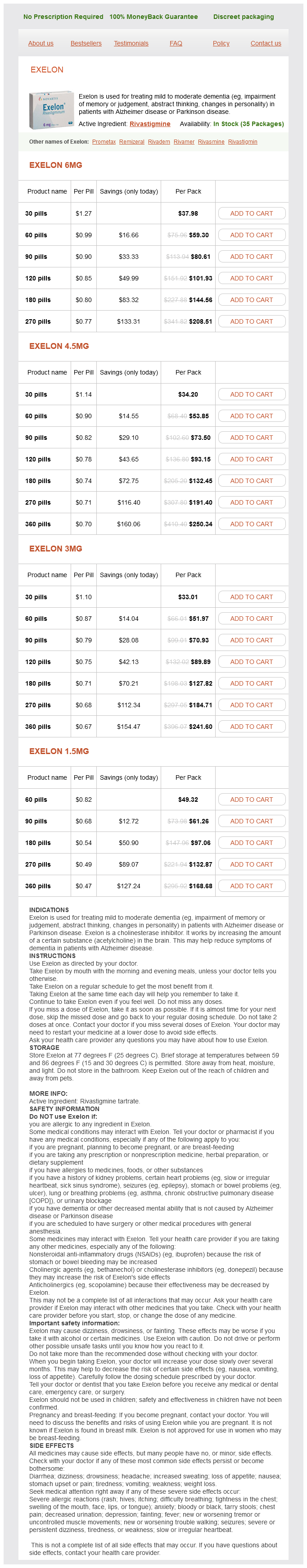
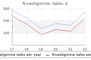
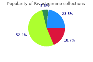
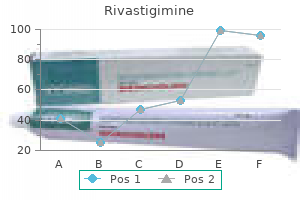
Rivastigimine
Buy rivastigimine 4.5mg with mastercard
In children medications you can take during pregnancy effective rivastigimine 6mg, a Salter-Harris type I fracture of the distal fibular physis cannot be confirmed radiographically so it must be suspected clinically based on tenderness over the physis. Ankle sprains are far more common than fractures in older children and adolescents. The Ottawa ankle rules have been validated as a decision tool for ordering ankle X-rays in adults. These rules probably work for children as well, but validation data for children have yet to be presented. The rules indicate that radiographs should be ordered if there is swelling or tenderness of either of the malleoli, or inability to bear weight after the injury. Fractures of the metatarsal shaft are usually the result of direct trauma to the foot resulting from a fall, bicycle, or sledding injury. Swelling, ecchymosis, and localized tenderness to the fifth metatarsal tuberosity suggests a fracture. Adequate alignment can be achieved by "buddy" taping the fractured toe to an adjacent stable toe. Name at least three fractures that are difficult to identify on X-rays and must often be diagnosed clinicallyfi Fractures in children heal more rapidly than those in adults because the pediatric bone has a thicker periosteum and more efficient remodeling. A fracture is described by its anatomic location, configuration, relationship of the fracture fragments to each other, and relationship of the fracture fragments to the surrounding tissue. External fixation refers to fixation of bones by splints, casts or transfixion pins. A cast is sometimes considered merely external support, rather than external fixation. Internal (or intraosseous) fixation is stabilization of the bone fragments by direct fixation to one another with surgical wires, screws, pins, rods, or plates. Itoman this is a 7 year old female who presents to the clinic with a chief complaint of left wrist pain. She was rollerblading with several friends, and was accidentally pushed from behind. She has mild discomfort upon palpation of her left knee, but she is able walk, stand, and jump without difficulty or discomfort. Her right wrist is normal, but tenderness is elicited upon palpation of her left distal radius. Radiographs reveal a non-displaced distal radius fracture of the left wrist without angulation. She is placed in a forearm sugar tong splint, and her mother is given instructions to follow-up with an orthopedic surgeon. Splints are used to temporarily immobilize fractures, subluxations, sprains or soft tissue injuries. Other indications for splinting include acute arthritis, severe contusions and abrasions, skin lacerations or burns across joints, tendon lacerations, tenosynovitis, animal bites, deep space infections, joint infections, and puncture wounds (1). The goal of splinting is immobilization to minimize pain and prevent further damage to nerves, vessels, muscle, skin, etc. Immobilizing tender joints, as seen in tenosynovitis, hemarthrosis, or acute arthritis, reduces pain and inflammation. Abrasions and lacerations that cross joints can be stretched open if the extremity is not immobilized. Immobilization of fractures reduces the risk of further displacement, minimizes hemorrhage, soft tissue damage, and risk of neurovascular injury. All injuries that present with immobility, pain with movement, swelling, reproducible pain on palpation, anatomic deformity, discoloration, or crepitus should be evaluated with appropriate radiographic studies (3). A Salter-Harris type 1 injury may not exhibit any radiographic evidence of a fracture, and may present like a sprain. All children who present with tenderness over the physis (growth plate) of a long bone should be presumed to have a Salter-Harris type 1 fracture injury and immobilized in an appropriate splint (3). The presence of a non-displaced Salter-Harris type 1 fracture is identified clinically during the follow up examination. Persistent tenderness several days after the injury implies the presence of a fracture (to be confirmed by additional radiographs which may show new born formation 7 to 10 days after the injury). The two categories of splints are classified based on their raw materials, plaster and fiberglass. Cardboard, aluminum and other semi-rigid or malleable materials can also be used for temporary splints. Plaster splints are made from gauze material impregnated with plaster of Paris, which is made from gypsum. When water is added, the gypsum-powder hardens as the calcium sulfate dihydrate molecules recrystallize (1). The reaction is exothermic and can possibly burn the patient, but most of the time, it just feels warm. Depending on the temperature of the water (hot water allows for a quicker set time) the plaster may take anywhere from 2-8 minutes to set. An upper extremity injury may require anywhere from 8-10 layers of plaster while the lower extremity may take 10-20 layers (4). Despite the large amount of material used, plaster is still relatively inexpensive (1). Excessive water will cause the crystallization to become unstable, making the splint soggy. The prepackaging reduces the steps needed to prepare the limb prior to splinting, but increases the cost. The padding also absorbs water and sweat well, minimizing the accumulation of moisture (1). Kinks, although small, may be a potential sight of irritation causing skin breakdown and pressure injury. The procedure for splinting should always start with a general inspection of the limb. Next, the limb should be rechecked for signs of compartment syndrome and neurovascular compromise. The splint width should be approximately half as wide as the circumference of the extremity. Since the splint is used to support the limb, the posterior surface is usually used as a measuring guide. A longer length will also allow for contraction of the plaster as it crystallizes (4). Optionally, stockinette (tube sock) can be rolled over the limb and cut to a length slightly longer than needed. The stockinette should look as if a long sock with an open hole has been placed over the extremity. Take time to smooth out the stockinette to prevent pressure spots and kinks at flexion creases. Also, make sure the stockinette is positioned so that there is extra material both proximal and distal to the area to being splinted. If stockinette has been used, then the cast padding is rolled over the stockinette. Each successive roll of cast padding around the extremity should cover the previous roll by approximately 50-60% (4). Make sure that the "extra" stockinette distal and proximal to the area being splinted is not covered with cast padding. The plaster will heat up as it hardens, and this may scare and burn a child but this is unlikely. While the plaster still soft, fold the proximal and distal ends of the plaster back over itself to provide a smooth edge. An optional cast padding layer can be applied over the splint to prevent the soggy plaster from Page 614 incorporating into the elastic wrap applied in the next step. Roll an elastic bandage over the outside of the extremity, usually in a distal to proximal fashion, securing the plaster to the extremity. Fiberglass splint materials come encased in cast padding material rather than as bare sheets of fiberglass. Once the limb has been inspected, and the proper splint width and length are selected, cut the length needed and place the fiberglass splint in water. Removed the excess water from the fiberglass splint by rolling it in a dry towel and applying pressure to remove water from the fiberglass.
Order rivastigimine visa
Lead was used in Legg-Calve-Perthes disease A hip disorder in household paint until 1978 symptoms lyme disease purchase rivastigimine amex, and it was also found in children that is due to interruption of the blood supleaded gasoline, some types of batteries, water ply to the head of the femur (the ball in the ballpipes, and pottery glazes. This still found in many older homes, and lead is somedisease is most common between ages 6 and 9, and times also found in water, food, household dust, and it tends to affect boys most commonly but is more soil. The symptoms calcium can help protect people against absorbing include hip and thigh pain, stiff hip, a limp, and lead. Over a period of 18 blood is removed and metals are filtered out to 24 months, the blood supply usually reestablishes through a machine, then reinfused into the patient. During this period, the bone is soft and liable Treatment cannot repair damage to the brain done to fracture under pressure, causing collapse of the by lead poisoning, but it may prevent further damhead of the femur. Also known as Legg disease, Legg-Perthes disease, Perthes dislearning disability One of several childhood ease, and avascular necrosis of the femoral head. The bacterium vival are prompt recognition of the disease, immethat causes it, now known as Legionella, thrives in diate use of appropriate antibiotics and drainage of the mist of condensers, air conditioners, and evapabscesses. Lennox-Gastaut syndrome A severe form of epilepsy that usually begins in early childhood. Early symptoms may lens the transparent structure inside the eye that include poor sucking ability and loss of head confocuses light rays onto the retina. Most light microimpairment of respiratory and kidney function, may scopes now have a turret that bears a selection of occur. Most light micronates in smooth muscle, the major structural comscopes are binocular, with one ocular lens for each ponent of most hollow internal organs and the walls eye. Leiomyosarcoma can occur almost anywhere in the body but is most frequently found lentigo maligna melanoma See melanoma, in the uterus and gastrointestinal tract. Today leprosy can be cured, particusis, the most common being cutaneous and visceral larly if treatment is begun early. Surgery can be perform of the disease causes skin sores and is usually formed to reconstruct damaged faces and limbs. Visceral leishmaleptin A hormone produced mainly by adipocytes niasis affects the internal organs of the body and can (fat cells) that is involved in the regulation of body be fatal. Deep in leptomeninges the two innermost layers of tisthe abscess, the anaerobic bacteria can flourish. The bacteria penetrate from the abscess into the sue that cover the brain and spinal cord. Also known as central nervous system, and other tissues of the acute lymphocytic leukemia. Neurological symptoms include leukemia, acute myeloid A quickly progressive facial grimacing, involuntary writhing, and repetitive malignancy in which there are too many immature movements of the arms and legs. The prognosis is blood-forming cells that are precursors to the granupoor, with death usually occurring in the first or locytes or monocytes in the blood and bone marrow. Also known as acute myelogenous leukemia and acute nonlympholethargy Abnormal drowsiness, stupor. Letterer-Siwe disease A form of Langerhans leukemia, blastic phase of A stage in chronic cell histiocytosis starting in infancy that involves myeloid leukemia in which 30 percent or more of proliferation of histiocytes in multiple organs such the cells in the bone marrow or blood are the maligas the skin, bones, and other organs. See also leukemia, chronic include rash, swollen glands, enlargement of the myeloid. Letterer-Siwe disease is the leukemia, chronic lymphocytic the most most severe form of Langerhans cell histiocytosis common form of leukemia in adults, in which lymand has a high mortality rate. Strictly may crowd out other blood cells in the bone marspeaking, leukemia should refer only to cancer of row, resulting in a shortage of red cells (producing the white blood cells (leukocytes), but in practice it anemia) and platelets (producing bleeding and can apply to malignancy of any cellular element in bruising). See also accelerated phase of leukemia; Treatments may include chemotherapy, monoclonal leukemia, blastic phase of; leukemia, chronic antibody therapy, and bone marrow transplantation. This phase may last from several which the high number of white blood cells found months to several years. In the next phase, the on a blood test resembles the numbers seen in accelerated phase, there are more blast cells in leukemia. For example, infectious mononucleosis blood and bone marrow and fewer normal cells. Treatment leukodystrophy A disorder of the white matter may include chemotherapy, biological therapy, and of the brain. In some nerve fibers rather than nerve cells themselves, and cases, bone marrow transplantation is done. Levocardia virtually always is associated with a conligament A tough band of connective tissue that genital heart disease. Parkinsonian symptoms in other disorders, restless legs syndrome, and herpes zoster. Levodopa conligament, lateral collateral knee See lateral verts to the neurotransmitter dopamine in the brain. Levoxine, Levoxyl, Synthroid) that is used as a thyroid hormone replacement drug to treat an underligament, posterior cruciate See posterior active thyroid gland (hypothyroidism). The abnormal areas on the skin in lichen lightheadedness A feeling that one is about to planus are typically flat-topped (hence the term faint. Lightheadedness is medically distinct from planus), itchy, and frequently have a polygonal or dizziness, unsteadiness, and vertigo. Lichen planus lights, flashing A sensation that is created when on the scalp may lead to hair loss. The causes the clear, jelly-like substance that fills the middle of of lichen planus are unknown. However, it can be the eye (vitreous humor) shrinks and tugs on the triggered by the use of certain drugs, such as thiretina. These flashes of light can appear off and on azide diuretics, phenothiazines, and antimalarials. Some of them are present at birth (congenital), and others are limb An arm or a leg. In lipodystrophy syndrome, the face, together as a package because of their location near arms, and legs become thin due to loss of subcutaone another on the same chromosome. See also cephalothoracic lipodylip One of the two fleshy folds that surround the strophy; protease inhibitor. The upper lip is separated from the nose by the philtrum, the area that lies lipoma A benign tumor of adipocytes (fat cells). Small blind pits lipomatosis, familial benign cervical See are sometimes seen at the corners of the mouth; cephalothoracic lipodystrophy. The lips may be abnorlipoprotein A molecule that is a combination of mally thin or thick. Lipids do not travel in the blood alcohol syndrome typically have a thin upper lip and by themselves, but they are carried through the flat philtrum. Most frequently seen in older adults (age 40 and above), liposarcomas are the most common of lipid A fat. Lipids are easily stored in the body liposuction the surgical suctioning of fat and serve as fuel. Among the well-known lipids are deposits from specific parts of the body, the most cholesterol, triglycerides, fatty acids, and steroids common being the abdomen, buttocks, hips, thighs (such as cortisone). A phospholipids are all compound lipids (lipids in hollow instrument called a cannula is inserted combination with other types of chemicals). A high-pressure vacuum is then applied to the cannula to suck out lipid profile A pattern of lipids in the blood. Lipid storage diseases result in the abnormal includes the local anesthetic lidocaine to numb accumulation of lipids in various organs.
Purchase cheap rivastigimine line
These are characterized by the absence or marked decrease in the number of intrahepatic interlobular bile ducts medicine syringe generic 6mg rivastigimine with visa, with normal sized arteries and portal veins in the triad. Intrahepatic bile duct paucity was previously referred to as "intrahepatic bile duct atresia" or "intrahepatic biliary atresia" (6, 8). These diseases are diagnosed by the presence of cholestasis and bile duct paucity on liver biopsy. The non-syndromic form describes a common pathology with various etiologies, which are still poorly understood. With the syndromic forms there are characteristic findings associated with the bile duct paucity (8). For example in Alagille syndrome, or arteriohepatic dysplasia, there are characteristic facies along with ocular, cardiovascular, vertebral, and kidney pathology. When a child presents with jaundice, the first step is to evaluate the total and fractionated bilirubin in each patient with jaundice. If the elevation is isolated to the unconjugated (indirect) fraction of bilirubin then significant liver pathology is unlikely (3). However, if the elevation is in the conjugated (direct) fraction or it is 20% or greater of an elevated total bilirubin then cholestasis more likely (6). If the presence of cholestasis is established, then the etiology must be found in a timely fashion. Panels of testing can quickly rule out or diagnose entities such as hypothyroidism, galactosemia, tyrosinemia, alpha-1-antitrypsin deficiency, and infectious diseases. The newer iminodiacetic compounds have greater concentrations in the bile then the original compounds. Findings include edema of the portal tracts, tortuous proliferation of bile ductules, fibrosis, along with intracellular and canalicular cholestasis. There is also a mixed infiltration of neutrophils and lymphocytes with a mild non-specific cholangitis (7). Of note is that there can also be ballooning of the hepatocytes and multinucleated giant cell formation. If an intact extrahepatic biliary system is not visible, then extrahepatic biliary atresia is evident. If an intact biliary tree is visible, then an intraoperative cholangiogram is done, in which the surgeon cannulates the bile duct and injects contrast to determine if the biliary ducts are patent. A patent biliary system visualized on the intraoperative cholangiogram rules out extrahepatic biliary atresia. If the intrahepatic bile ducts are obliterated or reduced in number, then bile duct paucity (formerly called intrahepatic biliary atresia), is present. An intact and patent extrahepatic biliary tree rules out extrahepatic biliary atresia and the presence of normal intrahepatic bile ducts rules out bile duct paucity. Page 344 Diagnostic algorithm of biliary atresia: 1) Direct hyperbilirubinemia prompts evaluation and lab work-up. A correctable lesion has fibrosis of the distal biliary tree with the proximal biliary tree and the intrahepatic bile ducts remaining patent. In these cases, excision of the fibrotic area and direct drainage into the bowel is possible. In the non-correctable lesions, the biliary system is fibrotic to the level of the porta hepatis. The Kasai procedure is the Roux-en-Y hepatoportoenterostomy where the porta hepatis is attached to a loop of bowel after resection of the fibrotic biliary system. This procedure basically anastomoses the liver directly to the bowel so that in theory, bile can flow from the liver into the bowel. The reason that the procedure succeeds is that at the porta hepatis there are microscopic bile ductules that have proliferated which communicate with the intrahepatic system. Some groups use frozen section biopsy during the laparotomy to examine the tissue at the porta hepatis using the size of the vessels as a marker for the likelihood of successful reestablishment of bile flow. However, there have been variations in the size of vessels required and this theory is not universally accepted. The success of the Kasai procedure depends largely on 2 factors: age at procedure and experience of the center it is performed at. To obtain maximum benefit from the Kasai procedure it should be performed before the patient is 3 months old, ideally less then 2 months. Establishment of bile flow is achieved in 80% of the patients who are less then 2 months, 50% between 2 and 3 months, and less then 10% if older then 3 months (12). Outcomes of the procedure and post-procedure survival are improved when the hospital does more then 5 procedures a year (1,5,13). If the child is diagnosed at an age greater then 3 months, the Kasai procedure has a low probability of success. Performing a Kasai procedure after this age is thus controversial, versus proceeding straight to liver transplantation, which is the treatment for a failed Kasai procedure. It is the general consensus that a patient should undergo the Kasai procedure even if they present at ages greater then 3 months if it is possible that bile flow can be established (12,14). While a post Kasai transplant is technically more difficult, there was no reported change in survival after transplantation in patients who underwent primary transplantation versus those who had a failed Kasai procedure prior to transplantation (11,13). Ascending cholangitis is the most common complication, occurring in 40-60% of Kasai procedures (1). The normal anatomy of an intact bile duct prevents bowel contents from refluxing up toward the liver. In the Kasai procedure, the bowel contents containing digestive enzymes have direct access to the existing bile ducts and hepatic tissue causing the cholangitis. An anti-refluxing valve can be surgically created within the duodenal segment anastomosed to the liver, but such alterations in the Kasai procedure and various medical regimens have not proven successful. There does appear to be an increased risk in the patients with established bile flow, probably due to an intact pathway for ascending bacteria. Prophylactic antibiotics with trimethoprim-sulfamethoxazole is designed to reduce bowel bacterial counts. Repeated episodes of cholangitis can lead to extensive liver damage and cirrhosis. This occurs due to the progressive inflammation and fibrosis of the intrahepatic biliary system and/or repeated episodes of cholangitis leading to cirrhosis. The portal hypertension that develops will have the sequelae of other forms of portal hypertension such as varices, ascites, hypertensive gastropathy, hypersplenism, and encephalopathy (1). The most common presentation is esophageal variceal hemorrhage occurring in 3060% of patients (1). Treatment relies on the same methods employed in adults for other forms of portal hypertension. After the Kasai procedure, the patient can be stratified into 1 of 3 prognostic groups at 4 to 6 weeks post operatively (10). The first group are the patients who produce adequate bile flow and are relieved of their jaundice. The second group has moderate bile flow but they remain jaundiced and will continue to survive postKasai, however they will eventually need liver transplantation later in life. This is considered a failure of the Kasai procedure and they will need liver transplantation in order to survive. While the Kasai procedure establishes bile flow, it does not necessarily halt the intrahepatic inflammation and fibrosis. Patients who undergo the Kasai procedure can survive with their native liver in 20-30% of cases, but the remainder will eventually need liver transplantation (2,10). Ten year survival ranges from 33% in older studies to 68% in more recent studies (2,13). Despite this, there are still many children who are untreated at 3 months of age (as high as 14-19% in some studies) (1). The best method so far, is to maintain a high clinical suspicion in the jaundiced patient. A liver biopsy shows hepatocellular ballooning and the presence of multinucleated giant cells. A patient presents to you with lightly colored stool; however, when the stool is broken up it is noticed that the center is clay colored. A 16 week old patient is diagnosed with biliary atresia, should he/she undergo a Kasai procedure if there are no contraindications or should the patient just wait for a liver transplantfi
Buy discount rivastigimine line
In the secondary form medications known to cause seizures buy rivastigimine in united states online, correction of the underlying lesion to alleviate external compression is associated with a good outcome (3). Congenital airway anomalies must be considered when evaluating stridor of infancy. The key is to separate life-threatening conditions from those which are self-limited. Classically, the stridor in laryngomalacia is: a) inspiratory b) expiratory c) biphasic 3. Anatomically, congenital subglottic stenosis is usually associated with what other airway malformationfi As the second most common laryngeal anomaly, vocal cord paralysis accounts for what percentage of laryngeal lesionsfi In general, bilateral vocal cord paralysis can be attributed to a nervous system problem, while unilateral vocal cord paralysis is usually caused by an injury to the nervous system. Sleep Disorders Sze Mei Chung this is a 4 year old boy who is brought to the office by his single mother with a chief complaint of screaming at night for about a year. He has been in good health otherwise with no recent history of otitis or respiratory infection. According to his mother, she would hear a chilling scream, rush to her son, and find him sitting up in bed, sweating with a glassy stare. There is no response when she talks to him, and when she tries to hug him, he usually resists. But when her son does answer after more vigorous shaking, he seems confused and disoriented. In the morning he would seem fine and not remember having any nightmares or screaming. He is referred to a sleep specialist who assesses the boy as having sleep terrors. His mother is taught to help avoid stresses and fatigue for her son during the daytime. His mother is told that diazepam may be prescribed if his problem worsen, but most of the time, children will outgrow this disorder. This includes age of onset, patterns of daytime sleepiness and napping, questions about snoring and apnea, sleep related behaviors such as talking and head banging, psychiatric assessment regarding separation anxiety and nightmares, relevant medical/neurological conditions such as headaches, and mental retardation, and family histories of sleep disorders. Limb actigraphy uses an instrument resembling a wrist watch that detects body movements continuously for 3 days. In cases of arousal disorders (sleep terror and sleep walking), having the parents record the episodes on a video camcorder may be more useful. Sleep disorders can be categorized into dyssomnias, parasomnias, and sleep disorders due to medical or psychiatric conditions (2). Dyssomnias can be broken down into 3 categories: intrinsic dyssomnias, extrinsic dyssomnias, and circadian dyssomnias (5). Intrinsic dyssomnias are due to causes within the body and include breathing related sleep disorders (sleep apnea) and narcolepsy. Sleep apnea occurs when air flow is completely stopped and is diagnosed when there are 5 apneas or 10 apnea-hypopnea episodes per hour of sleep. In general, hypopnea can be thought of as episode where airflow is reduced by one-half to two-thirds (6). Patients are not aware of their apneas but sometimes do wake up with a choking feeling. Central apnea results from no respiratory effort because of brainstem respiratory neuronal immaturity, which is commonly seen transiently in premature babies and newborns. In young children, the obstruction is most often due to enlarged tonsils and adenoids and not usually from severe obesity (2). Apnea is accompanied by increased thoracic and diaphragmatic respiratory effort without air exchange due to upper airway obstruction. Because of apneic episodes, the patient experiences many short arousals to restore adequate oxygenation. In toddlers, growth retardation similar to that seen in failure to thrive can be observed possibly associated with disruption of growth hormone secretion during fragmented sleep. Prader-Willi syndrome is a genetic disorder due to deletion of q12 in the long arm of the paternal chromosome 15. Initially, the infant presents with hypotonia followed by rapid weight gain after 1 year of age leading to morbid obesity (11,12, 13). In cases of Pickwickian and Prader-Willi syndromes, reducing weight is of primary importance and should be achieved as fast as is feasible (14). Examples of other conditions predisposing to obstructive sleep disorders are Down syndrome, craniofacial anomalies, mucopolysaccharidoses, and neuromuscular disorders. Regular sleeping and rising times are recommended with scheduled naps 2-3 times a day. Psychosocial counseling and support are important as narcolepsy is a debilitating life-long condition once diagnosed. For treating cataplexy, tricyclic antidepressants and clomipramine have been successful. Unlike intrinsic dyssomnias, extrinsic dyssomnias are due to external causes and includes protodyssomnias (an inability to fall asleep and stay asleep) of infancy and insomnias of childhood (2). Predisposing factors include previous behavior reinforcement patterns, child temperament. Currently, it is not known whether these protodyssomnias progress to true dyssomnias later on (2). Individualized bedtime habits such as reading or playing a quiet game can also help. The teenager then tries to make up for the lost sleep by sleeping long periods during the weekend. Thus, this irregular sleep pattern leads to biologic clock disruption over time resulting in circadian rhythm dyssomnias characteristic of delayed sleep phase syndrome. This syndrome presents with an inability to sleep and wake at a customary time, excessive daytime sleepiness, and many naps with no difficulty in maintaining sleep once asleep. Treatment consists of eliminating sleep debt and reinforcing appropriate bedtime and rise time. Chronotherapy (phase delay treatment) allows the biological clock to reset by delaying sleep and rising times by 2 hours each day until the time of sleep onset is shifted back to a more reasonable hour. Unlike dyssomnias in which the sleep process is disrupted, parasomnias represent behavioral intrusions upon ongoing sleep. Parasomnias are more likely to occur in males than females, and a patient suffering from one parasomnia is likely to exhibit another. Problems occur when this normally smooth transition is characterized by autonomic activity. In sleep terror, the child typically sits up, screams, and has a glassy, unseeing gaze associated with autonomic symptoms of palpitations, diaphoresis, and irregular breathing. This lasts for about 1 to 5 minutes until the child calms down and continues to sleep. A child in this condition is difficult to wake up and appears disoriented and confused when he does awaken. Unlike nightmares, the child does not recall the incident or any dreams in the morning (15,16). The body movements that occur during sleepwalking are purposeless and uncoordinated. Locking the doors and windows and installing alarms that alert the parents when the child rises from bed are some of the safety measures (17). Rather, having the parents videotape the episode of attack can be more diagnostic. As daytime stress and fatigue are known precipitators of arousal disorders, they should be avoided. In severe cases or if the onset occurs in adolescence, it is important to rule out sleep-related seizures. Examples of this category of parasomnias are rhythmic movements, nocturnal leg cramps, and sleep talking. Rhythmic movements include head banging, sleep starts (defined as sudden muscle jerks at sleep onset that may involve the limbs, neck, or entire body), and body rocking that usually occurs at sleep onset for <15 minutes duration. Prevalence decreases with age, and generally, measures to prevent self injury are sufficient. Onset occurs at 3-6 years of age and up to 50% of this age group experience nightmares severe enough to worry their parents.
Purchase rivastigimine 3 mg free shipping
All subjects with autism had abnormalities of eye human exposure because of their use in plastics and other movement or facial expression medicine numbers discount rivastigimine 1.5 mg with visa, or both. Phthalates leach into the environthe nerve dysfunctions in people with autism refect an early ment and expose humans through ingestion, inhalation, brain injury that not only afects the cranial nerves but also and dermal routes. When the concentration of phthalates has secondary efects on later brain development. Many cases in the urine of autistic subjects was calculated, there was of autism are initiated very early in gestation. The mean function in infancy through the pre-school years (Jacobson exposure was in the sixth week post conception. These outcomes can result in low triisyndrome (liver and brain damage after viral infection) in odothyronine (T3) levels in the fetal brain during the period 76 H. A cluster site of autism is and may produce morphological brain changes leading to Brick Township, New Jersey, a rural township close to several autism (Roman, 2007). Specifcally, Brick Township was documented to have four times more prevaOrganophosphate pesticides lence of autism than that of the entire country. Cross-sectional data from tetrachloroethylene, trichloroethylene, and trihalomethanes. Exposure to the pesticides could be prenatal, supports research suggesting that autism may be caused by direct, or from food, drinking water or residential pesticide a neural tube defect (Rodier, 2000). A primary action Autism has increased to epidemic proportions, afecting four of organophosphates is inhibition of acetyl cholinesterase times as many males and females. In addition, lar ratios in many other countries, a very signifcant threat to Chlorpyrifos, an organophosphate insecticide widely used future generations is evident. This review cites documentation until a few years ago to control insects in schools and homes of causes of autism, including genetic mutations/deletions, and still used extensively in agriculture, is a developmental viral infections. It is possible that autism results from more than one cause, Other environmental causes with diferent manifestations in diferent individuals that Perhaps, additional environmental causes could be responshare common symptoms. Integrating the data presented sible for documented unusual brain growth patterns in early here, a hypothesis is that autism is the result of genetic life in autism (Courchesne et al. The data suggest that defects, with the contributory efect of advancing age of the autism involves abnormal regulation of brain growth during parents, and/or infammation of the brain. The infammation early life, with an unusual developmental neuroanatomic could be caused by a defective placenta, an immature bloodphenotype characterized by hyperplasia of cerebellar white brain barrier, the immune response of the mother to a viral matter and neocortical gray matter at the youngest ages with or bacterial infection, a premature birth, encephalitis in the slowed growth afterward, slowed growth in the cerebellar child after birth, or a toxic environment. There is evidence of a reduced head size at birth neuro-infammation, autoimmune reactions, brain injury, and a sudden and excessive increase in head size between and autism. Other Conclusion investigators reported brain volumes were signifcantly larger for children with autism 12 years old and younger, Autism has been documented to be caused by genetic defects but those older than 12 (including adults) had brain voland/or infammation of the brain. The head circumference caused by a wide variety of environmental toxicants, infecwas increased in both younger and older groups of autistic tions, and co-morbidities in individuals genetically prone to individuals, suggesting that those subjects greater than age the developmental disorder. Brain volumes in autistic adolescents and adults were normal, perhaps, due Acknowledgements to a slight decrease in brain volume for the autistic subjects at the same time that normal individuals are experiencing a Robert B. Epidemiology studies have documented the presence edged for editorial assistance in the preparation of this manuof cluster sites of incidence of autism. Another report documents that in the location with the highest rate e author reports no conficts of interest. Immune therapy in autism: historinip/acip cal experience and future directions with immunomodulatory therapy. Brainstem, cerebellar and limbic neuroanatomical abnorof age on brain volume and head circumference in autism. Maternal and population cohort of children in South T ames: the Special Needs and paternal age and risk of autism spectrum disorders. Vaccines and autism: evidence does not support a causal caspase-3 activation, membrane damage, and cell death in cultured association. Neuroanatomic observations of the disorders: focus on critical windows of immune vulnerability. The lifetime distribution of the incremental societal costs of traits: A study of individuals with congenital adrenal hyperplasia. Some etiologic and prognostic factors comparison of autistic syndromes with and without associated neurologiin early infantile autism and psychosis. Diagnostic criteria and methionine cycle-transsulfuration and androgen pathway markers in classifcation. Autistic syndrome with onset at age 31 years: herpes disorder and viral infections. Rates of chromosome abnormalities at diferent maternal of measles, mumps, and rubella vaccination and autism. T imerosal neurotoxicity is associated with glutathione depletion: to 4th digit ratio and autism. Can autism be triggered by acetaminophen activation of the in childhood autistic disorder is not associated with urinary creatinine endocannabinoid systemfi Platelet monoamine oxidase activity in children with mumps-rubella vaccination, and autistic disorder: the results of a parent attention-defcit/hyperactivity disorder. The early development of Antibodies to myelin basic protein in children with autistic behavior. Serological association of measles containing vaccines and autistic spectrum disorder: a critical review of virus and human herpesvirus-6 with brain autoantibodies in autism. Abnormal measlesSpectrum Disorder: Interleukin-6 Signaling as a Key Mechanistic Pathway. Antibodies to neuron-specifc antigens in children with Developmental Disabilities Monitoring Network, United States, 2006. Issues in the classifcation of autism and Genetically determined low maternal serum dopamine beta-hydroxylase related conditions. In: Handbook of Autism and Pervasive Developmental levels and the etiology of autism spectrum disorders. Autism: transient in utero hypothyroxinemia related to in a large autism population. Psychiatric Association Meeting May, maternal favonoid ingestion during pregnancy and to other environNew Orleans. Vitamin a supplementation of vitamin a defcient measles autistic spectrum disorder and the measles, mumps, and rubella vaccine: patients lowers the risk of measles-related pneumonia in zambian chila systematic review of current epidemiological evidence. Phagocytosis and killing ability of Candida albicans by blood leucocytes Rossignol, D. This society promotes the public welfare and improves patient care through the translation of new technologies/therapies into life saving diagnostic and therapeutic procedures. The society achieves its mission through multi-disciplinary collaborations with government agencies, patient advocacy groups, educational institutes and industry as well as philanthropic organization. Gullapalli the Annual World Congress is a multi-disciplinary forum designed to James McDonald facilitate cross-pollination and dissemination of technological and medical advances and scientific discovery. The society promotes policies that support rapid, safe, and cost-effective translation of new technology into medicine. The Society builds transdisciplinary and translational consortia which break down traditional barriers that impede application of new technology to medical problems. Translational research applies cutting edge basic science and advanced technologies to clinical neurosciences. The Society examines emerging disciplines such as nanotechnology, image-guided therapy, stem cell therapy, multi-modality imaging, biophotonics, and biomaterial and tissue engineering for their application to the diagnosis, treatment, and rehabilitation from neurological diseases. The Society achieves its goals through meetings, fellowships, publications, international collaborations, consortiums, and policy forums. These scientific advances also have contributed to the large gap of knowledge amongst the scientists in different disciplines. One of the major challenges of 21st century for the scientific community is how to close such gaps of knowledge amongst multiple disciplines. The purpose of the annual meeting is to create an interactive environment, which foster cross-pollination of ideas and pave the way for birth of new treatment and diagnostic modalities in the field. Other Financial or Material Support None financially but I am coJames Welsh founder of Radion Global, a board member of Coqui Jane Tavyev Asher Research Support: Muscular Dystrophy Association Other Financial or Material Support J.
1.5mg rivastigimine visa
The physical properties of proton beams make it possible to concentrate the radiation dose on the target with delivery of fairly small doses to structures in front and behind the target medications 1800 buy rivastigimine 4.5mg without a prescription. If ongoing research projects result in reducing the cost of proton therapy equipment, it is possible that protons will have wider use in the treatment of childhood tumours in the future. In some tumour types and sites, radiotherapy is the main treatment, used as an adjuvant to surgery, and it has an important role as a palliative therapy in all tumours (Table 21. Leukaemias and lymphomas Leukaemias and lymphomas are haematological malignancies originating from the bone marrow, lymph nodes and reticuloendothelial system. Leukaemias are aggressive malignancies, and although leukaemic cells are extremely sensitive to radiation, these malignancies are systemic and are treated exclusively by chemotherapy. Another use of radiotherapy in leukaemia is total body irradiation as part of the bone marrow transplantation schedule. Radiotherapy is effective in sterilizing the bone marrow, and reducing the immunological response against grafted marrow. Wide field irradiation to cover all 330 331 lymph node areas on either side of the diaphragm using doses up to 45 Gy was standard treatment. Treatment was successful in most patients, with a price of serious late effects such as anatomic deformities, breast cancer at younger ages and secondary leukaemia. In a malignancy with a cure rate higher than 90%, it is extremely important to limit the radiation and volumes. Usually they do not have any connections to the known aetiological factors, and their incidence is fairly similar in different regions of the world. Surgery performed by a skilled neurosurgeon is always the first choice to obtain diagnosis and to remove the tumour. Surgical resection has been shown to improve survival, but it is not possible to remove tumours with microscopically clear margins and recurrence is inevitable in many aggressive tumours. In principle, radiotherapy is indicated in almost all high grade tumours immediately after surgery and in low grade tumours after recurrence. In low grade gliomas, radiotherapy is usually delayed and not used as the first line treatment until tumour progression. Despite these combined efforts, local tumour progression occurs in almost all patients with high grade gliomas, and long term survival rates are less than 20%. Diffuse glioma of the brain stem, sometimes referred to as pontine glioma, is a unique tumour of childhood. Surgery is not possible due to its location, and radiotherapy alone or combined with chemotherapy is the only choice in the treatment. Unfortunately, this is one of the most aggressive tumours among children; less than 10% of the patients survive more than two years. This tumour arises exclusively from the posterior fossa and grows into the fourth ventricle. It is a highly aggressive tumour which may spread to the whole brain and spinal cord via the cerebrospinal fluid. It requires careful planning and precise treatment delivery in order to keep radiation dose to the spinal cord and brain below tolerance limits. Neuroblastoma Neuroblastoma is a neuroendocrine tumour arising from neural crest elements of the sympathetic nervous system. It is the most common extracranial solid tumour in childhood, and the most commonly diagnosed malignancy among infants. Neuroblastoma usually arises in one of the adrenal glands, but can also develop in nerve tissues in the neck, chest, abdomen or pelvis. Maturation and spontaneous regression of neuroblastoma are possible, especially among infants. However, neuroblastoma frequently disseminates to bone marrow, bones, liver and lungs. Surgery and chemotherapy are effective in controlling this disease even in advanced stages, and today radiotherapy is used only in high risk patients with unresectable tumours. This is an embryonic tumour of the renal parenchyma predominantly affecting children under five years of age, most commonly during the first two years of life. However, patients are usually too young for large fields of abdominal irradiation, so radiotherapy is used after surgery with reduced doses of approximately 10 Gy to control microscopic disease. Rhabdomyosarcoma Rhabdomyosarcoma is a malignant tumour of the skeletal muscle originating from embryonic cells. Since skeletal muscle is distributed throughout the whole body, rhabdomyosarcoma may arise anywhere. The 334 therapeutic approach to a patient with rhabdomyosarcoma depends on various factors such as age, location and size of tumour, stage and histological subtype, and requires careful pre-treatment evaluation of the patient and individualization of the treatment. North American treatment protocols using radiotherapy in most patients are quite different from the European approach, where radiotherapy is reserved for unresectable tumours and recurrences. Retinoblastoma Retinoblastoma is a malignant tumour of the eye arising in the foetal retinal cells. It affects children under five years of age and may affect both eyes, suggesting a hereditary aetiology. Retinoblastoma, when diagnosed early, can be treated effectively, with very high rates of disease control with preservation of useful vision. There are several options for the early stage disease, including cryotherapy, laser ablation, surgery, radioactive plaque implantation and external irradiation. The role of radiotherapy in the primary management of retinoblastoma has decreased recently due to the effective use of other local treatment methods and concern about increased risk of secondary osteosarcoma among survivors. Limb sparing surgery and chemotherapy are the first choice for treatment of both types of bone tumour. Thus, the late treatment effects and squelae have 335 become a significant problem among the increasing number of survivors [21. Although currently it is possible to reduce the radiation dose to healthy tissues around the tumour using state of the art radiotherapy techniques, a considerable amount of healthy tissue is inevitably exposed at a minimal to moderate radiation dose. Acute toxicity occurs from the first weeks of the treatment period until several weeks after the completion of radiotherapy. However, occasionally they can be serious enough that various medications and a break of a few days of treatment may be needed for healing. Acute effects are mostly temporary and do not cause permanent impairment of tissues and organs. Late effects of radiotherapy are more serious than acute effects; they are usually progressive, irreversible and permanent, may cause organ insufficiency and may even be life threatening.
Generic rivastigimine 3 mg with mastercard
A good guideline may be to consider 8 pads or 12 tampons treatment jokes purchase rivastigimine without a prescription, well soaked, as the upper limit of normal; however even these estimates are user-dependent and may not correlate well with actual blood loss. A normal cycle interval ranges from 21-35 days, with less than 21 or more than 35 days considered abnormal (8). Until ovulatory cycles are established in the adolescent, endometrial proliferation occurs without progesterone regulation. The endometrium grows until the level of estrogen cannot sustain it, resulting in endometrial sloughing. In the adolescent, this results in cycles that are irregularly regular until regular ovulation is established. The time to develop ovulatory cycles is dependent on the age at which menarche occurs. The same study demonstrated that it may take 6-7 years to attain 90% or greater ovulatory cycles. Most importantly, although most menstrual cycles in early adolescence are anovulatory, truly abnormal bleeding is rare. Classification of Abnormal Uterine Bleeding in the adolescent (8): Menorrhagia (hypermenorrhea): Prolonged (more than 7 days) or excessive (>80 ml) of uterine bleeding occurring at regular intervals. Polymenorrhea: Regular episodes of uterine bleeding occurring at intervals of <21 days. Metrorrhagia: Uterine bleeding occurring at irregular intervals with variable amount of flow. Menometrorrhagia: Irregular and frequent bleeding, which may be excessive in amount and/or prolonged in duration. Oligomenorrhea: Irregular bleeding episodes occurring at intervals of 35 days to 6 months. Intermenstrual bleeding: Bleeding episodes occurring between regular menstrual periods. Hypomenorrhea: Decreased amount of uterine bleeding occurring at regular intervals. Dysfunctional uterine bleeding: Excessive uterine bleeding with no demonstrable organic cause. Although polyps, myomas, tumors, or endometriosis may be included in the differential, in contrast to the mature woman, diseases of the uterus are rarely the cause for irregular uterine bleeding in adolescents (10,11,12). A common clinical problem seen by physicians is the adolescent who presents with irregular intervals of bleeding that is normal in duration and amount of flow. For most of these adolescents, reassurance and observation are usually sufficient. The adolescent should be encouraged to keep a record of duration of menses, cycle interval, and amount of bleeding. Signs of chronic disease, polycystic ovary disease, endocrine abnormalities, or blood dyscrasias, if present, should be evident in the physical exam with corresponding symptoms obtained in the history. The evaluation of abnormal uterine bleeding in the adolescent also requires a thorough gynecological exam. If the bleeding is active, the site of bleeding should be Page 652 determined, as occasionally rectal or urethral bleeding may be mistaken for menstrual spotting. A speculum exam should be performed to inspect for signs of infection, trauma, foreign bodies, or evidence of contraceptive devices. Vaginal irrigation may be used to obtain these samples in the patient who will not tolerate a speculum exam. A bimanual pelvic exam is used to check for cervical motion tenderness, adnexal tenderness, and masses. A single-finger digital palpation is adequate for most adolescents, but if the hymenal orifice is still too small for a single-digit exam, a rectoabdominal bimanual palpation may be done instead. Surgical interventions, such as a hysteroscopy and D&C are diagnostic methods of last resort (13). After organic, systemic, and iatrogenic causes are ruled out, the abnormal bleeding may be diagnosed as dysfunctional uterine bleeding (8). Most adolescents with irregular bleeding in the first two years following menarche do not require long term management (10). If anovulation is the suspected etiology, the initial hormonal intervention should be progestin therapy to initiate a secretory change of the endometrium and produce a controlled withdrawal bleed. Progestin stops endometrial growth and organizes endometrial sloughing so that menses will occur following progestin withdrawal, rather than at random times. Estrogen treatment causes the regrowth of endometrium over raw, denuded areas where previous bleeding occurred. It is often clinically useful in controlling acute bleeding episodes, but progestin therapy is also required if the etiology of the bleed is anovulation. Combination estrogen/progestin oral contraceptives are the treatment of choice in adolescents, and also serve the dual benefit of preventing pregnancy if the adolescent is sexually active (8). Dysmenorrhea Dysmenorrhea is defined as cramping pain in the lower abdomen that occurs in conjunction with menstruation. If the pain is due to pelvic pathology or alterations in normal pelvic anatomy, the pain is classified as secondary dysmenorrhea, whereas primary dysmenorrhea occurs in the absence of any known pelvic pathology. Secondary dysmenorrhea is uncommon in adolescents, but primary dysmenorrhea is the most common gynecologic problem in young women, with reported rates as high as 75-90% (14,15,16). The incidence increases with sexual maturity, with one study reporting a 38% incidence at Tanner stage 3, increasing to 66% at Tanner stage 5. Dysmenorrhea also increases with chronological age from 39% in 12 year olds to 72% in 17 year olds. Symptoms of primary dysmenorrhea are usually noted beginning 1-3 years after menarche. Pain that begins within 6 months or 3 years after menarche is more indicative of secondary dysmenorrhea. Patients typically report intermittent, cramping suprapubic pain that may radiate to the lower back or thighs. The pain may begin a few days before menstruation and continue for as long as 7 days following the start of flow. More commonly, the pain begins a few hours after the start of menstruation, and lasts 24-48 hours. The pain is often accompanied by systemic symptoms including nausea and vomiting, fatigue, diarrhea, lightheadedness, and headaches. Often, there is a family history of dysmenorrhea, and the physical exam is completely normal (18). Due to the nature of the symptoms and the timing of the pain coincident with menses, a focused history and physical exam is usually sufficient to rule out non-gynecologic conditions of lower abdominal pain such as appendicitis, urinary tract infections, or inflammatory bowel disease. As with all women of child-bearing age, pregnancy must be excluded, along with the possibility of ectopic pregnancy. Any sexually active adolescent should have a speculum exam with cultures taken for Chlamydia trachomatis and Neisseria gonorrhoeae, and have a Pap smear. Secondary causes such as endometriosis, polyps, fibroids, or tumors are rare in adolescents, and a workup for these conditions are not usually indicated. These drugs act to inhibit prostaglandin synthetase, and have reported efficacy rates of 64-100%. In contrast, aspirin and acetaminophen were not shown to be superior to placebo in double-blind studies (19,20). Unfortunately, many adolescents self-treat for dysmenorrhea without consulting an adult. Of those that are self-treating, many take ineffective medications (aspirin or acetaminophen) or use less than the recommended dosages. Therefore, it is important for physicians to inquire about dysmenorrhea during routine visits to ensure that patients are being treated appropriately. Oral contraceptives are a second treatment option for dysmenorrhea that is highly effective (90%) and also serves the dual benefit of birth control for sexually active adolescents. For the roughly 10% of those who do not respond to these options, other alternatives exist ranging from laparoscopic surgery to acupuncture (16).
Cheap 3mg rivastigimine free shipping
Of the selected cancers treatment laryngomalacia infant order rivastigimine without prescription, lung cancer accounted for the highest average number of cancer-related deaths for male 9 Indigenous Australians (83 deaths per year), followed by head and neck cancer (31 per year), liver cancer (24 per year) and colorectal cancer (22 per year). Data is for New South Wales, Queensland, Western Australia, South Australia and the Northern Territory. Incidence and mortality rates were calculated according to the level of remoteness area of residence at diagnosis or death. The area of usual residence was then classifed according to Remoteness Area 2011 (see Appendix H). Very remote areas have the highest rate of cancer-related deaths Between 2012 and 2016, the age-standardised mortality rate for all cancers combined was highest in Very remote areas (195 deaths per 100,000 persons) and lowest in Major cities (157 per 100,000 persons) ure 9. Very remote areas also had the highest age-standardised mortality rate for cancer of unknown primary site (13 per 100,000 persons), head and neck cancers (13 per 100,000 persons) liver cancer (11 per 100,000 persons) and lung cancer (42 per 100,000 persons) (online Table S9. Major cities had the lowest age-standardised mortality rate for cancer of unknown primary site (8. Outer regional areas recorded the highest age-standardised mortality rates for colorectal cancer (23 per 100,000 persons), pancreatic cancer (10 per 100,000 persons) and kidney cancer (4 per 100,000 persons) (online Table S9. The index scores each geographic area by summarising attributes of the population, such as income, educational attainment, unemployment and jobs in relatively unskilled occupations. In the following paragraphs, a rising scale is used where socioeconomic group 1 represents people living in the lowest socioeconomic areas (that is, highest socioeconomic disadvantage) and socioeconomic group 5 represents people living in the highest socioeconomic areas (that is, most socioeconomic advantage). People living in disadvantaged areas had higher rates of cancer Between 2010 and 2014, the age-standardised incidence rate for all cancers combined was highest for those living in the 2 lowest socioeconomic areas and lowest for those living in the 2 highest socioeconomic areas ure 9. Cancer in Australia 2019 111 Between 2010 and 2014, the age-standardised incidence rates increased as advantage increased for breast cancer (113 per 100,000 females to 135 per 100,000 females) and prostate cancer (149 per 100,000 males to 180 per 100,000 males) (online Table S9. Between 2010 and 2014, some of the larger 5-year observed survival rate diferences occurred between the most and least socioeconomic disadvantaged for cervical cancer (79% compared with 61%), head and neck cancer (with lip) (69% compared with 59%), non-Hodgkin lymphoma (71% compared with 61%), kidney cancer (74% compared with 66%), colorectal cancer (63% compared with 56%) and prostate cancer (87% and 80%); for each of these cancers the people living in the most socioeconomically disadvantaged areas had the lowest 5-year observed survival rate. Cancer mortality rates were highest for those living in disadvantaged areas 9 Between 2012 and 2016, the age-standardised mortality rate for all cancers combined was highest among those living in the lowest socioeconomic areas (187 deaths per 100,000 persons) and lowest among those living in the highest socioeconomic areas (136 per 100,000) ure 9. When the size and age structure of the population in each state and territory were considered, the highest incidence rates of all cancers combined were in Queensland (534 per 100,000) and Tasmania (502 per 100,000). While the Northern Territory records the second lowest incidence of all cancers combined, it had the highest incidence of head and neck cancer (31 per 100,000 persons), liver cancer (13 per 100,000 persons), pancreatic cancer (14 per 100,000 persons), lung cancer (56 per 100,000 persons), and cancer of unknown primary site (18 per 100,000 persons). Queensland had the highest age-standardised rate for all cancers combined but of the selected cancers records the highest age-standardised rate only for melanoma of the skin (72 per 100,000 persons) (online Table S9. Northern Territory records the highest cancer mortality rate Between 2012 and 2016, the average annual number of deaths from all cancers combined ranged from 291 in the Northern Territory to 15,010 in New South Wales. After taking the size and age structure of the population in each state and territory into consideration, the mortality rate for all cancers combined was highest in the Northern Territory (212 per 100,000) followed by Tasmania (189 per 100,000). The mortality rates were lowest in the Australian Capital Territory (148 per 100,000) and Victoria (158 per 100,000) (Table 9. Due to the diferences in data sources and analysis approaches, mortality data in this chapter are not directly comparable with those published by individual state and territory cancer registries. In the latter data, the deaths may or may not have occurred in the state or territory indicated (see Appendix C for more details). Mortality data may not be comparable with mortality data published in state and territory cancer reports since the data shown in this report relate to the place of residence at the time of death, not the place of residence at the time of diagnosis, as shown in some state and territory reports. Tasmania had the highest age-standardised mortality rates for colorectal cancer (24 per 100,000 persons) and pancreatic cancer (11 per 100,000 persons) (online Table S9. A combination of visual data exploration and linear or log-linear regression was used to model the case rate as a function of the claim rate and/or year. At this point, there were incidence estimates available for each 5-year age group for each year from 2016 to 2018. Carcinoma in situ of the cervix includes only those cases where a malignant cervical cancer case was not recorded within the 4 months leading up to and after the date of diagnosis of the in situ tumour. All urban and rural areas in all states and territories were included but, non-private dwellings, Very remote areas and discrete Aboriginal and Torres Strait Islander communities were excluded. The survey collected a wide variety of data on areas such as body mass index, physical measurements (for example, measured waist circumference, weight, height), blood pressure, breastfeeding, smoking, fruit and vegetable intakes, dietary behaviours, alcohol consumption, exercise and sedentary behaviour. The key results provide information on the prevalence of long-term health conditions, health risk factors and demographic and socioeconomic characteristics. These included the standard life table for fatal burden, health states and disability weights for the non-fatal burden and relative risks, and Theoretical Minimum Risk Exposure Distributions for the risk factor attribution. Australian Cancer Database All forms of cancer, except basal and squamous cell carcinomas of the skin, are notifable diseases in each Australian state and territory. Australian estimates used in the international context are age standardised to the World Standard Population and are therefore not comparable with national data presented elsewhere. National Bowel Cancer Screening Program data Data from the National Bowel Cancer Screening Register were used to indicate both the number of people who participated in the National Bowel Cancer Screening Program and the number of bowel cancers detected through the program. Registration of deaths is the responsibility of each state and territory Registry of Births, Deaths and Marriages. However, in some instances, deaths at the end of each calendar year may not be registered until the following year. Thus, year of death information for the latest available year is generally an underestimate of the actual number of deaths that occurred in that year. The Data Quality Statement for the National Radiotherapy Waiting Times Database can be found at meteor. The database includes services that qualify for a Medicare Beneft under the Health Insurance Act 1973, and for which a claim has been processed by the Department of Human Services from February 1984 onwards. Cancer in Australia 2019 131 Appendix E: Defnition of cancer-related hospitalisations Hospitalisations related to cancer A separation is the term used to refer to the episode of admitted patient care, which can be a total hospital stay (from admission to discharge, transfer or death) or a portion of a hospital stay, starting or ending in a change of type of care (for example, from acute care to rehabilitation). The length of stay and the procedures carried out are then known and the diagnostic information is more accurate. Separation is the term used to refer to the episode of admitted patient care, which can be a total hospital stay (from admission to discharge, transfer or death) or a portion of a hospital stay, starting or ending in a change of type of care (for example, from acute care to rehabilitation). Patient day (or day of patient care) means the occupancy of a hospital bed (or chair in the case of some same-day patients) by an admitted patient for all or part of a day.