
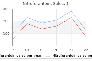
Nitrofurantoin
Purchase generic nitrofurantoin on line
Venous thromboembolism and mortality associated with recombinant erythropoietin and darbepoetin administration for the treatment of cancer-associated anemia antibiotics for acne and side effects cheap nitrofurantoin 50 mg without a prescription. Risks of venous thromboembolism and mortality associated with erythropoiesis-stimulating agents for the treatment of cancer-associated anemia. Erythropoietin to treat head and neck cancer patients with anaemia undergoing radiotherapy: Randomised, double-blind, placebo-controlled trial. Randomized, double-blind, placebo-controlled trial of erythropoietin in non-small-cell lung cancer with disease-related anemia. Darbepoetin alpha for the treatment of anemia in patients with active cancer not receiving chemotherapy or radiotherapy: Results of a phase iii, multicenter, randomized, double-blind, placebo-controlled study. Effects of erythropoietin receptors and erythropoiesis-stimulating agents on disease progression in cancer. Safety and efficacy outcomes with erythropoiesis-stimulating agents in patients with breast cancer: A meta-analysis. Consensus on the existence of functional erythropoietin receptors on cancer cells. Erythropoiesis-stimulating agents in oncology: A study-level meta-analysis of survival and other safety outcomes. Meta-analysis of epoetin beta and darbepoetin alfa treatment for chemotherapy-induced anemia and mortality: Individual patient data from japanese randomized, placebo-controlled trials. American society of hematology/american society of clinical oncology clinical practice guideline update on the use of epoetin and darbepoetin in adult patients with cancer. Prevalence of iron deficiency across different tumors and its association with poor performance status, disease status and anemia. Prevalence and management of cancer-related anaemia, iron deficiency and the specific role of i. Il-6 mediates hypoferremia of inflammation by inducing the synthesis of the iron regulatory hormone hepcidin. Impact of inflammation on ferritin, hepcidin and the management of iron deficiency anemia in chronic kidney disease. Management of anaemia and iron deficiency in patients with cancer: Esmo clinical practice guidelines. Anemia and iron deficiency in heart failure: Current concepts and emerging therapies. Clinical experience with ferric carboxymaltose in the treatment of cancer and chemotherapy associated anaemia. The effectiveness of intravenous iron for iron deficiency anemia in gastrointestinal cancer patients: A retrospective study. Iron deficiency across chronic inflammatory conditions: International expert opinion on definition, diagnosis, and management. Laboratory methodologies for indicators of iron status: Strengths, limitations, and analytical challenges. Regulation of iron homeostasis in anemia of chronic disease and iron deficiency anemia: Diagnostic and therapeutic implications. Hepcidin and hemoglobin content parameters in the diagnosis of iron deficiency in rheumatoid arthritis patients with anemia. Serum hepcidin in inflammatory bowel diseases: Biological and clinical significance. Hepcidin in tumor-related iron deficiency anemia and tumor-related anemia of chronic disease: Pathogenic mechanisms and diagnosis. Hepcidin is the major predictor of erythrocyte iron incorporation in anemic African children. Serum hepcidin levels predict response to intravenous iron and darbepoetin in chemotherapy-associated anemia. Toward worldwide hepcidin assay harmonization: Identification of a commutable secondary reference material. Intravenous iron optimizes the response to recombinant human erythropoietin in cancer patients with chemotherapy-related anemia: A multicenter, open-label, randomized trial. Addition of intravenous iron to epoetin beta increases hemoglobin response and decreases epoetin dose requirement in anemic patients with lymphoproliferative malignancies: A randomized multicenter study. Randomized, multicenter, controlled trial comparing the efficacy and safety of darbepoetin alpha administered every 3 weeks with or without intravenous iron in patients with chemotherapy-induced anemia. Randomized trial of intravenous iron supplementation in patients with chemotherapy-related anemia without iron deficiency treated with darbepoetin alpha. Darbepoetin alfa 300 or 500 mug once every 3 weeks with or without intravenous iron in patients with chemotherapy-induced anemia. The role of iron in the management of chemotherapy-induced anemia in cancer patients receiving erythropoiesis-stimulating agents. Intravenous iron supplementation for the treatment of chemotherapy-induced anaemia—Systematic review and meta-analysis of randomised controlled trials. Addition of iron to erythropoiesis-stimulating agents in cancer patients: A meta-analysis of randomized trials. Effect of intravenously administered iron sucrose on the prevention of anemia in the cervical cancer patients treated with concurrent chemoradiotherapy. Blood transfusion reduction with intravenous iron in gynecologic cancer patients receiving chemotherapy. Prevention of blood transfusion with intravenous iron in gynecologic cancer patients receiving platinum-based chemotherapy. Intravenous iron alone resolves anemia in patients with functional iron deficiency and lymphoid malignancies undergoing chemotherapy. Impact of ferric carboxymaltose on the evolution of hemoglobin and ecog performance status in iron-deficient patients with solid tumors: A 3-month follow-up retrospective study. Implementing a blood management protocol during the entire perioperative period allows a reduction in transfusion rate in major orthopedic surgery: A before-after study. Effect of single recombinant human erythropoietin injection on transfusion requirements in preoperatively anemic patients undergoing valvular heart surgery. Blood transfusion in major abdominal surgery for malignant tumors: A trend analysis using the national surgical quality improvement program. Iron therapy for the treatment of preoperative anaemia in patients with colorectal carcinoma: A systematic review. The feasibility and clinical efficacy of intravenous iron administration for preoperative anaemia in patients with colorectal cancer. Randomized clinical trial of preoperative intravenous iron sucrose to reduce blood transfusion in anaemic patients after colorectal cancer surgery. Pre-operative oral iron supplementation reduces blood transfusion in colorectal surgery—A prospective, randomised, controlled trial. Ferric carboxymaltose reduces transfusions and hospital stay in patients with colon cancer and anemia. The important role for intravenous iron in perioperative patient blood management in major abdominal surgery: A randomized controlled trial. Epidemiological and nonclinical studies investigating effects of iron in carcinogenesis—A critical review. Erythropoietin therapy after allogeneic hematopoietic cell transplantation has no impact on long-term survival. The effect of intravenous iron therapy on long-term survival in anaemic colorectal cancer patients: Results from a matched cohort study. Iron and innate antimicrobial immunity-depriving the pathogen, defending the host. Patient experienced clinical progression and no post-baseline tumor measurements were recorded. The liver also produces cholesterol, acids, and bile salts that get stored in the gallbladder until required to help break down ingested fats. More advanced liver damage leads to a feeling of fullness or pain in the upper right abdomen, itching, jaundice (yellowing of the skin and whites of the eyes), easy bruising, fatigue, and fluid retention. This is a condition whereby extra fat gets deposited in the liver, squeezing out normal liver cells. This can happen in other conditions as well, like diabetes, pregnancy, and obesity. No specific treatment is necessary as it does not cause any symptoms, but weight loss along with control of blood cholesterol levels will usually rid the liver of the extra fat and should be encouraged in any patient that has fat deposits.
Purchase nitrofurantoin 50 mg on line
The cause is excessive water intake infection 3 weeks after tonsillectomy nitrofurantoin 100 mg low cost, statement (c), causing euvolaemic hypotonic hyponatraemia. Tolvaptan is contraindicated in hyponatraemic urgencies, hypovolaemia, and liver disease. Practical Exercise 3: A 70 year old male was admitted with diarrhoea, vomiting for four days, lethargy, and inability to stand because of dizziness for two days. The cause is gastrointestinal and renal loss, statements (a), resulting in hypovolaemic hypotonic hyponatremia and hypokalaemia. Practical Exercise 4: A 76 year old male, chronic smoker, known case of hypertension admitted with increasing ankle swelling and shortness of breath. He felt unwell two months prior with intermittent central chest pains that lasted for a week; sought no medical advice. The cause is congestive cardiac failure, statement (e), resulting in hypervolaemic hypotonic hyponatremia. Diagnosis, Evaluation and Treatment of Hyponatremia: Expert Panel Recommendations. Clinical review: Practical approach to hyponatraemia and hypernatraemia in critically ill patients. Utility and limitations of the traditional diagnostic approach to hyponatremia: a diagnostic study. Consensus statement of the 1st International Exercise-Associated Hyponatremia Consensus Development Conference, Cape Town, South Africa. Hyponatraemia develops in very young or very old patients; those with altered mental state and dependent elderly are at particularly high risk[1–8]. Mechanism HrN is due to deficiency of body water in relation to existing Na stores Symptoms and Signs Primarily neurologic. Thirst; Neurologic symptoms vary – lethargy, confusion, Prominent when fits and coma in acute cases. Inability to drink; Renal ‘free’ water losses Excess salt (The failure of thirst – (rare). Renal ‘free’ water evident from is the commonest cause Conn’s S losses: Osmotic the history) 2. Prompt determination and treatment of the Acute (rare): treatment underlying cause Discontinue Na Only hypotonic b. Fluid replacement: enterally where possible, the s[Na] to 145 appropriate[4] safest route Monitor s[Na] every 1–2hrs Asymptomatic: Hypovolaemic1 Normovolaemic symptomatic: 1Symptomatic: Oral fluids a. Correct HrN fluids estimation with hypotonic equations, see fluids: text below Monitor s[Na] 4–6 hourly. The type of fluid depends on whether there is overall fluid depletion or sodium excess. The rate of hypernataemia correction relies on its duration: Rapid if acute Slow over 2–3 days if chronic at a maximum reduction of 10mmol/24hrs – rapid correction is potentially dangerous d. Add the 24-hour obligatory fluid losses and any further ongoing incidental fluid losses. Equations for estimating fluid replacement to correct hypernatraemia are given below. The response to correction should be guided by monitoring the clinical status and s[Na] levels. Re-measure s[Na] after 4-6hrs and adjust infusion rate if the correction is either too fast or too slow. Monitor s[Na] levels every 4–6hrs in patients with ongoing fluid losses and ‘Replace’. Acute Hypernatremia (#48) is rare[1–8]: Treatment: the goal is to rapidly reduce sNa1 to normal in,24hrs using the equations for estimating fluid replacement to correct hypernatraemia given below. Once s[Na] has reached 145mmol/l, the rate of infusion is reduced and continued until normal level (140mmol/l) is restored. Resuscitate; monitor fluids input and output and Replace any further ongoing water and electrolytes losses. Estimating fluid replacement to correct hypernatraemia: Using formula I and its derivative 2[4]: 1. Formula 1 (and its derivative formula 2) estimates the change in the s[Na] caused by the retention of 1 litre of any infusate. The required volume of infusate and hence the infusion rate, is determined by dividing the change in the s[Na] targeted for a given treatment period by the value obtained from formula 1. Formula 1 (and its derivative formula 2) permits a quantitative and flexible approach to the prescription of fluids that can easily accommodate different infusates and treatment periods[4]. Ensure adequate intake in ill patients, pre-emptive management of possible causes. None of the above A1: this is a case of pre-renal acute kidney injury and chronic hypernatremia secondary mainly to pure water loss. Hypernatraemia is caused by the un replaced free water loss secondary to fever and Lithium-induced nephrogenic diabetes insipidus, statement (d). He was unwell and confused, hence unable to compensate for the large urinary water loss, for few days prior to admission. A2: the required fluid volume for replacement is estimated using Formula 1, or conventional equation. Estimating fluid replacement using Formula 1: the desired water replacement in the first 24 hours to reduce s[Na] by ~ 10mmol/L using 5% dextrose is: 1L of 5% dextrose will reduce s[Na] by 4. Hyperglycaemia is deleterious; it will induce osmotic diuresis and worsen hypertonicity. This formula gives an estimation of the volume of additional fluid required to correct s[Na] to 140mmol/L. The volumes obtained from these equations are indicative: the correction needs good clinical acumen and close monitoring. Volume depletion versus dehydration: how understanding the difference can guide therapy. Fluid management of hypernatraemic dehydration to prevent cerebral oedema: a retrospective case control study of 97 children in China. This is the commonest electrolyte disorder in hospitalised patients; most cases are mild. S/S are prominent when the reduction in s[K] and Signs (S/S)[1–7] occurs rapidly or is large. Muscle weakness – ascending, can involve respiratory muscles, ileus, rhabdomyolysis secondary to decreased muscle blood flow. Glucose Intolerance; Metabolic Alkalosis; Polyurea S/S develop usually when s[K] is,3 and resolve with correction of HoK 31 C05-Hypokalaemia. Intracellular shift (redistributive HoK): insulin or bicarbonate treatment, theophylline, β2 agonists, periodic paralysis, chloroquine intoxication, rapid blood cell proliferation –. Metabolic alkalosis: indicates chronic depletion (see table) u[K]: if cause not obvious HoK: many patients, up to 40%, are also Mg deficient. Combined deficiency may potentiate the risk of cardiac arrhythmias – both are pro-arrhythmic. Redistributive HoK: cautious correction if serious complications present or imminent – paralysis or arrhythmia. If life threatening/intractable cardiac arrhythmia: double infusion rate, up to 40mmol/hr[3], and contact resuscitation team. Renal impairment: cautious K replacement – risk of overcorrection (hyperkalaemia) – contact renal team if severe renal failure or patient on dialysis Use pre-mixed infusion fluid. The second aim is to correct hypokalaemia and gradual correction of hypernatraemia over 2–3 days. Hyperkalaemia is the commonest electrolyte disorder causing cardiorespiratory arrest[8]. Signs (S/S) (s[K] in S/S are prominent when the increase in s[K] occurs rapidly or is large. The classification into mild, moderate and severe hyperkalaemia provides a guide to clinical decision-making.
Buy cheap nitrofurantoin 50 mg on-line
Three suture markers on aortic sewing cuff Holder/Rotator A holder/rotator is attached to each valve antimicrobial news discount 100 mg nitrofurantoin visa. This device may also be used to replace a previously implanted mitral or aortic prosthetic heart valve. Attempts to reuse the valve may result in valve malfunction, inadequate sterilization, or patient harm. The valve holder/rotator is intended for single use only and should be discarded after surgery. This could result in scratched or damaged valve components, leaflet fracture, or dislodgment. Precautions Valve Do not touch the prosthetic valve unnecessarily, even with gloved hands. This may cause scratches or surface imperfections that may lead to thrombus formation. Under the scan conditions defined above, the prosthetic valve is expected to produce a maximum temperature rise of less than or equal to 3. Potential Adverse Events Complications associated with replacement mechanical heart valves include, but are not limited to , hemolysis, infections, thrombus, or thromboembolism, valve dehiscence, unacceptable hemodynamic performance, hemorrhagic complications secondary to anticoagulation therapy, heart block requiring pacemaker implant, prosthetic failure, adjacent cardiac structure interference, heart failure, stroke, myocardial infarction, or death. The package includes: Sealed non-sterile outer tray Sealed sterile inner tray One (1) valve with identification tag, mounted on a plastic holder/rotator One (1) disposable support collar One (1) insert with Instructions for Use web address One (1) Medical Device Registration Form with attached Patient Identification Card and return envelope the sizer is supplied non-sterile. Clean and sterilize the sizer before each use following the reprocessing guidelines described in these instructions. Storage To minimize the possibility of contamination and to provide maximum protection, store valve in cool, dry area until needed. Valve Resterilization If resterilization of the valve is necessary, use only the recommended steam cycles and follow the instructions below. Recommended Sterilization Cycle Parameters Vacuum Cycle Pre-Vacuum Steam Pre-Vacuum Flash Purge Time: 6 minutes 6 minutes Pulses: 2 2 Pulse Pressure: 204. The sizer handle may be bent in any direction up to a 90° angle without degradation. When the flexible handle is exposed to the heat produced during autoclave sterilization, it will revert to its original shape. Alternatively, a Hegar dilator may be used to facilitate proper valve size selection. Consult the appropriate instrument Instructions for Use for full product description, and relevant cleaning and sterilization information. Instruments have been designed and tested for repeated use, however, if visual signs of deterioration become apparent, do not use the instrument, and contact Customer Service for a replacement. Directions for Use Removing the Native Valve Excise the native valve and prepare the annulus for the valve replacement. The cylindrical end of the sizer must pass readily without resistance through the annulus. Verify that the catalog number and serial number on the outer tray are identical to those on the box label. If the information is not identical, do not use the device, and contact Customer Service as soon as possible. Holding the outer tray by the bottom, present the inner tray to the sterile scrub nurse or surgeon. Complete the patient [medical device] registration information, as described in the “Patient Registration” section. Holding the inner tray with the cover upward, grasp the tab and pull back to completely remove the inner tray cover. Press the sterilized mechanical valve holder handle into the valve holder/rotator. To remove the valve from the inner tray, firmly lift the valve holder handle and the valve support collar. Prior to implanting the valve, remove the support collar from the valve by placing two fingers under the collar, pinching with the thumb and pulling back gently. Verify that the valve size and model number on the identification tag are identical to the valve size and model number listed on the packaging. Suturing techniques may vary according to the preference of the implanting physician and the needs of the patient. Align the valve such that the pivot guards are in the desired orientation in the annulus. Incorporate adequate sewing cuff material into each suture pass to maintain the valve in the desired position. It may be helpful to tag the sutures adjacent to pivot guards when inserting them. To prevent cutting the fibers in the sewing cuff, use of standard round or taper point needles is recommended. Cut the two retention sutures from the holder/rotator, and gently withdraw the holder/rotator from the valve. Maintain the holder/rotator in the sterile field for further use as a rotation instrument. Jude Medical™ Leaflet Tester, open the valve and inspect the area for any obstructive tissue. If visualization is inadequate, use the leaflet tester to confirm free leaflet motion. Test leaflet motion again, and if desired, rotate the valve using the holder/rotator handle (see “Valve Rotation”). If resistance is noted, the valve holder/rotator may not be properly seated in the valve, or the valve may be oversized. Properly seated valve holder/rotator 10 Postoperative Considerations Echocardiography is recommended to assess valvular competency and performance. This could result in scratched or damaged valve components, or leaflet fracture or dislodgment. Prophylactic antibiotic treatment should be considered for patients undergoing dental procedures. Acceptable forms of anticoagulants include intravenous unfractionated heparin or oral warfarin. Risk factors include first 3 months post-implant, younger age (< 6 months), small size (<6 kg), low flow state, unreliable oral intake, previous thromboembolism, and hypercoagulable condition. Use of low molecular weight heparin may be associated with an increased rate of complications. Intra-Operative Assessment the suggested method for assessing competence of the valve is with intra-operative Doppler echocardiography. Patient Registration A medical device registration form and return envelope are included with each device. Complete the identification card attached to the medical device registration form and provide it to the patient. After implantation, please complete all requested information and return the original form to St. Please disregard any request for patient information if this contradicts your local legal or regulatory requirements regarding patient privacy. Sizer Reprocessing Requirements these reprocessing instructions were validated for sizer model 905-15. However, if visual signs of deterioration become apparent, do not use the instrument and contact customer service for a replacement. Testing indicates that this instrument may undergo 100 cleaning/disinfection cycles when cleaned using the specified reprocessing method. Deviations from the specified method may result in reduced instrument life or inadequate cleaning and disinfection. After each reprocessing cycle inspect instruments for signs of cracking, crazing, or degradation that may affect function. Handle sizer sets in the same manner as other reusable instruments that require packaging or wrapping during autoclave sterilization. All instruments must be sterilized in a tray or container that is permeable to steam. Reprocessing Option 1 Manual Cleaning and Disinfection Method the following manual cleaning and disinfection method was validated: 1. Rinse the instrument in hot, running tap water for a minimum of 20 seconds to remove visible blood soil.
Buy cheap nitrofurantoin 100mg on line
In a patient with the sudden onset of sharp antibiotics for human uti cheap nitrofurantoin 50 mg fast delivery, centrally located chest pain radiating to the neck, shoulder, and trapezius ridge, aggravated by deep breathing and the recumbent posture, relieved when sitting up and leaning forward, the presence of a superficial, scratchy, grating, leathery-quality three-component pericardial friction rub, heard best along the left sternal border, generally increasing in intensity with inspiration, suggests acute pericarditis. The pericardial friction rub, although pathognomonic of pericarditis, may be missed on auscultation since it can be remarkably evanescent. When a rub is not heard initially in a suspected case of pericarditis, frequent, repeated auscultation using firm pressure on the diaphragm of the stethoscope with the patient sitting upright and leaning forward can be rewarding in its detection. As a rule, it is wise never to diagnose a “one-component pericardial friction rub. A pericardial effusion does not always cause the heart sounds to be reduced in intensity and may not eliminate the presence of a pericardial friction rub. In fact, acute pericarditis may be present without Curr Probl Cardiol, July 2008 393 significant (or any) pericardial effusion on echocardiography. Echocardi ography, therefore, is useful for confirming the diagnosis when it shows even a small pericardial effusion, but the absence of effusion does not exclude the diagnosis. In a patient with an underlying malignancy (especially lung, breast, lymphoma), nonpenetrating (eg, steering wheel) chest injury, or end-stage renal disease on dialysis, a new pericardial friction rub suggests pericar dial metastases or traumatic or uremic pericarditis, respectively. Constrictive pericarditis is characterized by a thick, rigid, scarred, pericardium that restricts filling of all four chambers of the heart. It is usually a chronic consequence of acute or viral pericarditis but may occur with carcinoma (especially breast and bron chogenic), prior radiation therapy to the chest for malignancy, and particularly following previous cardiac surgery. The presence of a high-pitched, early diastolic sound (pericardial knock), often becoming louder with inspiration (in the absence of a loud S1 or diastolic rumble) in the setting of unexplained, especially right sided heart failure with markedly distended neck veins, ascites, and edema should prompt the clinician to suspect the diagnosis of constrictive pericarditis. Although usually a chronic consequence of acute or viral pericarditis, nowadays, one should search specifically for these findings in the patient who has had prior radiation therapy to the chest for malig 179,180 nancy, and particularly after previous heart surgery. The Athlete’s Heart Clinical evaluation of the well-trained athlete may present a challenge to the clinician. Auscultatory findings that would be considered “abnor mal” in less well-conditioned individuals are not uncommon in healthy young athletes. The practitioner should be aware that the physiologic effects of training may produce changes in the cardiovascular system that can mimic pathologic heart disease (so-called “athletic heart syndrome”). Common auscultatory findings in highly trained athletes include a slow heart rate, a grade 1-2/6 (“innocent”) systolic murmur, wide splitting of S1 and S2, an S3 and S4 gallop, and a jugular venous hum. The distinction between the athlete’s heart and cardiac disease has important implications. The erroneous diagnosis of heart disease in a 394 Curr Probl Cardiol, July 2008 normal athlete may have unfortunate consequences, including limitation of physical activity or disqualification from participation in competition. On the other hand, with certain cardiac conditions, participation in competitive athletics carries the risk for sudden death. Determining which murmurs are pathologic and which are benign is perhaps the most challenging aspect of preparticipa 181-183 tion evaluation of the athlete. Conclusion this monograph is a timely reminder of the continued importance of cardiac auscultation in the contemporary practice of medicine. Despite the growing reliance on technological advances, cardiac auscultation remains a valuable and cost-effective clinical skill, often enabling the well-trained clinician to arrive at a rapid and accurate cardiac diagnosis, in many cases, without recourse to more elaborate and expensive “high-tech” investigative methods. It requires both continued interest on the part of the student and expert mentoring by experienced clinician teachers proficient in the art of auscultation. Once learned, cardiac auscultation must be practiced repeatedly to be performed skillfully. Traditional teaching methods, eg, textbooks and didactic classroom lectures, followed by a brief demonstration of heart sounds and murmurs, have yielded disappointing results. The best way to gain proficiency and greater accuracy and confidence in cardiac auscultation is to listen carefully to large numbers of actual patients under the expert guidance of an experienced clinician auscultator, obtaining “real-time” confirmation and immediate direct feedback. After decades of neglect and apathy, today’s medical trainees are now suffering the consequences, as there are few, if any, expert cardiac auscultators around to teach the finer points of this time-honored, but virtually lost, clinical art. These “high-tech” adjuncts can supplement deficiencies in clinical train ing and enable clinicians at any level to learn, practice, and perfect their cardiac auscultatory skills at their own time, convenience, and pace. Restoration of the lost art of cardiac auscultation may be a difficult task but is well worth the effort. The challenge for modern-day clinical educators, therefore, is to decide on what specific aspects of cardiac auscultation are most important and practical to teach and learn in today’s fast-paced health care environment. Does it have the characteristics of an “innocent” murmur or a “significant” murmur? Does it change with respiration, position (eg, standing or squatting), or certain physiologic maneuvers (eg, Valsalva)? Are there any other extra heart sounds present (eg, systolic click, ejection sound, opening snap)? Familiarity with these essentials of cardiac auscultation can dispel the panic and sense of intimidation often felt by today’s medical trainees when asked to listen to a patient’s heart. Only then will contemporary clinicians be willing to incorporate cardiac auscultation into routine clinical practice and teaching and pass along this time-honored art enthusiastically to the next generation of practicing physicians and other health care professionals. We must not allow cardiac auscultation to become a lost art or the stethoscope a medical relic. Auscultation of the heart remains the cornerstone of the cardiac clinical examination, and when skillfully performed, can lead to fewer misdiagnoses, foster improved patient trust and confidence, and provide more effective and economically sound cardiovascular care. Rediscovering the lost art of cardiac auscultation and restoring it to its rightful place and “time-honored” status may be a 184-227 tedious task, but it is well worth the effort (Fig 29). Dedication I wish to dedicate, in loving memory, this monograph on the lost art of cardiac auscultation to Dr. Proctor Harvey, who instilled in those of us fortunate enough to have been trained by him a love for clinical cardiology that has greatly enriched our professional lives. From the passion for cardiology that he instilled in us springs the inspiration to pass along his rich legacy and to carry on the teaching tradition that is his. Appropriate use of physical findings to reduce reliance on sophisticated and expensive methods. Cardiac auscultatory skills of internal medicine and family practice trainees: a comparison of diagnostic proficiency. The teaching of cardiac auscultation during internal medicine and family medicine training: a nationwide comparison. Physician-performed point-of-care echocardiography using a laptop platform compared with physical examination in the cardiovascular patient. Evaluation of the Patient with Heart Disease: Integrating the Physical Exam and Echocardiography. A systematic approach to the bedside differentiation of cardiac murmurs and abnormal sounds. Dynamic cardiac auscultation: bedside interventions useful in the differential diagnosis of heart sounds and murmurs. Diagnosis of left sided regurgitant murmurs by transient arterial occlusion: a new maneuver using blood pressure cuffs. In: Mitral Valve: Floppy Mitral Valve, Mitral Valve Prolapse, Mitral Valvular Regurgitation, 2nd edition. Postural changes in left ventricular and mitral valvular dynamics in the systolic click-late systolic murmur syndrome. Hemodynamic explanation of why the murmur of mitral regurgitation is independent of cycle length. Interobserver agreement by auscultation on the presence of a third heart sound in patients with congestive heart failure. The normal third heart sound and gallops, ejection sounds, systolic clicks, systolic whoops, opening snaps and other sounds. Audibility of the fourth heart sound: relationship to presence of disease and examiner experience. Prognostic importance of elevated jugular venous pressure and a third heart sound in patients with heart failure. Third heart sound revisited: a correlation with N-terminal pro brain natriuretic peptide and echocardiography to detect left ventricular dysfunction. Clinical significance of systolic murmurs: study of 1000 consecutive “non-cardiac” cases. Opinion—a probability-based management plan: what constitutes an adequate initial evaluation of the patient with a heart murmur? Prevalence of valvular regurgitation by Doppler echocardiography in patients with structurally normal hearts by 2-dimen sional echocardiography. Doppler echocardiographic evaluation of valve regurgitation in healthy volunteers. Doppler echocardiography: appropriate use in the evaluation of the patient with a heart murmur.
Buy line nitrofurantoin
Many pregnant women will experience deterioration of one class as pregnancy progresses antibiotics buy best order nitrofurantoin, and they should be warned about this. They may need to take more rest than usual during pregnancy, although it is also important for them to maintain their fitness as much as possible. Clinicians should be familiar with the appropriate questions to elicit symptoms accurately. For example, in response to the question ‘do you get short of breath climbing stairs? The correct question is ‘how many flights of stairs can you climb at a steady pace without having to stop because of shortness of breath? Most pregnant women complain of tiredness, and women with cardiac disease are no exception. This is why continuity of carer is so important, because sometimes deterioration in the woman’s condition is more apparent in her demeanour and the way she answers questions than in the precise answers she gives. A useful tactic is to call a woman to your consulting room yourself and watch how quickly she can walk from the waiting area to your consulting room, how short of breath this makes her, and what her pulse rate and rhythm is when she first sits down (a ‘mini exercise test’). The pulse rate is best measured using a stethoscope and auscultating the heart, because when the pulse becomes fast, irregular or faint, the radial pulse is often difficult to detect accurately. The woman’s blood pressure should be checked carefully using a manual sphygmomanometer. The woman should be seated comfortably, not talking, with an appropriately sized cuff placed on the correct arm (for example, the right arm is usually used in women with coarctation of the aorta, 80% of whom will also have a bicuspid aortic valve). The arm should be supported and held out at an angle so that the cuff is at the level of the left atrium. An excellent resource showing how the blood pressure should be taken correctly can be found at. Heart murmurs are graded from one (extremely soft) to six (the loudest one has ever heard). It is usual for a murmur to increase by one grade as pregnancy progresses because of the increase in cardiac output. A sudden increase in the loudness of a heart murmur can suggest the development of vegetations from endocarditis. For example, in a woman with Marfan syndrome, the appearance of a diastolic murmur can indicate dilatation of the aortic root with the onset of aortic regurgitation. This will usually require urgent intervention as it may lead to heart failure or aortic dissection. Women sometimes have persistent crackles in a localised area following previous surgery, and this should be recorded at the beginning of pregnancy so as not to be confusing later on. Sometimes women develop crackles as a result of poor lung expansion late in pregnancy, when the diaphragm is splinted by the enlarging uterus. Asking the woman to take several deep breaths and cough several times will usually cause such crackles to disappear. Any woman who complains of feeling suddenly less well, who develops ‘funny turns’ (any loss of consciousness is always significant in such women), a sudden increase in shortness of breath or new palpitations associated with other symptoms should always be assessed carefully by a cardiologist. In tertiary centres it is usually possible to obtain an emergency echocardiogram 24/7. Arterial blood gas measurement can be informative, as can a chest X-ray, taken with screening of the fetus. If the woman complains of chest pain, it is useful to take blood immediately for measurement of troponin I levels and repeat the test 24 hours later to assess whether there has been any significant myocardial damage. In tertiary centres, an exercise treadmill test is the first non-invasive test of choice to investigate the possibility of coronary artery disease, assuming the patient is well enough. A myocardial perfusion scan or coronary angiography can be considered if symptoms continue or worsen despite treatment. Pulmonary embolism should also be considered and blood taken for measurement of d dimer levels – if these are raised, anticoagulant treatment is probably the safest response. In doubtful cases, a ventilation/perfusion scan or computed tomography pulmonary angiography should be carried out, depending on local availability (bearing in mind that both expose the fetus to some radiation, particularly computed tomography scanning, although it is diagnostic in a higher proportion). Doppler examination of the leg vessels should be performed to identify any deep vein thrombosis. Dissection of the aorta should also be considered and may be detected on echocardiography, although magnetic resonance imaging is more sensitive, particularly for the thoracic aorta. Computed tomography scanning can also be used but exposes the fetus to a considerable radiation dose. Management of a woman who develops new symptoms is dependent on the nature of the underlying lesion and the results of urgent investigations of cardiac function. It is not possible to give a brief account of the various management strategies which will be necessary, because they vary depending on the underlying lesion. Women with cyanotic heart disease, valvular disease, aortic dissections or arrhythmias require very different management, and many women will have an almost unique combination of lesions, requiring management tailored to their individual diagnosis. This is why an experienced cardiologist used to seeing pregnant women should always be involved in their care, especially in emergencies. They should be given an estimate of their risks which is as accurate as possible, and this risk should be reassessed every five years (or more often if their condition deteriorates significantly). They should be advised whether specialist care from a high-risk pregnancy with heart disease team is advisable in the event of pregnancy. If so, they should be advised to see the appropriate high-risk team as soon as a pregnancy is confirmed, which will usually be by a urinary pregnancy test within two weeks of the missed period. Women who present initially to their general practitioner or community/local hospital midwifery service, and give a history of heart disease should be referred promptly to an appropriate high-risk pregnancy and heart disease team. At the initial assessment by the high-risk multidisciplinary team, a full clinical examination should be carried out and all recent investigations reviewed. An electrocardiogram should be taken and kept in the notes for future reference, in the event that there is any change in cardiac status. The woman should be asked to carry her notes with her at all times, in case of any emergencies. It is important to offer the woman a fetal nuchal translucency scan, as this is a significant indicator of recurrent cardiac disease in the fetus. Once this scan has confirmed a viable fetus without obvious abnormalities, a standard fetal anomaly scan at approximately 20 weeks of gestation, and a fetal cardiac scan at approximately 22 weeks of gestation, should be organised. Depending on her cardiac status, the woman should be seen by an appropriately experienced consultant obstetrician every two to four weeks until 20 weeks of gestation, then every two weeks until 24 weeks of gestation, and then weekly thereafter. Continuity of carer is of particular importance, because this makes it much easier to detect any deterioration in the woman’s condition. If the woman threatens to go into labour before 34 weeks of gestation, immediate assessment by the multidisciplinary team is important to assess the best management. In pregnancies that are progressing satisfactorily, a multidisciplinary team assessment at 32–34 weeks of gestation is important to plan care around the time of delivery and to establish optimum management. The woman should be given clear instructions about how to recognise the onset of labour. Once labour begins, she should immediately ring the labour ward to alert them that she is coming. She needs to make sure that they appreciate she is a cardiac patient so that they do not give her advice to wait at home, go for a bath, etc. On arrival at the labour ward, the woman should make herself known immediately to the labour ward staff. This is likely to include informing senior staff, usually consultants, of the woman’s admission. The majority of women with significant lesions will have epidural anaesthesia during labour, and a significant number will have an assisted vaginal delivery. All anaesthetics should be given by senior staff who are familiar with the delivery plan and have experience of pregnant women with cardiac disease. Following delivery, the woman should be transferred to a high-dependency area where she can be monitored closely for anything between 12 and 48 hours. She should not be transferred to a normal labour ward until she has been reviewed by senior staff (preferably consultants) who can determine whether she will be safe in an area where monitoring will be less intensive. Before discharge, a check should be made that the woman has appropriate appointments for obstetric and cardiac follow-up and that she is aware of her contraceptive options. At the postnatal check-up, the woman should be assessed for her recovery from giving birth. Her cardiac function should be checked by a cardiologist, and arrangements made for cardiological follow-up.

Generic nitrofurantoin 50mg visa
This principle implies an acceptable solu whilst recognizing the limitations of individual cate tion to the effective new organisation of the work of gorizations vyrus 985 c3 purchase nitrofurantoin 100 mg. Decisions on the reduction of must stand at the beginning of all decisions if realistic medical services or on the withholding of innovative, values for health are to be determined. Demographic new forms of therapy base mainly on the subjectivity development should not primarily be the main argu of the doctors involved and the ability of hospitals to ment for a limitation of fundamental services. Objective and safe support more attention must be paid to an existent co-morbid systems to help decision making lack completely. Although sidering the extent and prognosis of the illness and the their information according to present requirements individual situation of the patient, it is possible to dis must be regarded as being completely inadequate, they cover constellations which come into question for a could in future years really document the transparency limitation of therapy. The content of the quality reports must be extended to include real medical quality parameters. In future years it will not be possible for Acute and intensive medical care and the regular oper physicians to avoid dealing with economic questions ative treatment needed here make these areas most in the field of medicine. At the same time it is here that the borderline be physicians we should see it as a chance to bring in our tween life and death, medical–technical possibilities systems of value and our ideas of quality to this dis and ethical-human decisions lie very close to each oth cussion on economy or even to avoid erroneous trends. It is daily necessary to redefine what is responsible In so doing we should also make use of ethical thought from a humane point of view and what is possible to define the framework conditions of our decisions. It can be ethically justified is acute and intensive medicine that daily experiences that intensive care doctors are at present developing a the borderline situation between life and death, be system whereby reliable indicators can be developed tween what is medically possible and necessary and for ending further therapeutic measures so as to avoid which stands under high cost-pressure as one of the prolonged dying when primary hopeless prognosis has most expensive areas of any hospital. Equally the acceptance of the patient’s will tive co-operation of ethics and economy can form the can be justified in so far as this is explicitly formulat basis of a high quality and eligibility for financing in ed and the prevailing critical state of health allows tensive medicine in the interest of our patients. A positive side-effect is the possible Economy can help to develop strategies for ration reduction in costs in intensive medical treatment. It can identify and avoid what is superfluous unacceptable to undergo an intensive “triage” merely and unnecessary without having to ration. However, such decisions should be Economy continues to make ethics possible, that made in acknowledgment of the patient is: makes it possible again! Diener German Cochrane Centre, Institute for Medical Biometrics and Medical Informatics, University Hospital Freiburg, Germany Medical practice should be based on comprehensive 1. By appraising the randomiza based on valid results from sound, patient-oriented tion procedure (or the assessment whether it was done clinical research to reduce the misleading influence of at all), blinding of the patients and/or investigators and biases and the play of chance. However, this comparability of the study patients and study interven favourable approach is challenged from several sides. Hence, the steadily growing source of evidence is separated from the utilization of If the results are likely to be valid and if we can anti the existing evidence by a barrier often asterisked as cipate a more or less unbiased assessment of treatment the “Know-Do-Gap” (1). Secondly, much of the avail effect, the reader has to appraise the size of the treat able literature is of questionable quality (2). Since the “true effect” sult, information overload and poor quality necessitate can never be known the point estimate of the treatment efficient strategies to separate junk from valuable re effect observed in the study is the best we have. As search data and reliable assessment of the latter to suming that the true value lies somewhere in its neigh come to an overall conclusion that can be used for de bourhood, it is fundamentally important to know the cision making. Quality assessment (Critical Appraisal) precision of the point estimate, that is the confidence is an important step when preparing systematic re interval. A 95% confidence interval can be simply in views but also for the daily evidence-based medical terpreted as defining the range that includes the true practice (3). Will the results help me in caring for my the leading principles to judge whether trial results can patients? Randomization, blinding, concealing the sequence of treatment allocations, adequate treatment Having assessed internal validity and the size of the of drop-outs in the analysis are particularly relevant treatment effect, the clinical usefulness of the findings for bias protection, but there are others which also has to be interpreted. Thus, when assessing an article nal validity, generalisability or applicability are often dealing with therapeutical interventions, one can use mentioned (6). Relevance of the findings strongly de fully pose at least the three following questions (4): pends on external validity, which is the prerequisite for a reasonable application of the trial results to a defin able group of patients in a particular clinical setting in routine practice. Amongst other things, we have to de the role of sound scientific data for evidence-based decision making 37 3. Churchill were considered and whether the likely treatment ben Livingstone efits are worth the potential harms and costs. Die Epiduroskopie ermöglicht eine zielgerichtete Therapie betrof fener schmerzrelevanter Regionen. Auch eine Platzierung von Kathetern, lasergestützter Lösung von Narbenfeldern und Unterstützung bei weiteren invasiv-interventionellen Eingriffen erweitern bei Schmerzpatienten die vorhandenen therapeutischen Möglichkeiten. One of the essential elements is provision of ade quate postoperative pain relief without excessive seda tion. In addition, early resumption of bination with neuraxial techniques, either single shot spontaneous ventilation is of particular benefit in sin or with an indwelling catheter, and the use of alpha-2 gle ventricle physiology by improving cardiac output agonists such as clonidine or dexmedetomidine in the (8). The anesthetic technique preferred by us has been immediate postoperative period have been described an inhalation-based anesthetic technique supplement (5). Because a majority of patients presenting for car ed by very low dose narcotics, and caudal or spinal diac surgery in our institution are either on platelet in morphine. In addition, all patients undergo modified hibitors or anticoagulated for rhythm disorders, neu ultrafiltration. Close to 80% of patients leave the oper raxial techniques are mostly contraindicated (6). Ex At the Mount Sinai Medical Center we do not clusion from fast track are critical ill neonates, patients have a formal fast-track protocol for adult patients. Our results compare favor Patients who underwent mitral valve repair are mostly ably with published data (9, 10). Our main seda Multiple logistic regression revealed that not pro tion modality for fast track patients is an infusion of ceeding with planned extubation was associated with dexmedetomidine at a rate of 0. Early extubation in pediatrics has been advo quired re-intubation for respiratory depression. Ann Thorac Surg 29: 228-33 sessment of resource use in fast-track cardiac surgery 1-year af 8. Crti Care Med 30: 787-91 unit and hospital length of stay in cornary artery bypass pa 11. Critical ating room extubation after congenital heart disease surgery in Care Med 34: 1624-34 children. Retrospective review of all cases qualifying for early J cardiothoracic Vasc Anesth 19: 49-53 extubation from July 2002-May 2005 40 H. Mahla Risk of recent coronary artery stenting before noncardiac surgery: On the edge between stent thrombosis and surgical bleeding H. Clopi Cardiology for percutaneous coronary artery interven dogrel, a thienopyridin, inhibits platelet aggregation tion recommend that after implantation of a bare met by irreversible blockade of adenosin diphosphat medi al stent clopidogrel must be continued for 3 to 4 weeks ated platelet function. Therefore, we have to rely on recommendations of ago, who is scheduled for prostatic surgery task forces and expert opinions (16 19). Basically, when assessing the perioperative risk of patients with recent coronary artery stenting before noncardiac surgery we have to plot the risk of throm bosis vs. Dual antiplatelet regime can be stopped, changed or continued accord ing to this assessment. Figure 3 shows the algorithm for the preoperative management according to the ur gency of noncardiac surgical procedures. Depending on the calculation of the thrombosis/bleeding risk 3 options are possible: 1. Continue aspirin, stop clopidogrel this concept balances in many cases the risk bene fit ratio Fig. This option should be strictly restricted to high bleeding procedures, like urologic, intracranial and some types of tumor surgery. Anesthesiology 94: stents and dual antiplatelet therapy scheduled for non 367-68 cardiac surgery benefit from a close cooperation be 2. A re Interventions 63: 141-5 port of the American College of Cardiology/American Heart As 7. Guidelines for Percutaneous Coronary Inter with coronay artery stent: what should the anesthesiologist ventions. Jahrestagung der Deutschen Gesellschaft für Klinische Mikrozirkulation und Hämorheologie Myokardiale Mikrozirkulation Entzündungsreaktionen zuzüglich begleitender Veränderungen des Hämostasesystems und der Mikrozirkula tion können weder teleologisch noch klinisch getrennt voneinander betrachtet werden.

Order nitrofurantoin from india
If lameness at a walk is due to a sore muscle antibiotic colitis purchase nitrofurantoin no prescription, the lame ness might very well appear less pronounced at a trot. This is due to the masking effect of the muscle groups working strongly together at the faster gait. If lame in a front leg, the horse will raise his head as the lame leg strikes the ground. If lame in the hindquarters, the horse will drop his head as the lame leg strikes the ground. Any lameness that is still obvious at a trot could indicate that the problem lies deeper, for example, in a ligament or joint. The conformation check-up routine helps you determine abnor malities and understand certain aspects of the signs and symptoms manifested by the horse. The ground check routine contributes greatly to your evaluation and helps you decide the best course of action for your horse’s treatment. A solid knowledge of equine conformation is important as it helps you better understand the different criteriums of selection for move ment. So many breeds are available today that a classical under standing of proper conformation will assist you in evaluating muscular conditions and faulty gaits. This understanding will build up your confidence in your application of equine massage. The Head and Neck A horse uses his head and neck to keep the rest of his body in bal ance during motion. A long-necked horse with average head carriage will extend the leg and have a long stride. A short-necked horse with an average head carriage will bend the knees and have a short stride because of the short neck. A long neck gives a horse a mechanical advantage in balancing himself by making a wide range of adjustments along the length of his body during any athletic movements. The 7 cervical ver tebrae have considerable lateral and vertical flexibility; they act as a blueprint for the shape of the neck. The length of the neck varies from breed to breed because each breed has different sizes of vertebrae. The “S” curve is extreme in Saddlebreds, average in Thoroughbreds, and almost flat in Quarter Horses. The ligamentum nuchae (nuchal ligament) is a strong liga ment that runs from the poll to the withers, where the splenius muscle attaches to it. When the splenius muscle contracts it raises the neck; it lowers the neck as the muscle relaxes. The multifidus cervices and the rectus capitis lateralis muscles assist lateral rota tion of the head. Remember that during motion all the muscle groups of the body work at once, ever ready to assist the horse in any situation. Signs and Symptoms: When this muscle is tight the horse shows discomfort, pulling the head to that side, resisting sideways motion to the opposite side and continuously stretching the neck and head. At rest, the horse will have a tendency to keep his head low, continuously stretching it to relieve muscular tension. If the stress point is very tender the horse will flinch and perhaps try to pull away from the pressure. The whole muscle will feel tight from its origin all the way to its attachment on the skull. When both sides contract simultaneously, they extend the neck, bringing the head up (extension). When contracting unilaterally, the muscle turns the head and neck to the side (lateral flexion). Signs and Symptoms: When the muscle is tight, the horse shows discomfort by extending the neck or by pulling the head and neck to the affected side. At rest, the animal will have a tendency to keep the head low, continuously stretching it to relieve the muscular tension. If the stress point is very tender the horse will flinch, per haps trying to pull away from the pressure. This is a sign of exces sive tightness and stress; if you feel heat, suspect inflammation. It is located on the insertion tendon of the splenius muscle at the base of the skull. When both sides contract simultaneously during motion they bring the point of the shoulder up toward the head. When one side of the brachio cephalic muscle contracts, the horse will move his neck to that side. Signs and Symptoms: When the muscles are tight, the horse shows discomfort by stretching his neck upwards or to the oppo site side during rest periods. During motion, the horse is fine on straight lines, but on circles he will be off. If the stress point is very tender the horse will flinch, perhaps trying to pull away from the pressure. This is a sign of excessive tightness and stress; if you feel heat, sus pect inflammation. Stress Point 3 will be felt as a rigid knot three quarters of the way down from the poll, with flinching at the point of shoulder. They attach from the first cervical vertebra and run down and attach to the middle of the anterior edge of the scapula (shoulder blade). In action, the horse resists movement to the direction opposite to the muscle with the stress point. If the stress point is very tender the horse will flinch, perhaps trying to pull away from the pres sure. Stress Point 4 will be felt as a rigid knot in front of the ante rior (closer to the neck) edge of the shoulder blade. Other Tension Areas in the Neck the ligamentum nuchae: Runs from the poll all the way to the withers and provides a strong attachment support for all neck muscles. Muscle squeezing applied along its length will do won ders to relax this strong ligament. The serratus cervicis: Found on both sides of the neck, this mus cle attaches on the upper edge of the scapula and runs forward to attach on the cervical spine. Its contraction causes the scapula to move forward during retraction of the foreleg. The scalene muscles: Found on both sides of the neck, these muscles attach along the cervical vertebrae and run upward to attach to the base of the skull. The contraction of one scalene muscle will cause the head to rotate to the corresponding side. The intervertebral muscles: these small muscles run on each side of the vertebral column and attach on every second vertebra. Their contraction causes the neck to rotate on itself (torque) as well as assists lateral flexion of the neck. Stretching of the neck muscles (see chapter 8) will help bring these deep muscles into relaxation. The Shoulders Powerful, flexible, pain-free shoulders are essential for peak ath letic performance. A horse uses his shoulders to stretch the legs forward while extending the front legs and to fold the legs up tight in front of the body when jumping. Visually noticeable points are: the upper edge of the scapula (by the withers), the scapular spine, the point of shoulder (head of humerus), and the point of elbow (head of the ulna). The scapula is attached by a muscular sling that supports the thorax and reduces concussion from the front legs. The slope of the scapula and the angle formed by its junction with the humerus provides shock absorption and has much to do with the smooth ness of gait of a riding horse. Good flexibility and muscle power at the shoulder joint will ensure a high level of performance from the forelegs during jumping and galloping. Long, sloping shoulders are valued because the shoulder joint can go higher and the humerus can become almost vertical—the best combination for a longer stride. After each training session, follow with complete stretching exercises for the foreleg. Watch for discomfort, resistance, or restriction in the range of motion of that limb. Its contraction con tributes to the spinal extension and to lateral flexion of the horse’s body.