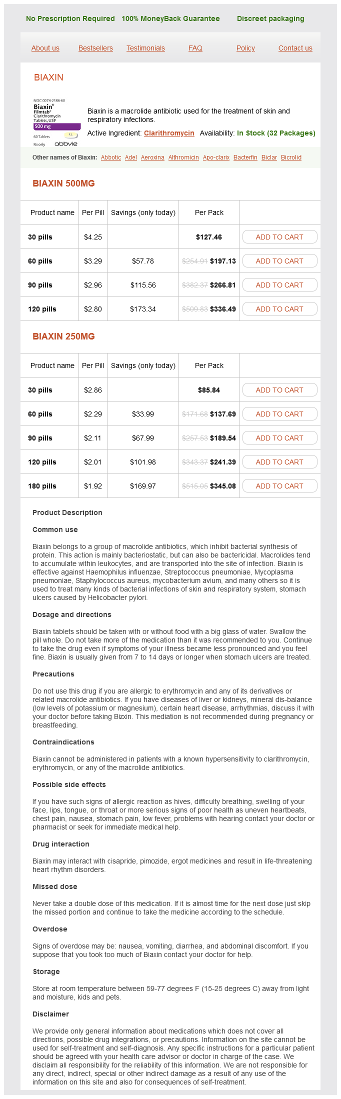
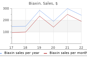
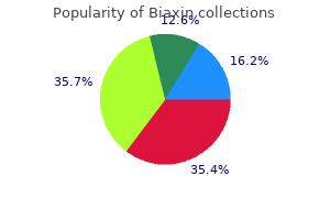
Biaxin
Order biaxin online pills
Contraindications Include thrombocytopenia or coagulation defect gastritis not responding to omeprazole order biaxin 250mg with mastercard, raised intracranial pressure, and signicant cardiorespiratory compromise, as positioning may risk cardiorespiratory arrest (see also b p. Exchange transfusion can be performed by either both withdrawing and then infusing blood via a single central venous catheter. This replaces 790% of total blood volume and should be performed over a minimum of 2hr. Simultaneously, the same volume of fresh warmed blood is infused into the patient via the other venous catheter using a high rate ow infusion pump. Cyanosis Distinguishing between respiratory and cardiac causes of cyanosis is important (see b pp. Heart failure Heart failure may be manifested by symptoms of poor tissue perfusion alone. Causes of heart failure In children the most common cause of heart failure is congenital structural defects of the heart. The location where a murmur is best heard may give a clue to the underlying aetiology. It arises due to the rapid ow and turbulence of blood through the great vessels and across normal heart valves. It does not signify the presence of any underlying cardiac abnormality or any other pathology. The latter is caused by deoxygenated blood gaining abnormal access to systemic side of the circulation via the left side of the heart or the aorta. The defect is usually isolated, found incidentally, and 3 times more common in girls. This is achieved usually by insertion of an occlusion device at cardiac catheterization or by open heart surgery. It is located in the lower atrial septum and is associated with a three leaet mitral valve. Those with larger defects are predisposed to recurrent chest infections and to heart failure. Without surgical repair congestive cardiac failure may develop in infancy/early childhood. This is performed at 3mths of age, before the pulmonary hypertension causes pulmonary vascular disease (Eisenmenger syndrome). It is dened as a duct still being present 1mth after the date that the child should have been born. Clinical features Tetralogy of Fallot presents in early infancy with the following. Duration ranges from a few minutes to hours; severe episodes result in syncope and occasionally convulsions/hemiparesis. Treatment Severe tetralogy of Fallot with worsening cyanosis in early neonatal period requires prostagladin E infusion; (see b p. Denitive surgery to repair the underlying heart defects is carried out from 4mths of age onwards. Long-term follow-up (up to 30yrs) suggest that improved quality of life is maintained and most are able to lead unrestricted lives. Cardiac conduction defects, including complete heart block, are seen post-operatively and require treatment. Pulmonary venous blood returning to the left side of the heart is retuned directly to the pulmonary circulation via a connecting pulmonary artery. Clinical features Infants usually present in the rst few hours or days with worsening ductdependent cyanosis. This is a medical emergency and early diagnosis and intervention are required to avoid severe hypoxia. Treatment Once diagnosed care is needed to maintain body temperature as hypothermia will worsen the metabolic acidosis of hypoxaemia. Before cardiac surgery, systemic arterial oxygenation can be improved with prostaglandin E infusion (see b p. There is a large defect often from the middle of the atrial septum down to the middle of the ventricular septum. In addition there is not a separate mitral and tricuspid valve, but there is a common atrioventricular valve of 5 leaets guarding the atrioventricular junction. Clinical features Most patients with Down syndrome are screened for congenital heart disease with an echocardiogram. The clinical features will vary depending on other associated cardiac abnormalities. Treatment In an emergency, duct patency is achieved with prostaglandin E infusion (see b p. It is due to thickening of the aortic valves, although subvalvular (subaortic) stenosis is also an important form of obstruction. A supravalvular form of aortic stenosis is also recognized, which may be sporadic or familial. Clinical features Are dependent on the severity of obstruction and age at presentation. In the older child sudden unexpected syncope and chest pain on exertion may occur. Management Surgical or balloon dilatation is indicated if symptomatic, or if a high resting pressure gradient of >64mmHg is present; avoidance of competitive sports recommended if severe. Prognosis Mild to moderate stenosis is compatible with normal activities, but patients require monitoring because worsening obstruction and signicant pressure gradients may develop, which will predispose to heart failure when very severe. Treatment Severe pulmonary valve stenosis requires treating by transvenous catheter balloon dilatation. In the majority (98%) of cases, it is usually distal to the origin of the left subclavian artery at the level of the ductus arteriosus. Most often, this occurs in the neonatal period and the infants present at 48hr old when the duct closes. CoA is seen more often in boys than girls (2:1), although it is common in Turner syndrome (b p. Complications include premature coronary artery disease, congestive cardiac failure, hypertensive encephalopathy, and intracranial haemorrhage. Older children or adolescents require stent insertion at cardiac catheter or surgical resection. The left ventricle is small and non-functional and the right ventricle maintains both pulmonary and systemic circulations. Most infants will appear sick (greyish-blue colour) with poor peripheral perfusion and weak peripheral pulses.
Discount biaxin 500 mg on-line
The study concluded that Libyan diabetic patients had a significantly higher prevalence of gallstones than non-diabetics (~2 gastritis diet ����������� buy biaxin australia. Female diabetic patients were significantly more affected than males and the prevalence significantly increased with age particularly in males. In a large Danish study published in 1991, an age and sex stratified random population sample (ages 30, 40, 50 and 60 years) of Danish origin was followed up with ultrasonography after five years (Jensen, 1991). The study also showed that gallstones could have disappeared due to dissolution or spontaneous passage in 4. The incidence was related to age and gender, although the difference between women and men decreased with increasing age. Jorgensen, (1989) analysed the presence of gallstones diagnosed by ultrasonography in relation to relative weight, weight change since age 25, slimming treatment, physical activity, smoking, consumption of coffee, and diabetes mellitus in a cross sectional study. A random sample of 4581 men and women of Danish origin (aged 30, 40, 50, and 60 years). The study established a significant association in women with high body mass index, history of slimming treatment, and weight gain since the age of 25 of more than 5 body mass index units with gall stones. In men, history of slimming treatment and smoking were significantly associated with gallstones. A total sample of 2,584 residents in the Yaeyama District of Okinawa, Japan, were investigated in 1984 to determine the prevalence of gallstone disease and its associated factors by Nomura, et al. The results further indicated that age and fatty liver were significant predictors of gallstone disease. Subjects were assigned to one of four groups based on age (18-30, 31-40, 41-50, and 23 University of Ghana ugspace. Following a structured interview of each study subject, an ultrasound examination was carried out and a blood sample obtained for laboratory study. The prevalence of gallbladder disease correlated positively with age, reaching a maximum of 13. In all, 433 cholecystectomies and 179 cases of newly symptomatic, unremoved gallstones, diagnosed by ultrasonographic examination or x-ray films, were reported during the four-year follow-up. The age-adjusted relative risk for very obese women, who had a Quetelet index of relative weight greater than 32 kg per square meter, was 6. Also, high energy intake, as compared with a low energy intake was associated with an increased incidence of symptomatic gallstones and an alcohol intake of at least 5 g per day was associated with a decreased incidence as compared with abstention. The study concluded that there is a strong association between obesity and symptomatic gallstones Risk of symptomatic gallstones in women with severe obesity was quantified by Stampfer, et al. The study established a monotonic increase in gall stones disease risk with obesity. Prevalence of gallbladder disease and associated risk factors among American Indian populations using ultrasonography of the gallbladder and standardized diagnostic criteria was established by Everhart, et al. Participants included individuals aged 47 years and older, of 13 American Indian tribes or communities in Arizona, Oklahoma, and South and North Dakota. Gall bladder disease was the sum of gallstones (determined by ultrasound examination) and cholecystectomy (determined by ultrasound and self-report). Also, American Indian heritage, and waist circumference were associated 25 University of Ghana ugspace. Age, American Indian heritage, diabetes, and parity were associated with gall bladder disease among women. Body mass index was not independently associated with gall bladder disease in either sex. With a comparison of their demographic characteristics and biochemical parameters, ultrasonography was done. Jorgensen, (1989) examined the relationship between occurrence of gallstone disease diagnosed by ultrasonography and complaints about abdominal pain and discomfort. The results of the study indicated low (0 to 25%) predictive values of various complaints about pain and discomfort, in particular, pain was not associated with size, number or motility of the stones. The study concluded that in a random population it is difficult to define the symptoms specific for gallstones and thereby to distinguish between symptomatic and asymptomatic gallstones. Cancer diagnosis was confirmed by either biopsy, cholecystectomy, or at the 27 University of Ghana ugspace. Furthermore, elevated body mass index, high total energy intake, high carbohydrate intake (after adjustment for total energy intake), and chronic diarrhea were also found to be associated with gallbladder cancer. The study concluded that the findings are consistent with a major role of gallstones, or risk factors for gallstones, in the cause of gallbladder cancer. Most studies document an elevated risk associated with childbearing and parity (Novacek, 2006; Angelico et al. However, with regard to pregnancy two large studies, one in Germany and one in France could not verify these findings (Walcher et al. Geography and particularly ethnicity play an enormous role in the prevalence of gallstone disease and also the type of stone that forms: cholesterol gallstones predominate in the developed countries of the Western world; brown pigment stones in the bile ducts are more common in Asia (Salam et al. Prevalence is inordinately high in American Indians and their admixtures, and also Northern Europeans; somewhat lower in European and American whites (Everhart et al. Mexican Americans are also at heightened risk when compared to White Americans; however, this risk is directly related to the degree of Amerindian admixture (Everhart, 2001). The lowest frequencies (<5%) occur in sub-Saharan Black Africans 28 University of Ghana ugspace. The majority of gallstones in developed countries consist predominantly of cholesterol (>85%), where as the remainder constitutes black pigment stones composed of calcium bilirubinate (Tazuma, 2006). The situation differs in East Asia where brown pigment stones are located in bile ducts, predominately associated with parasitic infestation. In developed countries, however, these bile duct stones arise in association with the inflammation and infection that result from biliary strictures and malignancies (Tazuma, 2006). The frequency of hepatolithiasis, as a proportion of all bile duct stones, is as high as 20% in China and Taiwan, yet as low at 2% to 3% in Japan, Singapore, and Hong Kong (Shod, Tanaka, Sagar, 2003). The stone type curiously has recently shifted in developing Asian countries from pigment to cholesterol stones. The basis for this change may reflect a decreased rate of chronic biliary infections and consumption of a more Westernized diet (Shaffer, 2005). Familial studies reveal an increased frequency: a nearly 5 times elevated risk in the relatives of gallstone patients. These rates are even higher in monozygotic twins at 12% and dizygotic twins at 6% (Gilat et al, 1983). Yet spouses of affected patients do not have any increased risk, thereby eliminating a shared environment as the basis i. In a Swedish twin study, genetic effects accounted for 25%, shared environmental influences for 13% and unique environmental effects for 62% of the phenotypic variance (Grjibovski et al. In fact, stone formation is a complex interaction of genes and environmental factors, particularly diet-gene interactions (Rudkowska & Jones, 2008). The stone type also changes with age: initially being composed predominantly of cholesterol (corresponding to an increased cholesterol secretion into and saturation of bile) but in late life tending to be black pigment stones. Further, symptoms and complications increase with age, leading to more frequent cholecystectomies (Volzke et al. The aboriginal populations of South America also have an exceedingly high prevalence of gallstones: 49. The explanation to the above facts is that the female gender has a most compelling association with gallstone disease, especially during the fertile years. Women are almost twice as likely as men to form gallstones; the gap narrows following menopause after which men begin to catch up (Einarsson et al. A new 4th generation progestin, drospirenone, used in some oral contraceptives may further heighten the risk of gallstone disease and cholecystectomy; however, the increased risk is quite modest and not likely to be clinically meaningful (Etminamm, et al. During pregnancy when female sex hormones are endogenously raised, biliary sludge (particulate material that is composed of cholesterol, calcium bilirubinate, and mucin) appears in 5% to 30% of women. Resolution frequently transpires during the post-partum period: sludge disappears in two-thirds; small (<1 cm) gallstones (microlithiasis) vanish in one-third, but definitive gallstones become established in ~5% (Maringhini, et al. Biliary hyper secretion of cholesterol, which is an important determinant in gallstone formation, is profoundly exacerbated by obesity. Rapid weight loss is also associated with an increased risk of developing gallstones (Torgerson et al. After bariatric surgery such as Roux-en-Y gastric bypass (but not gastric banding) with rapid weight loss, approximately 40% of the patients form stones.
Syndromes
- Foul-smelling vaginal discharge
- After swimming, place 1 or 2 drops of a mixture of 50% alcohol and 50% vinegar in each ear to help dry the ear and prevent infection.
- Remain still and rest when you have symptoms.
- Low blood pressure
- Barotrauma
- Eyes that move quickly from side to side (nystagmus)
- Does your own snoring ever wake you up?
- Open aortic valve surgery -- done by making a large cut in your chest
- Decrease in contrast sensitivity -- even with 20/20 vision, objects may appear fuzzy or gray
- Headaches
Buy biaxin 500 mg fast delivery
A 29-year-old construction worker fell 15 ft from a roof and broke his right humerus gastritis symptoms patient uk discount biaxin 250mg with visa, as depicted in the accompanying radiograph. In a failed suicide gesture, a depressed student severs her radial nerve at the wrist. A few hours later he begins to experience intense pain, swelling, and weakness in the ipsilateral hand. A 39-year-old man presents with an isolated fracture of the tibia after being hit on the leg with a car. The patient is stable and a radiograph of the leg shows a tibial shaft fracture with severe dislocation. Plate fixation Questions 453 to 456 For each description, select the type of fracture or dislocation with which it is most likely to be associated. A patient has tenderness over the forearm after being attacked by gang members with baseball bats. Questions 457 to 460 For each description, select the type of bone disease with which it is most likely to be associated. A patient with elevated parathyroid hormone levels presents with brown tumors in the long bones. A genetically determined disorder in the structure or processing of type I collagen may require your patient to wear various orthoses to protect himself. A patient presents with cortical thickening and cortical expansion of the bone with sclerotic areas. Questions 461 to 463 For each description, select the type of lesion with which it is most likely to be associated. A radiograph shows a circumscribed, densely sclerotic mass limited to the metaphysis of the lower femur. X-ray demonstrates an aggressive lesion with a permeative pattern of bone lysis and periosteal reaction. The knee is held in slight extension and while holding the thigh steady with one hand the tibia is pulled anteriorly with the other hand. The posterior drawer test is used to evaluate for a posterior cruciate ligament injury and is essentially the reverse of the Lachman test with a slightly flexed knee. Excessive valgus laxity (outward deviation of the leg) is associated with a medial cruciate ligament injury while excessive varus laxity can be seen with a lateral cruciate ligament injury. Debridement requires meticulous removal of all foreign material and resection of all nonviable tissue from the wound to reduce the bacterial count. The wound is aggressively explored because the area of injury is always larger than expected from merely observing the wound. Irrigation with copious amounts of saline is performed with repeat debridements 48 to 72 hours later to assess for further necrosis. Given the significant amount of muscle damage in open fractures, fasciotomies are liberally performed during debridement. Compared to cast and splints, internal or external fixation allows greater access to wound care and is the preferred method of stabilization. The pathogenesis is thought to involve a period of ischemia in the proximal femoral epiphysis followed by revascularization. Plain radiographs show a small and denser-thannormal femoral head on the affected side. Slipped capital femoral epiphysis is a disorder involving dissociation between the epiphysis and metaphysis of the proximal femur. The physical examination is most remarkable for limitation of internal rotation of the hip. Developmental dysplasia of the hip involves a spectrum of disorders with differing degrees of instability of the hip and underdevelopment of the acetabulum. The most definitive study to differentiate between the 2 diagnoses is synovial fluid aspiration. In septic arthritis, synovial fluid aspirate demonstrates bacteria and white blood cells with high neutrophils on differential count. A bone scan would target the area of inflammation but not give any information as to the source of the inflammation. It is bounded laterally by the powerful deltoid muscle; superiorly, the acromion process precludes upward dislocation. However, anteriorly and inferiorly the pectoralis major and the long head of the biceps do not completely stabilize the glenohumeral joint; in this region the articular ligaments and joint capsule provide the major structural support. Thus, the joint is not strongly supported in its anteroinferior aspect and consequently anterior (or anteroinferior) dislocations are the most common glenohumeral dislocations. The humeral head is driven anteriorly, which tears the shoulder capsule, detaches the labrum from the glenoid, and produces a compression fracture of the humeral head. Most glenohumeral dislocations result from a posteriorly directed force on an arm that is partially abducted. Posterior dislocation is much rarer and should raise the possibility of a seizure as the precipitating cause. Clinical suspicion and physical examination are important in diagnosis of glenohumeral joint dislocations; diagnosis can be confirmed by radiologic plain films (anteroposterior, scapular lateral, and axillary views). The radial nerve runs in a groove on the posterior aspect of the humerus as it courses into the forearm compartment and is therefore at high risk of injury. If the nerve injury is apparent before any manipulation has been done, the fracture should be reduced; the nerve injury should be observed, since the nerve function will likely improve with time. If the nerve injury is present only after reduction, immediate surgical exploration is warranted because the nerve might be trapped in the fracture site. At this level of the arm, the ulnar and median nerves are well protected by muscle. The posterior interosseous nerve is a distal branch of the radial nerve and may be injured in fractures near the radial head, but it is in no danger from injuries at the level seen in this radiograph. The dorsum of the hand from the radial aspect of the fourth digit over the thumb, including the thenar pad and thumb web, becomes insensate after severance of the radial nerve at the wrist. Radial injuries more proximally would impair extension of the wrist and digits as well as forearm supination. The cast needs to be immediately removed and the upper extremity needs to be thoroughly assessed. If the symptoms do not improve with removal of the splint then surgical decompression with fasciectomy is warranted. Compartment syndromes result from increasing pressures in the fascial compartments. Capillary blood flow is compromised first resulting in loss of oxygen delivery to tissues and increased extremity edema because of increased capillary permeability. Arterial flow is the last to be compromised and therefore pulse changes are a late finding and normal pulses do not rule out a compartment syndrome. Extreme pain (out of proportion to the injury), pain on passive extension of the fingers or toes, pallor of the extremity, motor paralysis, and paresthesias are all components of the syndrome. The diagnosis can be confirmed by measuring intracompartmental pressures, but, whenever physical findings or symptoms are suspicious, the patient should be taken immediately to surgery. Observation, repeated imaging, and elevation would waste precious time and are not appropriate measures for a patient who is suspected of having a compartment syndrome. Surgical fixation, as compared to closed treatment, provides better control of alignment, allows motion of the foot and ankle, and the possibility of earlier weight bearing. Multiple large clinic trials have demonstrated that unreamed nailing strategies have higher incidence of nonunion and malunion as opposed to fracture fixation with reamed cannulated nails. Plate fixation of tibial fractures is generally reserved for periarticular injuries too proximal or distal for intramedullary nailing. Closed treatment is the method of choice for tibial shaft fractures of minor severity and dislocation. Dislocation of the radial head with a fracture of the proximal third of the ulna is known as Monteggia deformity. It is most often repaired with internal fixation of the ulna with closed reduction of the radial head.
Purchase biaxin on line
Add 8334 Pulp cap direct Services/procedures 8301 Possibility of intended to treat diseases of deleting gastritis diet ������ order generic biaxin on-line, the dental pulp and their evidence plus sequelae. Does not 8333 include diagnostic evaluation and necessary radiographs/ diagnostic images. Limitation: Intra-operative radiographs/ diagnostic images are limited to three on a single canal tooth and five on a multi-canal tooth for Page 61 of 109 each completed endodontic therapy. Obturation of root Canals 8335 8328 8336 8337 Root canal therapy Codes 8329, 8338, 8339 and 8338 anteriors and premolars 8340 (endodontic treatment 8329 first canal completed at a single visit) 8339 may not be used with codes 8340 8332, 8333 and 8334 8631 (endodontic preparatory 8633 visits and re-preparation of previously obturated canal). Colonic lavage 88081 Enema administration 88082 Aspiration of stomach/gastric 88083 lavage. Any urinary tract procedure 88090 including catheterisation, bladder stimulation and emptying. Condom catheter application, 88091 penile dressing, catheter care including bag change or catheter removal. Incontinence management (30 88093 minutes) this fee includes intermittent catheterisation, external sheath drainage, taking of history, providing literature and teaching. Complex stoma a poorly 88201 constructed, non-sited stoma requiring convexity or build up. Moderate stoma a fairly well 88202 constructed, sited stoma which may require straight forward convexity or build up. Consumables used, and charged 88301 according to rule 05 Page 68 of 109 Equipment hire per day, charged 88302 according to rule 06. Treatment of extensive 88041 wounds/burns requiring extensive nursing management. Rehabilitation may be needed by an individual who experiences difficulties in mobility, vision, hearing, speech, swallowing, or cognition. Interventions may include services provided by rehabilitation professionals such as physiotherapists, occupational therapists, speech and language therapists, orthotic and prosthetic technicians, psychologists, physical and rehabilitation medicine doctors. The Policy Framework and Strategy for Disability and Rehabilitation services in South Africa outlines comprehensive and integrated disability and rehabilitation services within the broader health and developmental context to facilitate improved access at all levels of health care(39). The different interventions include training, exercises, and compensatory strategies, education, support and counselling. This visit includes elements of the first assessment visit, as well as further discussions around goals of care, planning of future care, preferences around end-of-life issues, preferences regarding hospitalization and resuscitation etc. Communication between members of a team, including patient and family, is an often under-estimated and under-valued aspect of a good and comprehensive palliative care service. The proposed set of services and interventions are a first step towards defining services that should be available in a primary health care setting. The next step will be to cost and prioritise the services according to the principles in annexure A. Mortality and causes of death in South Africa, 2016: Findings from death notification [Internet]. Mental illness in the Western Cape Province, South Africa: A review of the burden of disease and healthcare interventions. Guidelines for Maternity Care in South Africa: A manual for clinics, community health centres and distrcit hospitals. Exercise or other physical activity for preventing pre-eclampsia and its complications. Pelvic floor muscle training for prevention and treatment of urinary and faecal incontinence in antenatal and postnatal women. Flexible sigmoidoscopy versus faecal occult blood testing for colorectal cancer screening in asymptomatic individuals. Effects of a long-term vitamin D and calcium supplementation on falls and parameters of muscle function in community-dwelling older individuals. Exercise Interventions for Preventing Falls Among Older People in Care Facilities: A Meta-Analysis. Framework and Strategy for Disability and Rehabilitation Services in South Africa 2015-2020. In its first deliberations, the Committee set about defining the priority setting process in order to gain a common understanding of the scope of work. Priority setting was understood to be a process of rationing or resource allocation, a multifaceted and complex problem faced by decision makers in the health system (1). From the outset, it was understood that priority setting was a complicated process that needed to be guided by a set of principles that were agreeable to multiple stakeholders. Resource allocation is value based and there is not one way of defining a successful priority setting process, with different disciplines offering different solutions on how priority setting can be done: health economics for example, focuses on efficiency, while policy approaches focus on legitimacy and evidence-based medicine primarily focuses on effectiveness (2). As such, it was important to firstly agree on a definition of priority setting and then set common values and principles that would guide the process. The selection of an essential package of services should thus be based on rational principles that ensure an equitable distribution of resources. However, balancing the gap between resources and opportunities in healthcare is a difficult process, and if not handled carefully, resource allocation can have a detrimental effect on both providers and consumers. It is thus hoped that embarking on a priority setting process that is accompanied by acceptable values and norms will ensure delivery of a package that benefits all stakeholders (1). The Principles for Priority Setting Recommended by the Committee the Committee consulted widely and reviewed relevant literature to assess the common principles of priority setting adopted globally. The principles evaluated are given in Annexure A, showing how they have are applied in various settings, and from these, the following five core principles were adopted by the Committee: 1. Clinical and Cost Effectiveness; Efficiency 1) Health Needs Burden of disease is the total and cumulative health, social or economic impact of a disease or range of diseases in a population. Access relates to the opportunity to obtain and appropriately use quality health services. Availability of health services (or physical access: whether appropriate services are available at the right place and time to meet population needs. Affordability of services (financial access): ability to pay in the context of the household budget. Acceptability of health services (cultural access: fit between provider and patient attitudes towards and expectations of each other. A robust definition of access is needed that is acceptable to both patients and providers and all stakeholders. Data to measure the various indicators of access may also not be readily available to inform a priority setting process. Those in need should have access to all needed quality health services without financial hardship. The main challenge with these concepts of financial risk protection is that an issue such as catastrophic expenditure, is difficult to measure, often requiring huge investments in population-based surveys to identify at risk households. Sustainability is the long-term ability to mobilize and allocate sufficient resources for activities that meet individual or population health needs.
Biaxin 500mg cheap
Because dehydrogenases are depleted in the area of ischemic necrosis (they leak out through the damaged cell membranes) gastritis symptoms diet buy generic biaxin 500mg line, an infarcted area is revealed as an unstained pale zone (while old scarred infarcts appear white and glistening) (Fig. Subsequently, by 12 to 24 hours, an infarct can be identified in routinely fixed gross slices owing to a red-blue hue caused by stagnated, trapped blood. Progressively thereafter, the infarct becomes a more sharply defined, yellow-tan, somewhat softened area that by 10 days to 2 weeks is rimmed by a hyperemic zone of highly vascularized granulation tissue. The histopathologic changes also have a fairly predictable sequence (summarized in Table 12-5 and Figure 12-16). Using light microscopic examination of routinely stained tissue sections, the typical changes of coagulative necrosis become detectable variably in the first 4 to 12 hours. An additional but sublethal ischemic change may be seen in the margins of infarcts: so-called vacuolar degeneration or myocytolysis, involving large vacuolar spaces within cells, probably containing water. This potentially reversible alteration is particularly frequent in the thin zone of viable subendocardial cells. Note the myocardial hemorrhage at one edge of the infarct that was associated with cardiac rupture, and the anterior scar (arrowhead), indicative of old infarct. A, One-day-old infarct showing coagulative necrosis along with wavy fibers (elongated and narrow), compared with adjacent normal fibers (at right). Widened spaces between the dead fibers contain edema fluid and scattered neutrophils. C, Nearly complete removal of necrotic myocytes by phagocytosis (approximately 7 to 10 days). E, Well-healed myocardial infarct with replacement of the necrotic fibers by dense collagenous scar. Figure 12-17 Temporal sequence of early biochemical, ultrastructural, histochemical, and histologic findings after onset of severe myocardial ischemia. For approximately 30 minutes after the onset of even the most severe ischemia, myocardial injury is potentially reversible. Thereafter, progressive loss of viability occurs that is complete by 6 to 12 hours. The benefits of reperfusion are greatest when it is achieved early, with progressively smaller benefit occurring as reperfusion is delayed. A, Schematic illustration of the progression of myocardial ischemic injury and its modification by restoration of flow (reperfusion). Hearts suffering brief periods of ischemia of <20 minutes followed by reperfusion do not develop necrosis (reversible injury). Late reperfusion may still have a beneficial effect on reducing or preventing myocardial infarct expansion and left ventricular remodeling. B, Large, densely hemorrhagic, anterior wall acute myocardial infarction from patient with left anterior descending artery thrombus treated with streptokinase intracoronary thrombolysis (triphenyl tetrazolium chloride-stained heart slice). This is the characteristic appearance of markedly ischemic myocardium that has been reperfused. D, Fibrinous pericarditis, showing a dark, roughened epicardial surface overlying an acute infarct. E, Early expansion of anteroapical infarct with wall thinning (arrow) and mural thrombus. The left ventricle is on the right in this apical fourchamber view of the heart. The shape of the left ventricle (to the right) has been distorted by the right ventricular enlargement. It is therefore not surprising that these normally delicate structures suffer cumulative damage complicated by formation of calcific deposits (composed of calcium phosphate mineral), which may lead to clinically important disease (see Chapter 1). The most frequent calcific valvular diseases, illustrated in Figure 12-22, are calcific aortic stenosis, calcification of a congenitally bicuspid aortic valve, and mitral annular calcification. Each comprises primarily dystrophic calcification without significant lipid deposition or cellular proliferation, a process distinct from but with some features of atherosclerosis. A, Calcific aortic stenosis of a previously normal valve having three cusps (viewed from aortic aspect). Note that the commissures are not fused, as in postrheumatic aortic valve stenosis (see Fig. C and D, Mitral annular calcification, with calcific nodules at the base (attachment margin) of the anterior mitral leaflet (arrows). A, Long axis of left ventricle demonstrating hooding with prolapse of the posterior mitral leaflet into the left atrium (arrow). C, Opened valve with pronounced hooding from patient who died suddenly (double arrows). A, Acute rheumatic mitral valvulitis superimposed on chronic rheumatic heart disease. Small vegetations (verrucae) are visible along the line of closure of the mitral valve leaflet (arrows). Previous episodes of rheumatic valvulitis have caused fibrous thickening and fusion of the chordae tendineae. B, Microscopic appearance of Aschoff body in a patient with acute rheumatic carditis. The myocardial interstitium has a circumscribed collection of mononuclear inflammatory cells, including some large histiocytes with prominent nucleoli and a prominent binuclear histiocyte, and central necrosis. C and D, Mitral stenosis with diffuse fibrous thickening and distortion of the valve leaflets, commissural fusion (arrows), and thickening and shortening of the chordae tendineae. C, Histologic appearance of vegetation of endocarditis with extensive acute inflammatory cells and fibrin. Figure 12-27 Diagrammatic comparison of the lesions in the four major forms of vegetative endocarditis. Histologically, they are composed of bland thrombus without accompanying inflammatory reaction or induced valve damage. Should the patient survive the underlying disease, organization may occur, leaving delicate strands of fibrous tissue. This may be related to some underlying disease, such as a cancer, and, in particular, mucinous adenocarcinomas of the pancreas. Endocardial trauma, as from an indwelling catheter, is also a well-recognized predisposing condition, and one frequently notes right-sided valvular and endocardial thrombotic lesions along the track of a Swan-Ganz pulmonary artery catheter. The lesions are small single or multiple, sterile, granular pink vegetations ranging from 1 to 4 mm in diameter. The lesions may be located on the undersurfaces of the atrioventricular valves, on the valvular endocardium, on the cords, or on the mural endocardium of atria or ventricles. Histologically the verrucae consist of a finely granular, fibrinous eosinophilic material that may contain hematoxylin bodies (the tissue equivalent of the lupus erythematosus cell of the blood and bone marrow, see Chapter 6). An intense valvulitis may be present, characterized by fibrinoid necrosis of the valve substance that is often contiguous with the vegetation. A, Nearly complete row of thrombotic vegetations along the line of closure of the mitral valve leaflets (arrows). A, Characteristic endocardial fibrotic lesion involving the right ventricle and tricuspid valve. Movat stain shows underlying myocardial elastic tissue black and acid mucopolysaccharides blue-green. B, Calcification with secondary tearing of a porcine bioprosthetic heart valve, viewed from the inflow aspect. Far less frequently observed is disease whose cause is intrinsic to the myocardium. Myocardial diseases are a diverse group that includes inflammatory disorders (myocarditis), immunologic diseases, systemic metabolic disorders, muscular dystrophies, genetic abnormalities in cardiac muscle cells, and an additional group of diseases of unknown etiology. However, a major advance in our understanding of myocardial diseases, previously considered idiopathic, has been [96] [97] [98] the demonstration that specific genetic abnormalities in cardiac energy metabolism or structural and contractile proteins underlie myocardial dysfunction in many patients. Moreover, myocardial disease of diverse and even unknown etiologies may have a similar morphologic appearance. Therefore, our discussion avoids the controversies associated with classification schemes and emphasizes clinicopathologic, etiologic, and mechanistic concepts. Without additional data, the clinician encountering a patient with myocardial disease is usually unaware of the etiology. Hence the clinical approach is largely determined by one of the following three clinical, functional, and pathologic patterns (Fig. Within the hemodynamic patterns of myocardial dysfunction, there is a spectrum of clinical severity, and overlap of clinical features often occurs between groups.
Purchase discount biaxin
Dysphagia (difficulty in swallowing) is encountered both with deranged esophageal motor function and with diseases that narrow or obstruct the lumen gastritis diet v8 purchase biaxin 500mg with amex. Pain and hematemesis are sometimes evoked by esophageal disease, particularly by those lesions associated with inflammation or ulceration of the esophageal mucosa. The clinical diagnosis of esophageal disorders often requires specialized procedures such as esophagoscopy, radiographic barium studies, and manometry. The most common is ectopic gastric mucosa in the upper third of the esophagus ("inlet patch"), occurring in up to 2% of individuals. The acid secretions of the ectopic gastric mucosa or pancreatic enzymatic secretions can produce localized inflammation and discomfort. These are usually duplication cysts, containing double smooth muscle layers and derived from the lower esophagus in 60% of cases. Rarely, bronchial or parenchymal pulmonary tissue may arise from the upper gut and is denoted bronchogenic cyst or pulmonary sequestration, respectively. Lastly, impaired formation of the diaphragm may permit herniation of abdominal viscera into the thorax. When severe, this lesion is incompatible with life, since the lungs are severely hypoplastic at the time of birth. This condition is to be distinguished from hiatal hernias, to be discussed presently. Because they cause immediate regurgitation when feeding is attempted, they are usually discovered soon after birth. Absence (agenesis) of the esophagus is extremely rare; much more common are atresia and fistula formation (Fig. In atresia, a segment of the esophagus is represented by only a thin, noncanalized cord, with a proximal blind pouch connected to the pharynx and a lower pouch leading to the stomach. It rarely occurs alone, but is usually associated with a fistula connecting the lower or upper pouch with a bronchus or the trachea. Associated anomalies include congenital heart disease, neurologic disease, genitourinary disease, and other gastrointestinal malformations. Atresia sometimes is associated with [3] the presence of a single umbilical artery. Aspiration and paroxysmal suffocation from food are obvious hazards; pneumonia and severe fluid and electrolyte imbalances may also occur. Well-developed webs rarely protrude more than 5 mm into the lumen, with a thickness of 2 to 4 mm. When an upper esophageal web is accompanied by an iron-deficiency anemia, glossitis, and cheilosis, the condition is referred to as the Paterson-Brown-Kelly or Plummer-Vinson syndrome, with an attendant risk for postcricoid esophageal carcinoma. Esophageal rings are concentric plates of tissue protruding into the lumen of the distal esophagus. One occurring above the squamocolumnar junction of the esophagus and stomach is referred to as an A ring. One located at the squamocolumnar Figure 17-1 Esophageal atresia and tracheoesophageal fistula. Gross view demonstrating longitudinal lacerations extending from esophageal mucosa into stomach mucosa (arrow). A, A view of the everted esophagus and gastroesophageal junction, showing dilated submucosal veins (varices). B, Low-power cross-section of a dilated submucosal varix that has ruptured through the mucosa. Numerous eosinophils within the squamous epithelium, elongation of the lamina propria papillae, and basal zone hyperplasia are present. A, B, Gross view of distal esophagus (top) and proximal stomach (bottom), showing A, the normal gastroesophageal junction (arrow) and B, the granular zone of Barrett esophagus (arrow). C, Endoscopic view of Barrett esophagus showing red velvety gastrointestinal mucosa extending from the gastroesophageal orifice. Microscopic view showing squamous mucosa and intestinal-type columnar epithelial cells (goblet cells) in a glandular mucosa. Some alcoholic drinks contain significant amounts of such carcinogens as polycyclic hydrocarbons, fuel oils, and nitrosamines, along with other mutagenic compounds. Nutritional deficiencies associated with alcoholism may contribute to the process of carcinogenesis. Alcohol and tobacco cannot be invoked as risk factors in many high-incidence regions of the world. The presence of carcinogens, such as fungus-contaminated and nitrosamine-containing foodstuffs in China, may play a significant role in the extraordinary high incidence of carcinoma in this region. Dietary deficiencies in vitamins and essential metals have been documented in China and South Africa. Based on the above considerations, dietary and environmental factors have been proposed to increase risk, with nutritional deficiencies acting as promoters or potentiators of the tumorigenic effects of environmental carcinogens. For example, methylating nitroso compounds in the diet and in tobacco smoke may be the reason for the broad spectrum of p53 point mutations present in over half of esophageal cancers. This is in keeping with the concept that stepwise acquisition and accumulation of genetic alterations ultimately give rise to cancer. Finally, the chronic esophagitis so commonly observed in persons living in areas of high incidence may itself be the result of sustained exposure to the carcinogens listed earlier. This chronic esophagitis results in an increased epithelial cell turnover, which, over a length of time in a continuously carcinogenic environment, progresses to dysplasia and eventually to carcinoma. The rate of progression along the chronic esophagitis-dysplasia-cancer sequence may well be modified or modulated by genetic or racial factors. Like squamous cell carcinomas arising in other locations, those of the esophagus begin as apparent in situ lesions (intraepithelial neoplasm or carcinoma in situ). When they become overt, about 20% of these tumors are located in the upper third, 50% in the middle third, and 30% in the lower third of the esophagus. Early lesions appear as small, gray-white, plaque-like thickenings or elevations of the mucosa. In months to years, these lesions become tumorous masses and may eventually encircle the lumen. Three morphologic patterns are described: (1) protruded (60%), a polypoid exophytic lesion that protrudes into the lumen; (2) flat (15%), a diffuse, infiltrative form that tends to spread within the wall of the esophagus, causing thickening, rigidity, and narrowing of the lumen; and (3) excavated (ulcerated, 25%; Fig. The fortunate patient is found at the stage of superficial esophageal carcinoma, in which the malignant lesion is confined to the epithelial layer (in situ) or is superficially invading the lamina propria or submucosa (Fig. Several histologic variants may be seen, such as verrucous squamous cell carcinoma, spindle cell carcinoma, and basaloid squamous cell carcinoma. Irrespective of their degree of differentiation, most symptomatic tumors are quite large by the time they are diagnosed and have already invaded the wall or beyond. The rich lymphatic network in the submucosa Figure 17-8 Large ulcerated squamous cell carcinoma of the esophagus. Figure 17-9 Squamous cell carcinoma of the esophagus: low-power microscopic view showing invasion into the submucosa. A, Gross view of an ulcerated, exophytic mass at the gastroesophageal junction, arising from the granular mucosa of Barrett esophagus. The gray-white esophageal mucosa is on the top, and the folds of gastric mucosa are below. A, Gross view showing punctate erosions in an otherwise unremarkable mucosa; adherent blood is dark due to exposure to gastric acid. B, Low-power microscopic view of focal mucosal disruption with hemorrhage; the adjacent mucosa is normal. Nevertheless, infected persons are at increased risk for the development of peptic ulcer disease and possibly gastric cancer. Extensive molecular studies suggest that the bacteria cause gastritis by stimulating production of pro-inflammatory cytokines and by directly injuring epithelial cells (discussed later). The underlying mechanisms contributing to this difference are not completely clear, but host-microorganism interplay appears to be critical. Noninvasive tests include a serologic test for antibodies, fecal bacterial detection, and a urea breath test. The current treatment regimens [22] include antibiotics and hydrogen pump inhibitors. Prophylactic and therapeutic vaccine development is still in the early research stage, but it holds the promise to eradicate or at least greatly reduce the worldwide prevalence of H. In the most severe cases, production of intrinsic factor is lost, leading to pernicious anemia.
Kanzo (Licorice). Biaxin.
- Are there any interactions with medications?
- Are there safety concerns?
- What other names is Licorice known by?
- Dosing considerations for Licorice.
- Muscle cramps, arthritis, lupus, infections, hepatitis, infertility, cough, stomach ulcers, prostate cancer, weight loss, atopic dermatitis (eczema), chronic fatigue syndrome (CFS), and other conditions.
- How does Licorice work?
Source: http://www.rxlist.com/script/main/art.asp?articlekey=96849
Discount biaxin 250 mg on-line
Purchase cheapest biaxin and biaxin
Practical and clinical skills the specialty of General Surgery requires assessed and documented numbers for "Practical and clinical skills"(see Appendix 2) gastritis diet ������ purchase biaxin us. Candidates for the qualification must demonstrate Skills in each of the above areas of responsibility and be able to present a complete and signed logbook. For each intervention/endoscopy/operation performed by the candidate as principle surgeon (the principle surgeon is the person who performs the majority of the essential steps of the procedure) 2 credit points are given. For each intervention/endoscopy/operation performed by the candidate as assistant 1 credit point is given. At least 50% of the total number of 1500 credit points have to achieved as principle surgeon. This means, that a total of 750 interventions/procedures/endoscopies/operations (categories A + B + C) are the minimum requirement, when they are all performed as principle surgeon. Operations (total) n=500 Head & Neck n=25 Thorax n=25 Abdomen n=400 Soft Tissues & Musculo-Skeletal System n=25 Vessels & Nerves n=25 For pragmatic reasons provisional arrangements are provided (see below: "Provisional arrangements") to enhance the qualification until complete European harmonisation of surgical training is achieved. These provisional arrangements allow a range of different compensations to consider various national and/or individual situations. Numeric deficits in one or more items have to be compensated by higher numbers in other items in order to reach the total minimum n=250credit points for each Category. Within the 5 subcategories the particular total number has to be reached at least to 75%. Numeric deficits in one or more subcategories have to be compensated by higher numbers in other groups in order to reach the total minimum n=1000credit points. Category A: Interventions & Procedures If the candidate is not able to present a detailed log-book on category A "Interventions and Procedures" a formal confirmation signed by 2 independent experts about the candidates experience in this category may be accepted. In this case the minimum number (n=250 credit points) for category A has to be added to category C in order to reach total n=1500credit points. Category B: Flexible Endoscopy If flexible endoscopy is not performed by the General Surgeon in a specific country, category B may be omitted for the individual candidate. In this case the minimum number (n=250 credit points) for category B has to be added to category C in order to reach total n=1500 credit points. Experienced and practicing specialized surgeons with at least 10 years of continuous service in formally recognized posts can apply for an Honorary Diploma and be exempted form the examination process. Applications should be submitted via e-mail to the headquarters of the Section of Surgery: office@uemssurg. A complete application should include (all documents in English): 1) A letter from the applicant highlighting the reasons he is worthy of an Honorary Diploma. Emphasis should be given to clinical experience as well as research and educational achievements. The letters must analyse the overall achievements of the applicant and give emphasis to his/ her suitability as a trainer in surgery; this needs to be measured against the well established criteria of the Country where the applicant is practicing. The General Assembly of the Division finally decided regarding the acceptance or not of the application (majority of at least 2/3 of the members). A successful applicant is awarded the title of Honorary Fellow of the European Board of Surgery. The application has to be submitted to the Division at least 4 weeks prior to the business meeting. The Committee will visit the Centre on site and meet with the Chairman, the Faculty and the Trainees and review on site the work of the Centre in all the domains of the original application. The visit will last one day and it will be strictly professional with no social programme. The Centre applying for accreditation needs to cover the travel and accommodation expenses of the members of the Committee. An effort is made so that the members are from countries close to the applying Centre to minimize the expenses. Following the visit, the Committee will produce a report and scoring of the Centre in the same domains of the initial application. Each domain will be scored from 0-3: 0: insufficient/ absent 1: sufficient 2: good 3: excellent A minimum score of 36 (75%) is required for the Committee to give a positive recommendation to the Division. This will be presented at the next business meeting of the Division where a vote will be taken by all members regarding the approval of the application; 2/3 majority needed for approval. Preoperative Management Physical examination Tests of respiratory, cardiac, renal and endocrine function Electrocardiography and interpretation Management of associated medical conditions. Intraoperative Care Patient positioning Prevention of nerve and other injuries in the anaesthetised patient Principles of general and regional anaesthesia Care and monitoring of the anaesthetised patient 3. Postoperative Management Pain control Post-operative monitoring Post-operative complications Prevention, recognition and management of complications Techniques of venous access Assessment and maintenance of fluid and electrolyte balance Blood transfusion-indications, hazards, complications, plasma substitutes Respiratory failure-recognition and treatment Nutritional support-indications, techniques, total parenteral nutrition 4. Harmonic) Lasers-principles and precautions Explosion hazards relating to general anaesthesia and endoscopic surgery Procedures Drainage of body cavities and retentions Sampling of body fluids and/ or body excretions for laboratory investigation, interpretation of results Local and regional anaesthesia Excision of cysts and benign tumours of skin and subcutaneous tissues 6. Critical Surgical illness and Intensive Care Medicine the applied basic science relevant to the clinical assessment of critically ill patients and to the understanding of disorders of function caused by haemorrhage, shock and sepsis. Traumatology and Emergency Surgery the applied basic science relevant to the clinical assessment of more or less severely injured patients and to the understanding of disorders of function caused by trauma, haemorrhage and shock. Haematopoietic and Lymph reticular Systems the anatomy, physiology and pathology of the haemopoietic and lymphoreticular systems appropriate to the understanding of clinical signs and special investigations. Musculo-skeletal System Musculo-skeletal anatomy, physiology and pathology relevant to the clinical examination of the locomotor system and to the understanding of disordered locomotor function with emphasis on the effects of trauma. Common disorders of infancy and childhood Metabolic and degenerative bone disease: osteoporosis and osteomalacia Bone and joint infections including those related to prostheses Principles of joint replacement Amputations Principles of orthotics and rehabilitation of the amputee Diagnosis and treatment of common fractures Diagnosis and treatment of common injuries and disorders of joints Hand infections and injuries Principles of tendon repair Common disorders of the foot Cervical and shoulder pain Back pain and sciatica Differential diagnosis of arthritis Peripheral nerve lesions Nerve regeneration-principles of nerve repair Malignant disease of bone and soft tissues 11. Head & Neck Conditions Upper airway obstruction Epistaxis Mucosal cancers of the oral cavity, pharynxand larynx Parotid gland tumours Submandibular gland tumours Cervical lymphadenopathy Procedures Tracheostomy Cricothyroidotomy Resection of lip/tongue lesions Parotidectomy Modified radical neck dissection 12. Abdominal Wall and Alimentary Tract the surgical anatomy of the abdomen and its viscera and the applied physiology of the alimentary system, relevant to clinical examination, to the interpretation of special investigations, to the understanding of disorders of function and to the treatment of abdominal disease. Endocrine the surgical anatomy, applied physiology and pathology of the endocrine glands relevant to clinical examination, to the interpretation of special investigations, to the understanding of disordered function and to the principles of surgical treatment of common endocrine disorders. Vascular System the surgical anatomy and applied physiology of the vascular system relevant to clinical examination, to the interpretation of special investigations and to the understanding of the disorders of function caused by diseases and injuries of the blood vessels. Special techniques used in the investigation of vascular disease Limbischaemia: acute and chronic-arterial embolism Gangrene Aneurysms. Thoracic the surgical anatomy and pathology of the heart, great vessels, air passages, chest wall, diaphragm and thoracic viscera and the applied cardio-respiratory physiology relevant to clinical examination, interpretation of special investigations and understanding of disorders of cardio-respiratory function caused by disease, injury and surgical intervention. Paediatric Please refer to the curriculum and requirements of the Section of Peadiatric Surgery 19. Plastic Surgery Please refer to the curriculum and requirements of the Section of Plastic, Reconstructive and Aesthetic Surgery 20. Urology the surgical anatomy, applied physiology and pathology of the urinary system, relevant to clinical examination, to interpretation of special investigations, to the understanding of disordered function and to the principles of the surgical treatment of urinary disease and injury. Gynaecology Conditions Gynaecological causes of acute abdominal pain Ectopic pregnancy Pelvic inflammatory disease Incidental ovarian mass/cyst Endometriosis Benign and malignant ovarian neoplasms Benign and malignant uterine neoplasms Cystocoele Rectocoele Procedures Hysterectomy Salpingo-oophorectomy Caesarian section Repair cystocoele Repair rectocoele Surgery of infiltration endometriosis 23. Central Nervous System the anatomy and physiology relevant to clinical examination of the central nervous system, to the understanding of its functional disorders, particularly those caused by cranial or spinal trauma, and to the interpretation of special investigations. Oncology the applied basic sciences relevant to the understanding of the clinical behaviour, diagnosis and treatment of neoplastic disease. Radiology Principles of diagnostic radiography, Sonography, Computed Tomography and Magnetic Resonance Imaging and related techniques Principles and handling of contrast media Diagnostic and therapeutic interventional radiological methods Interventional radiological implantation of prostheses and stents into vessels, organs and other structures X-ray guided detection of foreign bodies Sonographically guided identification of unpalpable lesions Sonographic "Doppler" investigation of abdominal and limb vessels Sentinel lymph node marking and detection Security measures in Radiology 26. Diabetic foot, defects of the skin and soft tissue, compartment n=10 syndrome, amputations) 5. The evolving process of the Board Examinations is paralleled by the European ambition towards harmonization and standardization of medical education, specialist training and qualification. Frequency of Board Examination, location and language are subject of continuous development. Date, Location & Language the Board Exam takes place at least annually mainly in cooperation with surgical meetings. Date, location and languages(s) of the Board Exam are to the discretion of the committee. This and further details about the next Board Exam(s) are published on the Board website.
Buy biaxin 500 mg without a prescription
The area distal to the trachea on the transverse view is obscured by air in the trachea gastritis diet �������� buy biaxin overnight. The longitudinal (A) and transverse (B) sonograms reveal an enlarged thyroid gland that contains an irregularly shaped anechoic structure (arrow). A B Feline hyperthyroidism is typified by diffuse but not massive enlargement of one or both thyroid glands. Sonography has been used to define size and determine if cystic areas are present within the glands (Fig. It cannot be evaluated without contrast administration and almost always requires a dynamic examination using cinefluoroscopy or videofluoroscopy. The events must be examined slowly and repeatedly before a definite diagnosis can be reached. An esophagram with a single radiograph or series of radiographs may identify the site. Therefore, patients with oral or pharyngeal dysphagia should be referred to hospitals that have fluoroscopic equipment. Retention of food or barium in the oral pharynx suggests an oropharyngeal disorder. Contrast or food in the nasal pharynx may indicate the failure of the soft palate to close the nasal pharynx or it may indicate incoordination between pharyngeal muscle contraction and dorsal movement of the soft palate. Abnormal cricopharyngeal muscular activity may produce a cricopharyngeal achalasia, or failure of the muscle to relax; chalazia, or failure to contract with resulting sphincter incompetence; or incoordination between the pharyngeal muscle contraction and cricopharyngeal muscle relaxation. Aspiration of food or barium may indicate a lack of normal laryngeal movement, incoordination between the pharyngeal 236 Small Animal Radiology and Ultrasonography Fig. Within the ovoid thyroid gland (black arrows), there is a hypoechoic nodule near its anterior pole (white arrows). Both liquid barium and barium-impregnated food should be used when studying animals with oropharyngeal disorders. Again, because of the complex nature of swallowing, static radiographs rarely produce a definite diagnosis. Am J Vet Res of volume and x-ray attenuation of the lungs of beagles in various 1985; 46:1479. J Am Anim Hosp Assoc weight on echocardiographic values in four breeds of dogs of differ1985; 21:161. Vet Radiol Ultrasound 1994; indices of the nonracing, healthy Greyhound to reference values 35:310. Schille S, Skroczki M: M-mode echocardiographic reference values tion of the thyroid gland of hyperthyroid cats: comparison to in cats in the first three months of life. Vet Clin North Am Small Anim Pract 1998; the dog: technique, anatomy and imaging planes. Vet Radiol Ultrasound 2000; orthopedic injury associated with traumatic rib fracture in cats: 75 41:320. Nyska A, Perl S, Bargai U: Invagination of the stomach into the thodiaphragmatic hernia. J Am mesothelial remnant as an aid to the diagnosis of feline congenital Anim Hosp Assoc 1982; 18:147. J Am Anim Hosp Assoc 1996; and radiographic findings of peritoneopericardial diaphragmatic 32:409. J Am Vet Med Assoc 1992; bral actinomycosis: diagnosis and long-term therapy in 3 dogs. Comp Cont Educ Pract Vet 1993; problem in two cases of bronchoalveolar carcinoma in the dog. J Am and pulmonary thromboembolism in a dog with autoimmune Vet Med Assoc 1997; 210:1628. J Am Anim tures of anterior mediastinal lymphosarcoma in the cat: a review of Hosp Assoc 1993; 29:78. Okin R: Pseudomembranous tracheitis resembling collapsed tramegaesophagus in dogs. Tidholm A: Retrospective study of congenital heart defects in 151 Anim Pract 1989; 30:311. J Small Anim Pract 1985; Congenital heart abnormalities in the cat: a description of 16 cases. J Am Vet Med Assoc 1986; the dog: a retrospective study of radiographic, epidemiologic, and 188:874. Vet cinefluorographic analysis of experimentally-induced and spontaRadiol Ultrasound 1995; 36:157. Ackerman N: A right to left shunting patent ductus arteriosus with esophagitis in 2 dogs. Vet Radiol Ultrasound 1997; characterization of an unusual form of subvalvular aortic stenosis in 38:94. Am J the conotruncal septum in Keeshond dogs: pathologic and genetic Cardiol 1978; 41:746. Dilated cardiomyopathy in the Newfoundland: a study of 37 cases J Am Anim Hosp Assoc 1984; 20:271. J Am Vet Med Assoc 1989; diomyopathy in Great Danes and results of a pedigree analysis: 17 164:71. Aust Vet J 1997; characterization of an unusual form of subvalvular aortic stenosis 75:862. An prognostic factors in Doberman Pinschers with end-stage carechocardiographic study. Churchill Livingstone, New York, evaluation of cardiomyopathy and thromboembolism in the cat. J cardiography and radiography of cats with dilatation cardiomyopaAm Anim Hosp Assoc 1999; 35:21. J Vet Intern Med associated with developing dirofilariasis in experimentally infected 1990; 4:117. Sottiaux J, Franck M: Pulmonary embolism and cor pulmonale in a diomyopathy associated with hyperthyroidism due to thyroid adecat. Venco L: Ultrasound diagnosis: left ventricular thrombus in a cat microfilaria: a problem in diagnosis. J Vet Intern worm-infected dogs: a radiographic, electrocardiographic and Med 1990; 4:118. J Am Vet sitologic observations of trypanosomiasis in dogs infected with Med Assoc 1997; 210:1635. Venco L, Calzolari D, Mazzocchi D, et al: the use of echocardiogravalve in a dog with infective endocarditis. Vet Radiol Ultrasound phy as a diagnostic tool for detection of feline heartworm 1998; 39:436. Jacobs G, Patterson D, Knight D: Complete interruption of the aorAm Vet Med Assoc 1990; 197:1501. J Vet Med A Physiol Pathol Clin Med giosarcoma using nonselective angiography in a dog. Cornelius L, Mahaffey M: Kinking of the intrathoracic caudal vena with visceral leishmaniasis. Vacirca G, Mantelli F, Ferro E, et al: Pericardial effusion with feline Ultrasound 1992; 33:187. Ciorba A, Vitellozzi G, Avalos E: Study of pulmonary changes in lung tumors in the dog: a review of 36 cases. Venco L, Calzolari D, Morini S: Pulmonary thromboembolism in a lomas in two dogs and literature review. Clercx C,Coignoul F,Jakovljevic S,et al: Tuberculosis in dogs: a case J Am Vet Med Assoc 1980; 176:36. Novoa C, Pickering X, Sanchez B, et al: Pathology and diagnosis of with lymphosarcoma. Kohn B,Arnold P,Kaser-Hotz B,et al: Malignant histiocytosis of the sis in five dogs. Moise S, Wiedenkeller D,Yeager A, et al: Clinical, radiographic, and seminated granulomatous disease associated with Aspergillus fumibronchial cytologic features of cats with bronchial disease: 65 cases gatus and Candida species infection in a Golden Retriever.
Buy biaxin 500 mg on line
Aloe cream also increased the overall percentage of healed patients and there were no significant adverse reactions reported (Syed et al 1996b) gastritis symptoms dizziness biaxin 500 mg discount. It has been shown to improve different parameters of gastrointestinal function in normal subjects, such as colonic bacterial activity, gastrointestinal pH, stool specific gravity and gastrointestinal motility (Bland 1986). Besides this indication, there is still a need for scientific validation to establish which gastrointestinal conditions are most receptive to treatment with aloe. Both treatments were administered for 1 month with a follow-up period of 3 months. Aloe induced clinical remission in 30% of subjects compared to 7% for placebo and symptom improvement in 37% compared to 7% for placebo. The Simple Clinical Colitis Activity Index and histological scores also decreased significantly for patients on the aloe treatment, but not for those receiving placebo. In one trial aloe juice consisting of 80% gel or placebo was given in a trial of 40 patients who were recently diagnosed with type 1 diabetes at the dose of 1 tablespoon twice daily. From day 14 the blood sugar levels in the aloe group began to fall significantly compared with the control group and Aloe vera 27 continued to steadily drop during the period of study (P < 0. A single blind, placebo-controlled trial found that oral aloe gel was more effective in reducing blood sugar levels when combined with glibenclamide than glibenclamide alone in 72 patients with type 2 diabetes. Hypersensitivity manifested by generalised nummular eczematous and papular dermatitis, and presumably by contact urticaria, developed in a 47-year-old man after 4 years of using oral and topical aloe. Avoid in patients with known hypersensitivity to aloe or with nausea, vomiting or signs and symptoms of gastrointestinal obstruction. Avoid excessive use and long-term use (more than 2 weeks), as potassium losses may occur, which may alter cardiac electrophysiology. A case study of depression of thyroid hormones in a woman taking Aloe vera juice has been reported (Pigatto & Guzzi 2005). The patient consumed 10 mL daily for 11 months and laboratory testing showed reduced levels of thyroxine and triiodothyonine. Levels returned to normal progressively after discontinuing the aloe juice and the patient achieved full clinical remission after 16 months. Reduced serum levels of the thyroid hormones T3 and T4 have been reported for Aloe vera in vivo (Kar et al 2002). There is good scientific evidence that aloe may be of benefit in these conditions; however, the chemical composition of Aloe vera products will vary depending on Aloe vera 29 geographical and processing factors. Improvement occurs within several weeks with the condition continuing to improve with use. Avoid chronic use of laxative preparations that contain highly irritant compounds, known as anthraquinone glycosides, in the latex. Protection in mice by fraction F1 against infections by Listeria monocytogenes, Yersinia pestis, Candida albicans and Plasmodium berghei. Influence of Aloe vera on collagen characteristics in healing dermal wounds in rats. A review on the relationship between aloe vera components and their biologic effects. Randomised double-blind placebo-controlled trial of aloe vera for irritable bowel syndrome. Aloe vera, hydrocortisone, and sterol influence on wound tensile strength and antiinflammation. Effects of Aloe vera and sucralfate on gastric microcirculatory changes, cytokine levels and gastric ulcer healing in rats. The stimulation of postdermabrasion wound healing with stabilized aloe vera gel-polyethylene oxide dressing. Evaluation of antioxidant potential of aloe vera (Aloe barbadensis miller extracts). Adverse reactions to vitamin E and aloe vera preparations after dermabrasion and chemical peel. In vitro evaluation of the synergistic antiviral effects of acemannan in combination with azidothymidine and acyclovir. Relative efficacy of three medicinal plant extracts in the alteration of thyroid hormone concentrations in male mice. Acemannan purified from Aloe vera induces phenotypic and functional maturation of immature dendritic cells. Chemical and biological characterization of a polysaccharide biological response modifier from Aloe vera L. Glucomannan minimizes the postprandial insulin surge: a potential adjuvant for hepatothermic therapy. Double-blind placebo-controlled pilot trial of acemannan in advanced human immunodeficiency virus disease. A novel angiogenic factor derived from Aloe vera gel: beta-sitosterol, a plant sterol. Retardation of wound healing by silver sulfadiazine is reversed by Aloe vera and nystatin. The effect of aloe vera gel/mild soap versus mild soap alone in preventing skin reactions in patients undergoing radiation therapy. A double-blind, placebo-controlled study of a commercial Aloe vera gel in the treatment of slight to moderate psoriasis vulgaris. Decreased mortality of Norman murine sarcoma in mice treated with the immunomodulator, Acemannan. Antioxidant effect of Aloe vera gel extract in streptozotocininduced diabetes in rats. Aloe vera for preventing radiation-induced skin reactions: a systematic literature review. Acemannan-containing wound dressing gel reduces radiation-induced skin reactions in C3H mice. Comparative evaluation of aloe vera in the management of burn wounds in guinea pigs. Effectiveness of Aloe vera on the antioxidant status of different tissues in irradiated rats. Purification and characterization of a glutathione peroxidase from the Aloe vera plant. Effect of water extracts of aloe and some herbs in decreasing blood ethanol concentration in rats. Sennosides and aloin do not promote dimethylhydrazine-induced colorectal tumors in mice. Chemomodulatory action of Aloe vera on the profiles of enzymes associated with carcinogen metabolism and antioxidant status regulation in mice. Therapeutic effects of Aloe vera on cutaneous microcirculation and wound healing in second degree burn model in rats. Prevention of ultraviolet radiation-induced suppression of contact and delayed hypersensitivity by Aloe barbadensis gel extract. Management of psoriasis with Aloe vera extract in a hydrophilic cream: A placebo-controlled, double-blind study. Effects of low molecular constituents from Aloe vera gel on oxidative metabolism and cytotoxic and bactericidal activities of human neutrophils. Effect of aloe vera gel to healing of burn wounds: a clinical and histologic study. Evaluation of aloe vera gel gloves in the treatment of dry skin associated with occupational exposure. Antioxidant, free radical scavenging and anti-inflammatory effects of aloesin derivatives in Aloe vera. Systematic review of herbs and dietary supplements for glycemic control in diabetes. The role of thromboxane in experimental inadvertent intra-arterial drug injections. Activation of a mouse macrophage cell line by acemannan: the major carbohydrate fraction from Aloe vera gel. Isolation, structure elucidation, antioxidative and immunomodulatory properties of two novel dihydrocoumarins from Aloe vera.