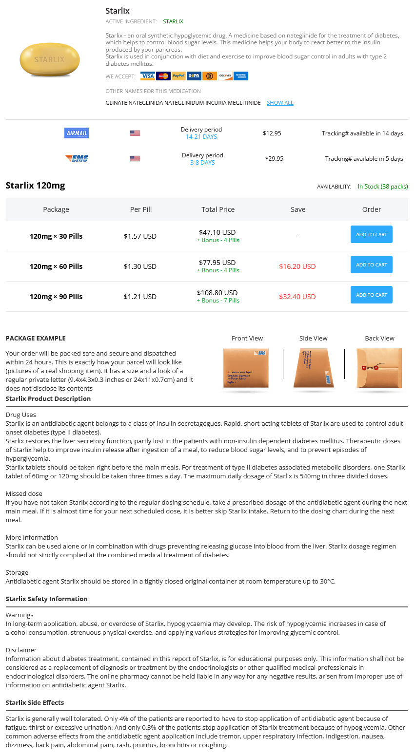
Starlix
Buy starlix with mastercard
Some patients may unknowingly take a similar medication young living antiviral buy 120mg starlix with visa, not recognizing the name of the drug. When in doubt, subspecialty consultation may be required to confirm need for a given medication. Physical examination Since drug reactions may involve virtually any organ system, a careful physical examination is recommended. Cutaneous manifestations are the most common presentation for drug allergic reactions. Characterization of cutaneous lesions is very important in regard to determining the etiology, further diagnostic tests, and management decisions. The clinical manifestations of these cutaneous reactions have been discussed earlier. Drug allergic reactions can also affect other organ symptoms, and a complete physical examination is typically appropriate. Additionally, drug reactions may cause a wide array of physical abindentities including mucous membrane lesions, lymphade-nopathy, hepatosplenomegaly, pleuropneumonopathic abindentities, and joint tenderness/swelling. Routine laboratory evaluation appropriate to the clinical setting may be useful for the evaluation of a patient with suspected drug reaction depending upon the history and physical exam findings. A complete blood count with a differential cell count and a total platelet count may help to exclude the possibility of cytotoxic reactions. While eosino philia is often suggestive of a drug allergic reaction, most patients with drug allergic reactions do not have eosinophilia, and therefore, the absence of eosinophilia clearly does not exclude a drug allergic etiology. Immune complex assays lack sensitivity for serum sickness, and similarly hypocomplementemia may not be present either. In cases of suspect anaphylaxis, a diagnosis of anaphylaxis may be made by detecting a rise in serum total tryptase levels above baseline or in serum mature tryptase (a. Occasionally, biopsies of involved organs may define specific histopathologic lesions. Skin biopsies may be of value in the diagnosis and management of drug allergic reactions but are typically not helpful for implicating a particular drug. In complex cases where multiple drugs are involved without a clear-cut temporal relationship, a skin biopsy may be useful in suggesting a drug-induced eruption. Skin biopsies are useful in differentiating vasculitis, bullous diseases, and contact dermatitis. However, there are no absolute histologic criteria for the diagnosis of drug-induced eruptions, and a skin biopsy may not definitively exclude alternative etiologies. Furthermore, features suggestive of drug exanthems such as interface dermatitis with vacuolar alteration of keratinocytes, foci of spongiosis, and tissue eosinophilia are not specific and may be seen with other cutaneous diseases. A liver biopsy helps to differentiate between cholestatic and hepatocellular drug reactions but does not identify the specific cause. Membranous glomerulonephritis initiated by deposition of immune complexes in the kidney can be readily identified by immu-nofluorescent stains for IgG, IgM, and complement in renal biopsy specimens. Drug-specific testing in drug allergy While the history and physical examination are very useful tools in the assessment of drug allergic patients, by themselves they are often not adequate to confirm or negate a true drug allergy. Modalities for testing for specific drug allergic reactions include skin tests, in vitro tests, and drug challenge. Drug challenge is the gold standard for confirming a drug allergic reaction, while skin tests and in vitro tests may indicate sensitization but have the potential for false-positive and false-negative results. Immediate epicutaneous (prick) and intradermal skin testing Drug skin testing using epicutaneous (prick) and intradermal methods may be used for immediate type I immunologic drug reactions. In the case of IgE-mediated drug reactions, demonstration of the presence of drug-specific IgE is usually taken as sufficient evidence that the individual is at significant risk of having a type I reaction if the drug is administered. Penicillin is the only low-molecular-weight agent for which validated testing has been documented. Skin testing to most drugs is limited by knowledge regarding degradation products and/or metabolites and how they are conjugated with body proteins. Skin testing to native formulations of the drug may therefore lack the important immunogenic epitopes and lead to inadequate negative predictive value. In the case of type I drug allergic reactions, skin testing to penicillin, cephalosporins, platinum-based chemotherapeutics, certain perioperative agents, and insulin may offer the best diagnostic utility and will be discussed in more detail in the sections corresponding to specific drugs. Delayed skin testing Skin testing using both intradermal and patch tests has been utilized for certain delayed immunologic drug reactions. The negative predictive values for these techniques have not been well established, and therefore, a negative test does not preclude a drug allergy. The technique for performing delayed intradermal skin tests is similar to intradermal testing for immediate reactions, with intra-dermal injection of 0. However, tests are read after 24 hours or later and considered positive when there is an infiltrated erythematous reaction. When directly compared, intradermal drug tests appear to be more sensitive than patch tests in most circumstances. Patch testing has also been utilized in delayed immunologic drug reactions in a similar fashion as intradermal tests. Nonirritating concentrations have not been firmly established for drug patch tests. Typically, drug patch testing is performed starting with 1% concentration in petrolatum, going up to a 10% concentration. In vitro tests Several different in vitro tests have been utilized in drug allergy including tests for specific IgE, lymphocyte transformation tests, basophil activation tests, and a number of investigational tests that are not commercially available. Most in vitro tests have been evaluated in IgE-mediated reactions and, when compared to skin tests, are not as sensitive. Overall, commercially available in vitro tests for drug allergy require further study to determine if they are clinically useful. Immunoassays for other drugs have been even less well studied and generally lack adequate controls to validate the testing. The lymphocyte transformation test has been studied as an in vitro correlate of drug-induced cellular reactions. This test is used primarily in research studies as a retrospective test and is not clinically available in most medical centers. There is considerable disagreement among investigators about the value of this assay in evaluating drug allergies because neither its positive nor negative predictive values have been systematically investigated. The lymphocyte transformation test has recently become commercially available for selected drugs, but there are no published studies using these assays, either alone or in comparison with previous independent assays. Further data are required to determine if there is any clinical utility for commercially available lymphocyte transformation tests. Further confirmatory studies, especially with commercially available tests, are needed before it can be accepted as a diagnostic tool. Drug challenge Graded dose challenge, drug provocation, and test dosing are all terms used to describe a procedure to determine if a patient will have an adverse reaction to a particular drug by administering lower than therapeutic doses over a period of time with observation for reactions. If a patient has a negative drug challenge, they can be considered not allergic to that drug. The rationale for starting with a lower dose is based on the concept that a smaller dose of allergen will result in a less severe and more easily treated reaction. Valid diagnostic tests are not available for most drugs, and therefore, it is not possible to be certain that a patient is not allergic to a drug. Importantly, drug challenges are intended for patients who, following a full evaluation, are deemed to be at low risk for being allergic to the given drug. Furthermore, the benefit of treatment with the drug should outweigh the risk of performing the drug challenge. Common indications for drug challenges include (1) excluding a drug allergy in patients with unconvincing histories of drug allergy, (2) exclude cross-reactivity of structurally related drugs, and (3) to reassure patients with histories of multiple adverse reactions to drugs. Drug challenges are usually contraindicated in several types of drug reactions such as autoimmune diseases. The starting dose for graded challenge is generally higher than for induction of drug tolerance procedures, and the number of steps in the procedure may be two or several. The time intervals between doses are dependent on the type of previous reaction, and the entire procedure may take hours or days to complete.
Buy 120 mg starlix visa
It is felt that the more rapid the onset of symptoms after exposure to allergen antiviral elixir generic starlix 120mg visa, the more severe the event. Perhaps the most common atypical presentation is cardiovascular collapse with shock in the absence of other symptoms or signs. A significant percent of such patients express neurologic manifestations including seizures and muscle spasms. Asthmatic children with food allergies can have episodes that begin initially with only asthma. Of course, respiratory reactions and cardiovascular reactions are responsible for the majority of fatalities. Shock and myocardial infarction due to coronary artery vasospasm are also causes of fatal reactions. Duration of anaphylaxis Anaphylactic episodes can be uniphasic, biphasic, or protracted. Uniphasic events usually have a rapid onset, and symptoms subside within an hour or two and do not return. Biphasic events are characterized by a recurrence of symptoms after resolution of the initial episode. The majority of these appear within 8 hours after resolution of the initial symptoms, but such events can be delayed as long as 24 hours and rarely even longer. Because of the clinical significance of biphasic reactions in terms of the suggested length of patient observation after an initial resolution of symptoms, it is important to be aware of the risk factors for biphasic events. Factors that have been cited that increase the risk of a biphasic event are the presence of hypotension during the first phase, the failure to administer epinephrine or a delay in its administration, and an event due to an ingested (vs. Foods are probably the most common cause of anaphylaxis overall and are certainly the most common cause in children. Of these, milk, egg, wheat, soy, peanut, and tree nut produce the majority of reactions in children, and shellfish, fish, and peanut are the most common food offenders in adults. However, overall, in adults, medications rival food as the most common cause of anaphy lactic events. It is important to note that in published series of adults experiencing anaphylaxis, the majority of events are idiopathic. These include children with spina bifida and genitourinary abnormalities, workers with occupational exposure to latex, and health care workers. Latex-induced anaphylaxis can occur in a variety of situations including direct contact with latex (this is usually via gloves and also condoms) or by aerosolization of latex antigen adhered to corn starch from the powder of latex gloves. Latex reactions in this setting have also been reported due to the administration of a drug through a latex port. Latex anaphylaxis has become less common as the use of powdered latex gloves in health care settings has declined. It still, however, remains a problem in the other two groups of at risk individuals. Unfortunately, there is no standardized skin test for latex available in the United States today. If the in vitro test is positive, there is a high clinical likelihood that latex sensitivity is present. Patients with a diagnosis of latex allergy should wear a medical identification bracelet. If there is any chance of exposure, they should also carry an automatic epinephrine injector. Patients with latex sensitivity should be instructed to notify all their health care providers including dentists of their sensitivity. Surgical procedures should be done in latex-free operating rooms, and precautions should also be taken during dental visits. Exercise-induced anaphylaxis Exercise-induced anaphylaxis has been reported due to almost any form of exercise including jogging, racket sports, aerobics, dancing, brisk walking, and weight lifting. Cessation of the exercise usually rapidly improves symptoms, but if exercise is continued, symptoms will progress. Fatal reactions to exercise-induced episodes are probably rare, but there has been at least one death. The most common cofactor is the ingestion of a specific food to which the patient is allergic. Neither the exercise nor the specific cofactor alone will produce an event, but if exposure is associated with exercise, the reaction will occur. An exercise challenge can be performed, but for reasons unknown, responses can be inconsistent. When a patient does have exercise-induced anaphy-laxis, it is important to identify cofactors on the basis of history, and allergy skin testing is also recommended when the history is suggestive. In the latter condition, the event is induced by elevation of body temperature sufficient to cause sweating. However, other triggers that raise body temperature such as a hot shower can also produce an event. Cholinergic urticaria is characterized by pinpoint wheals that can progress and coalesce to giant hives. In exercise-induced anaphylaxis, giant hives usually appear as one of the first signs. The patient should always have immediate access to autoinjectable epinephrine whenever they exercise. In those patients where a cofactor has been identified, exposure to this cofactor should be avoided when the patient is exercising. There is no definite evidence that oral antihistamines or corticosteroids will have a beneficial effect. Idiopathic anaphylaxis A group of patients (in fact as many as 60% of cases in adults) will experience repeated episodes of anaphylaxis without any identifiable cause. Regardless of how intensively such patients are evaluated, no cause can be determined. The symptoms are identical to those that are found in other causes of ana-phylaxis. Mast cell degranulation is certainly involved, since patients do exhibit elevated tryptase and also increases in urinary histamine metabolites. The diagnosis of idiopathic anaphylaxis is clinical and relies upon the exclusion of other causes. Therefore, patients should receive a careful evaluation with emphasis on the history and events surrounding the episodes. Selective skin testing to foods (sometimes employing fresh foods rather than commercial extracts) and/or tests for serum-specific IgE to foods are indicated. Patients with episodes of idiopathic anaphylaxis should be evaluated with a baseline serum tryptase, and if the tryptase level is elevated, a bone marrow biopsy should be considered. Patients have been treated with daily oral corticosteroids, H1 antagonists, and a combination of H1 and H2 antagonists. The studies have for the most part shown that such daily treatment can be helpful but most often does not control the episodes completely. Particular care must be taken with chronic administration of oral corticosteroids because of side effects. Radiocontrast media the vast majority of radiocontrast media reactions are probably due to the direct effect of radiocontrast on mast cells and basophils and do not appear to be IgE mediated. Anaphylactic episodes to radiocontrast media have declined markedly since the advent of agents that are iso-osmolar. It does happen, however, that atopic individuals are more prone to these events, not patients with shellfish allergy in particular, but those with atopy in general. The reason for this has not been completely established, but it is hypothesized that atopic individuals have a lower threshold for degranulation (not only to radiocontrast but to other direct-acting mast cell secretagogues as well).
Order starlix 120 mg with mastercard
A bladder pacemaker can be inserted beneath the skin to help the nerves that control the bladder anti viral meningitis purchase 120 mg starlix otc. The anterior segment of the eye which consists of the cornea, conjunctiva, trabecular meshwork, anterior chamber, iris, and crystalline lens is vulnerable to direct trauma. The worst outcome is often seen in the combined anterior and posterior segment injuries with the possibility of losing all useful vision. Ocular injuries are divided into open globe and closed globe injuries, however, there may be an overlap in their classification based on the causative agent or inflicting object involved. An open globe injury (an injury penetrating into the globe) involves a full thickness wound of the corneoscleral wall which may result from penetrating or blunt eye trauma. Open globe injuries include lacerations which are further divided into penetrating injuries, perforating injuries and intraocular foreign bodies. Closed globe injuries are commonly due to blunt trauma whereby the corneoscleral wall of the globe remains intact (a partial thickness corneal wound) however, intraocular damage may be present. Ruptures are caused by blunt objects with the actual wound being produced by an inside out mechanism. If the inflicting object is blunt, it can result in either a contusion or a rupture (open globe). Occasionally, an exit wound may be created by the object while remaining partially intraocular. Lacerations to the eyelids and the conjunctiva commonly occur from sharp objects but can also occur from a fall. Lacerations may occur in one of two ways: i) lacerations without prolapse of tissue when the eyeball has been penetrated anteriorly but without prolapse of the intra-ocular contents; and ii) lacerations with prolapse when a small portion of iris prolapses through a wound, or uveal tissue has been injured. Corneal lacerations can involve the iris and crystalline lens forming a cataract whereby management depends on the duration and extent of the incarceration. Corneal lacerations frequently result in prolapse of the iris with distortion of the pupil. Ocular lacerations are treated in different ways depending on whether or not there is tissue prolapse. However, the surgeon has to explore the extent of the wound first and then determine the status of the crystalline lens whether to remove it or not with the aid of a slit lamp. A crystalline lens can only be removed following a water tight closure of the laceration. If the wound is extensive and loss of intra-ocular contents has been great enough and the prognosis for useful function is hopeless, enucleation/evisceration is indicated as a primary surgical procedure. A precaution must be taken for a self-sealing wound because when an edematous cornea subsides a wound leak may develop. Therefore, Seidel test is indicated for evaluation of a corneal wound leak to determine whether aqueous is being emitted or not. The patient needs to be referred to the nearest ophthalmologist for surgical repair as soon as pos sible to restore the anatomy or structural integrity of the globe irrespective of the extent of the injury and the initial visual acuity. If a delay in specialist care is anticipated, a systemic oral antibiotic and tetanus prophylaxis should be administered to avoid development of endophthalmitis. However, it is advisable to wait until repair of the laceration has been completed before adding medications because these could be toxic to the retina. When considering the tear film disorders, the ocular surface microenvironment of the eyelids, conjunctiva, cornea and tear films need to be evaluated. Any alteration in this environment has the potential to cause a tear film disturbance. Signs of dry eyes are as follows: Tear film signs: it may show presence of stingy mucous and particulate matter. Marginal tear strip is reduced or absent (normal height is 1mm) Conjunctival signs; it becomes lusterless, mildly congested, conjunctival xerosis and keratinization may occur. Corneal signs; it may show punctate epithelial erosions, filaments and mucous plaques. Signs of causative disease such as posterior blepharitis, conjunctival scarring diseases (trachoma, Stevens Johnson syndrome, chemical burns, ocular pemphigoid) and lagophthalmos may be depicted. The following treatment modalities can be employed, namely; use of artificial tears, preservation of the existing tears by reducing evaporation and decreasing drainage, and treatment of causative disease of dry eye. Surgical treatment is in the form of temporary or permanent punctual occlusion to assist with tear preservation. It is evidenced by redness of the conjunctiva associated with discharges which may be watery, mucoid, mucopurulent or purulent. Infective conjunctivitis; bacterial, chlamydial, viral, fungal, rickettsial, spirochaetal, protozoal, parasitic 2. Keratoconjunctivitis, which is associated with disease of skin and mucous membrane 5. The condition is common in children and normally starts off in one eye before transmitting itself to the other eye thereby making unilateral bacterial conjunctivitis uncommon. Bacterial conjunctivitis is highly contagious and commonly occurs as an epidermics. Irrigation of the conjunctival sac with sterile warm saline once or twice a day iii. Health education information particularly on how to control the spread of infection ii. Follow up visits to detect changes in visual acuity, development of new symptoms 3. It is a preventable disease usually occurring as a result of carelessness at the time of birth. Curative management entails; Saline lavage hourly till the discharge is eliminated. It is a self-limiting condition and the disease does not persist to adult life except in very few cases. Uveitis may be classified in many ways but a simple classification is on the basis of anatomy, clinical features and aetiology (Kanski, 2003). Main symptoms of acute anterior uveitis are pain, photophobia, redness, lacrimation, and decreased vision. Since the outer layers of retina are in close contact with the choroid and also depend on it for the nourishment, the choroidal inflammation almost always involves the adjoining retina leading to chorioretinitis. Cycloplegic drugs they are very useful and most effective during acute phase of iridocyclitis. Examples are 1% atropine eye drops/ ointment, 2% homatropine or 1% cyclopentolate eye drops. Corticosteroids it provides potent effect especially in antigen-antibody reaction. Examples are 60-100mg of Prednisolone or equivalent quantities of other steroids like dexamethasone or betamethasone. Non-steroidal anti-inflammatory drugs aspirin can be used where steroids are contraindicated, naproxen in patients with ankylosing spondilitis, phenylbutazone and oxyphenbutazone in uveitis associated with rheumatoid disease. Immunosuppressive drugs these should be used only in desperate and extremely serious cases of uveitis. Azithromycin or tetracycline or erythromycin to treat patients with chlamydial infection C. Physical measures Hot formentation is very soothing, reduces pain and increases circulation. The use of dark goggles gives a feeling of comfort by reducing photophobia, lacrimation, blepharospasm. Specific treatment As effective as the non-specific treatment is, in most of the cases, it does not cure the disease, resulting in relapse. These enter the orbit from the infected frontal, maxillary, ethmoidal or sphenoidal sinuses. Orbital cellulitis is a potentially life-threatening and vision-threatening condition.

Purchase starlix 120mg fast delivery
The middle aqueous layer is elaborated by the major and minor lacrimal glands and contains water-soluble substances (salts and proteins) hiv infection timeline starlix 120 mg with mastercard. The deep mucinous layer is composed of glycoprotein and overlies the corneal and conjunctival epithelial cells. The epithelial cell membranes are composed mainly of lipoproteins and are therefore relatively hydrophobic. Mucin is partly adsorbed onto the corneal epithelial cell membranes and is anchored by the microvilli of the surface epithelial cells. This provides a new hydrophilic surface for the aqueous tears to spread over, which is wetted by a lowering of surface tension. Immunoglobulins IgA, IgG, and IgE as well as lysozymes make up the remaining 40% of total protein. IgA predominates and differs from serum IgA in that it is not only transudated from serum but is produced by plasma cells located in the lacrimal gland. In certain allergic conditions such as vernal conjunctivitis, the IgE concentration of tear fluid increases. Other tear enzymes may also play a role in diagnosis of certain clinical entities, for example, hexosaminidase assay for diagnosis of Tay-Sachs disease. Primary Sjogren syndrome, an immune mediated disorder of the lacrimal and salivary glands, characteristically manifesting as dry mouth as well as dry eyes, is the most important specific disease entity (see previous section on Conjunctiva). Etiology and Diagnosis of Dry Eye Syndrome 266 Etiology Many of the causes of dry eye syndrome affect more than one component of the tear film or lead to ocular surface alterations that secondarily cause tear film instability. Histopathologic features include loss of conjunctival goblet cells, abnormal enlargement of nongoblet epithelial cells, increased cellular stratification, and increased keratinization. Clinical Findings Patients with dry eyes complain most frequently of a scratchy or sandy (foreign body) sensation. Other common symptoms are itching, excessive mucus secretion, inability to produce tears, a burning sensation, photosensitivity, redness, pain, and difficulty in moving the lids. On gross examination, the eyes may appear normal, but on careful slitlamp examination, subtle indications of the presence of chronic dryness and irritation are found. The most characteristic feature is interruption or absence of the tear meniscus at the lower lid margin. Tenacious yellowish mucus strands are sometimes seen in the lower conjunctival 267 fornix. The bulbar conjunctiva loses its normal luster and may be thickened, edematous, and hyperemic. The corneal epithelium shows varying degrees of fine punctate stippling in the interpalpebral fissure. When performed without anesthesia, the test measures the function of the main lacrimal gland, whose secretory activity is stimulated by the irritating nature of the filter paper. Tear Film Break-Up Time Measurement of the tear film break-up time may sometimes be useful to estimate the mucin content of tear fluid. Deficiency in mucin may not affect the Schirmer test, which quantifies tear production, but may lead to instability of the tear film, resulting in its rapid break-up. This process ultimately damages the epithelial cells, which can then be stained with rose bengal. Damaged epithelial cells may be shed from the cornea, leaving areas susceptible to punctate staining when the corneal surface is flooded with fluorescein. Baring of the corneal epithelium following formation of a dry spot in the tear film. The tear film break-up time is measured by applying a slightly moistened fluorescein strip to the bulbar conjunctiva and asking the patient to blink. The tear film is then scanned with the aid of the cobalt filter on the slitlamp while the patient refrains from blinking. The time that elapses before the first dry spot appears in the corneal fluorescein layer is the tear film break-up time. Normally it is over 15 seconds, but it will be reduced appreciably by the use of local anesthetics, by manipulating the eye, or by holding the lids open. Tear film break-up time is reduced in eyes with aqueous tear deficiency and is always shorter than normal in eyes with mucin deficiency. Ocular Ferning Test A simple and inexpensive qualitative test for the study of conjunctival mucus is performed by drying conjunctival scrapings on a clean glass slide. In patients with cicatrizing conjunctivitis (mucous membrane pemphigoid, Stevens-Johnson syndrome, toxic epidermal necrolysis, erythema multiforme, diffuse conjunctival cicatrization), ferning of the mucus is reduced or absent. Impression Cytology Impression cytology is a method by which goblet cell densities on the conjunctival surface can be counted. In normal persons, the goblet cell population is highest in the infranasal quadrant. Loss of goblet cells has been documented in trachoma, mucous membrane pemphigoid, Stevens-Johnson syndrome, and avitaminosis A. Fluorescein Staining Touching the conjunctiva with a dry strip of fluorescein is a good indicator of wetness, and the tear meniscus can be seen easily. Both dyes will stain all desiccated nonvital epithelial cells of the conjunctiva and to a lesser extent the cornea. Tear Lysozyme Assay Reduction in tear lysozyme concentration usually occurs early in the course of Sjogren syndrome and is helpful in diagnosis. Tears can be collected on Schirmer strips and assayed, usually by spectrophotometric methods. Tear Osmolality 270 Hyperosmolality of tears has been documented in dry eye syndrome and in contact lens wearers and is thought to be a consequence of decreased corneal sensitivity. Reports claim that hyperosmolality is the most specific test for dry eye syndrome. Hyperosmolality may be found even when Schirmer test and staining with rose bengal and lissamine green are normal. Lactoferrin Tear lactoferrin is low in patients with hyposecretion of the lacrimal gland. Complications Early in the course of dry eye syndrome, vision is slightly impaired. In advanced cases, corneal ulceration, corneal thinning, and perforation may develop. Secondary bacterial infection occasionally occurs, and corneal scarring and vascularization may result in marked reduction in vision. Treatment the patient should understand that dry eye syndrome is a chronic condition and complete relief is unlikely except in mild cases when the corneal and conjunctival epithelial changes are reversible. Artificial tears, particularly preservative-free tears in more advanced cases, are the mainstay of symptomatic treatment. More prolonged duration of action can be achieved with drop preparations containing a mucomimetic such as methylcellulose, polyvinyl alcohol, or polyacrylic acid (carbomers), by using petrolatum ointment during the day and particularly during sleep, or with a hydroxypropyl cellulose (Lacrisert) insert. Mucomimetics, which also include sodium hyaluronate and autologous serum, are particularly indicated when there is mucin deficiency. If there is tenacious mucus, mucolytic agents (eg, acetylcysteine, 10% or 20% one drop six times daily) may be helpful.
Starlix 120mg
Which of the following instruments is designed for carving all interproximal tooth used primarily to remove debris from tooth surfaces What length needle antiviral neuraminidase inhibitor order starlix 120 mg with amex, measured in inches, is normally used for mandibular injections Extensions on the wide #2 matrix bands is designed with which of the following are known by which term Which of the following types of matrix bands is most commonly used in restorative 1. What type of cavity is present when exchange between the dentist and the three or more surfaces are involved Dental material is exchanged between the tooth structure in a cavity preparation, the dentist and the assistant in what zone Stubborn particles of debris may be removed from a cavity preparation by which 16-37. Which of the following materials may of the following materials dampened with be used to remove roughness or overhanging water or hydrogen peroxide What composite shade will appear if the dentinal tubules to help prevent tooth becomes dehydrated What instrument will the dentist use to bring any excess mercury from the amalgam 1. Glass ionomer cement will bond directly with which of the following tooth surfaces The film badge should be placed behind fluoresce the lead-lined barrier at least what number of feet from the tube head Never hold the tube head or the tube head cylinder of the X-Ray machine during exposure 4. At approximately what angle should the ankle be when taking an oblique radiograph What X-Ray machine settings should preferred method and recommended for you use when exposing a maxillary occlusal routine use when taking periapical radiograph on an adult When taking radiographs, which of the following factors should you consider before 17-26. When exposing a maxillary posterior occlusal radiograph, you should use what 17-22. The occlusal film packet contains two illumination used in the darkroom when X-Ray films. The processing solutions used in the darkroom, you should leave the penny on automatic processor are the same as those the X-Ray film for at least what number of used in the manual processing procedure. The cleaning of the roller transports and the solutions in the automatic processor are accomplished at what minimum intervals Which teeth can be identified radiographically by a large white region caused by the bone of the nasal septum You should wait what number of seconds between films before inserting another film into the automatic processor The developer and fixer solutions in the automatic processor should be changed at what minimum frequencies What dose refers to the least amount of that deals with the study of which specialty The most common factor influencing the with the preparation, dispensing, and proper amount of drug given to a patient is What is the proper dose in milliliters of Practice of Pharmacy ampicillin for an 8-year old child if the adult dose is 15 ml The minimum and maximum amount of a drug required to produce the desired effect 1. What term is used to define a milligrams of medication for a child medication that is placed under the tongue Increase the dosage because of her weight and further increase because of her sex 18-17. Decrease of dosage because of her sex but an increase because of her weight 18-18. Aluminum acetate, an astringent, is often used to treat which of the following 18-26. Macrolides are effective against which sulfonamides is in the treatment of which of of the following organisms Silver sulfadiazine is used almost substitute for penicillin when penicillin is exclusively in the treatment of Milk or milk products may interfere with the absorption of which of the following drugs Which of the following is the vitamin involved in absorption and use of calcium 1. Which of the following is equal to one in the urine and are stored in the body in one-hundredth of a liter What chapter of the Manual of the 54 grams of the compound is silver nitrate, Medical Department gives guidance on what is the percentage strength of silver pharmacy operations and drug control Fill the prescription as written the patient is the definition of which of the following terms A tourniquet is normally applied before preferred source for blood specimens to aid in the process of venipuncture. When performing a finger puncture, the first drop should be wiped away to avoid 19-6. The part of the microscope on which the prepared specimen is placed for examination is called the. The best urine specimen for screening purposes is that taken during which of the 1. False critical patients in a mass casualty situation when delay of blood products would cause a critical delay The specific gravity of a liquid is the weight of the substance as compared to an 1. Is used in patients that are in holding a compress danger of developing dehydration 2. The amount of normal saline inch roller bandage infused largely depends on the needs of the patient. An airway of proper size is measured from the tip of the earlobe to the corner of the mouth 3. Treatment of life threatening injuries takes precedence over decontamination procedures. None of the above developed a labeling system for indicating the health, flammability, and reactivity hazards of chemicals. Casualties in a non-tactical environment after the device is sitting between the teeth whose injuries are critical but who will and properly aligned between the printed require only minimal time or equipment are A patient with a skin assessment of pale obstruction, inhalation burns, or massive and cool and whose blood pressure dropped maxillofacial trauma who cannot be briefly would be consider to be in what type ventilated by other means are candidates for of shock When performing a needle chest decompression, what is the preferred size of 21-15. Approximately how long does it take for the needle required to adequately death to occur from massive hemorrhage
Simaruba. Starlix.
- What is Simaruba?
- How does Simaruba work?
- Dosing considerations for Simaruba.
- Diarrhea, malaria, water retention, fever, stomach upset, causing abortion, and other uses.
- Are there safety concerns?
Source: http://www.rxlist.com/script/main/art.asp?articlekey=96387
Purchase generic starlix line
Blood Supply of the Lacrimal Gland the arterial supply is by the lacrimal branch of the ophthalmic artery and infraorbital branch of the maxillary artery hiv infection how early symptoms starlix 120 mg cheap. The venous drainage is by the lacrimal vein which opens into the superior ophthalmic vein. Lymphatic Drainage the lymph vessels join the conjunctival and palpebral lymphatics and pass to the preauricular nodes. It is slightly alkaline and consists mainly of water, small quantities of salts, such as sodium chloride, sugar, urea, protein and lysozyme, a bactericidal enzyme. The Tear Film the fluid which fills the conjunctival sac consists of 3 layers namely: 1. Functions the surface of the eyeball must remain wet for comfort and normal functioning. The tear film spreads over the surface of corneal epithelium by gravity, capillary action and blinking of the eyelids. It contains protective substances such as lysozyme, immunoglobulin, lactoferrin, compliments. The oiliness of this mixed fluid delays evaporation and prevents drying of the conjunctiva and cornea. When a foreign body or other irritant enters the eye, the secretion of tears is greatly increased and the conjunctival vessels dilate. Etiology It is a rare condition occurring in association with mumps, influenza, infectious mononucleosis, etc. Symptom There is marked pain, redness and swelling in the upper and outer angle of the orbit along with excessive watering of the eye. A tender swelling is present at the outer part of the upper lid spreading towards the temple and cheeks. Etiology There is failure in canalization of the nasolacrimal duct, the lumen being blocked by epithelial debris. There is epiphora or continuous watering of the eyes usually evident in 2nd week of life. This constitutes the treatment of congenital nasolacrimal duct block up to 6-8 weeks of age. Then bring the thumb Massage with thumb downward pressing towards the ala of the nose. Massage increases the hydrostatic pressure in the sac and helps to open up the membranous occlusions. It should be carried out at least 3 times a day to be followed by instillation of antibiotic drops. Broad-spectrum antibiotic eyedrops are instilled frequently after expressing the contents of the sac by pressure over the sac area. Probing of Nasolacrimal Duct If there is no improvement after three months, probing of the nasolacrimal duct is performed through the upper punctum under general anesthesia. Great care is taken to avoid injury to the walls of the duct as it may cause fibrosis or infection. The probe is then rotated towards the middle line and pushed down the nasal duct till it reaches the floor of the nose. This procedure will Lacrimal probe in correct position cure most congenital cases. Etiology It usually occurs as an acute exacerbation of the chronic dacryocystitis. It is caused by pyogenic pathogens such as Pneumococcus, Staphylococcus, Streptococcus, etc. There is marked swelling, redness and tenderness of the skin over the sac and adjacent area. Fluctuation is present on palpation over the sac area when there is abscess formation. Hot compresses, systemic antibiotics, analgesics and anti-inflammatory are effective. In case of lacrimal abscess, a vertical incision is given over the sac area in the lower part to facilitate the drainage by gravity. In case of lacrimal fistula, excision of the fistulous tract and removal of sac is done. It is of clinical importance as hypopyon or even panophthalmitis may occur after intraocular surgery. The stagnant sac content gets infected by pyogenic bacterias such as Pneumococcus, Streptococcus, Staphylococcus and rarely by a fungus called rhinosporiodosis. Pathogenesis Chronic dacryocystitis There are two main factors resulting in a vicious cycle: 1. There is constant epiphora or passive overflow of tears over the lid margin which is aggravated by exposure to wind. There is swelling, pain and redness at the site of the sac (mucocele) in cases of acute or recurrent infection. Persistent congestion of the neighbouring conjunctiva, caruncle and skin may be seen. Lacrimal abscess may occur following probing or spontaneously due to pyogenic infection. X-ray is taken immediately and after 10-15 minutes to find out the size of the sac and site of obstruction. Subtraction macrodacryocystography with canalicular catheterization is a more accurate diagnostic technique. Radioactive tracer containing sulphur is instilled into the conjunctival sac and its excretion is visualized by gamma camera. In recent cases Repeating syringing of the nasolacrimal duct and frequent instillation of antibiotic drops is indicated in recent cases. Conjunctival sac is anaesthetized by frequent instillation of topical xylocaine or other suitable local anaesthetic. Lacrimal sac is syringed out 2-3 times using 24-26 gauge cannula attached to 5 cc syringe filled with normal saline and antibiotic drops are instilled.
Buy online starlix
Many people are unaware of their risk status; opportunistic and other forms of screening by health care providers are therefore a potentially useful means of detecting risk factors signs of hiv infection symptoms order 120mg starlix mastercard, such as raised blood pressure, abnormal blood lipids and blood glucose (18). The predicted risk of an individual can be a useful guide for making clinical decisions on the intensity of preventive interventions: when dietary advice should be strict and specic, when sug gestions for physical activity should be intensied and individualized, and when and which drugs should be prescribed to control risk factors. Such a risk stratication approach is particularly suitable to settings with limited resources, where saving the greatest number of lives at lowest cost becomes imperative (19). In patients with a systolic blood pressure above 150 mmHg, or a diastolic pressure above 90 mmHg, or a blood cholesterol level over 5. Therefore, targeting patients with a high risk is the rst priority in a risk stratication approach. Thus the use of guidelines based on risk stratication might be expected to free up resources for other compet ing priorities, especially in developing countries. It should be noted that patients who already have symptoms of atherosclerosis, such as angina or intermittent claudication, or who have had a myocardial infarction, transient ischaemic attack, or stroke are at very high risk of coronary, cerebral and peripheral vascular events and death. Risk stratication charts are unnecessary to arrive at treatment decisions for these categories of patients. Thus, it seems reasonable to assume that the evidence related to lowering risk factors is also applicable to people in different settings. Complementary strategies for prevention and control of cardiovascular disease In all populations it is essential that the high-risk approach elaborated in this document is comple mented by population-wide public health strategies (Figure 1) (11). Population-wide strategies will also support lifestyle modication in those at high risk. The extent to which one strategy is emphasized over the other depends on achievable effectiveness, cost-effectiveness and resource considerations. If resources allow, the target population can be expanded to include those with moderate levels of risk; however, lower ing the threshold for treatment will increase not only the benets but also the costs and potential harm. People with low levels of risk will benet from population-based public health strategies and, if resources allow, professional assistance to make behavioural changes. Ministries of health have the difcult task of setting a risk threshold for treatment that balances the health care resources in the public sector, the wishes of clinicians, and the expectations of the public. This threshold would apply to about 3% of men in the population aged between 45 and 75 years. Ministries of health or health insurance organizations may wish to set the cut-off points to match resources, as shown below for illustrative purposes. In a state-funded health system, the government and its health advisers are often faced with making decisions about the threshold at which drug and other interventions are affordable. In many health care systems, such decisions must be made by individual patients and their medical practitioners, on the basis of a careful appraisal of the potential benets, hazards and costs involved. Countries that use a risk stratication approach have tended to reduce the threshold of risk used to determine treatment decisions as the costs of drugs, particularly statins, have fallen and as adequate coverage of the population at the higher risk level has been achieved. In low-income countries, lowering the threshold below 40% may not be feasible because of resource limitations. Nevertheless, use of risk stratication approaches will ensure that treatment decisions are transparent and logical, rather than determined by arbitrary factors or promotional activity of pharmaceutical companies. Risk prediction charts: Strengths and limitations Use of risk prediction charts to estimate total cardiovascular risk is a major advance on the older practice of identifying and treating individual risk factors, such as raised blood pressure (hypertension) and raised blood cholesterol (hypercholesterolemia). Since there is a continuous relationship between these risk factors and cardiovascular risk the concept of hypertension and hyperlipidemia introduces an arbitrary dichotomy. The total risk approach acknowledges that many cardiovascular risk factors tend to appear in clus ters; combining risk factors to predict total cardiovascular risk is consequently a logical approach to deciding who should receive treatment. The risk charts and tables produced use different age categories, duration of risk assessment and risk factor proles. The current New Zealand (43) and Joint British Societies charts (40, 41) are similar in concept. Risk scores have different accuracy in different populations, tending to overpredict in low-risk populations and underpredict in high-risk populations. The threshold for high risk is dened as a risk of death of 5% or greater, instead of the composite fatal and non-fatal coronary endpoint of 20%. The evidence that underpins the use of risk factor scoring and management comes from a range of sources. There is now increasing evidence that cardiovascular risk factors are associated with clinical 10 Prevention of cardiovascular disease events in a similar way in a wide range of countries (31). Finally, there is strong evidence from clinical trials that reducing the levels of risk factors has benecial effects. Risk factor scoring and management have now been widely taken up in cardiovascular prevention guidelines in most high-income countries (36, 41, 43, 44). The risk factors included in current scoring systems are drawn from those used in the original Framingham score. It is possible that, as more epidemiologi cal data become available for low and middle-income countries, a new generation of risk scoring systems may emerge that have greater predictive accuracy. Older age and male sex are powerful determinants of risk; consequently, it has been argued that the use of the risk stratication approach will favour treatment of elderly people and men, at the expense of younger people with several risk factors and women. However, while younger people gain more life years if they have a non-fatal event, older people are a lot more likely to die from an event. When discounting is taken into consideration, the quality adjusted life years gained by preventing events in young people are very similar to those gained in old people (Table 3) (50). Concern about the metabolic syndrome, characterized by central obesity, elevated blood pressure, dyslipidaemia, and insulin resistance (51, 52), has raised the question of whether identifying people with this syndrome should be a priority. There is, as yet, insufcient evidence to justify using metabolic syndrome as an additional risk prediction tool (63, 64). There is insufcient evidence from randomized trials to support more specic management of dyslipidaemias (81). In summary, the great strength of the risk scoring approach is that it provides a rational means of making decisions about intervening in a targeted way, thereby making best use of resources available to reduce cardiovascular risk. Alternative approaches focused on single risk factors, or concepts such as pre-hypertension or pre-diabetes, have been popular in the past, often because they represented the interests of specic groups in the medical profession and professional societ ies. Such an approach, however, leads to a very large segment of the population being labelled as high risk, most of them incorrectly. If health care resources were allocated to such false-positive individuals, a large number of truly high-risk individuals would remain without medical attention. It enables the intensity of interven tions to be matched to the degree of total risk (Figure 2). Further research is required to validate existing subregional risk prediction charts for individual populations at national and local levels, and to conrm that the use of risk stratication methods in low and middle-income countries results in benets for both patients and the health care system. These charts are intended to allow the introduction of the total risk stratication approach for management of cardiovascular disease, particularly where cohort data and resources are not readily available for development of population-specic charts. The charts have been generated from the best available data, using a modelling approach (Annex 5), with age, sex, smoking, blood pressure, blood cholesterol, and presence of diabetes as clinical entry points for overall manage ment of cardiovascular risk. Some studies have suggested that diabetic patients have a high cardiovascular risk, similar to that of patients with established cardiovascular disease, and so do not need to be risk-assessed.
Best purchase for starlix
The Salmeterol Multicenter Asthma Research Trial: a comparison of usual pharmacotherapy for asthma or usual pharmacotherapy plus salmeterol hiv infection timeline discount starlix 120 mg with visa. Randomized comparison of strategies for reducing treatment in mild persistent asthma. Not only does the lung participate in systemic immunopathologic processes, but it is also capable of initiating local immune responses that may be beneficial or adverse to the host. With the exception of asthma, primary and secondary immunologic lung diseases are discussed in this chapter according to their presentation, immunologic features, pathologic features, diagnostic criteria, differential diagnosis, treatment, and prognosis. The result is a diffuse pulmonary process consisting of reticulonodular or alveolar processes (or both) with poorly formed granulomas. Hypersensitivity pneumonitis develops in only 5% to 15% of the exposed population, and the majority of patients are nonatopic and nonsmokers. The antigenic materials may be of animal, vegetable, fungal, bacterial, or chemical origin. In general, there is no age, sex, or significant geographic predilections other than those related to specific occupational exposures. This is only a partial list, and new sources from occupational exposure, homes, and hobbies are reported annually. Despite this long expanding list, these inciting antigens have striking similarities in their clinical, radiographic, and pathologic outcomes. Acute hypersensitivity pneumonitis occurs when exposure is heavy but intermittent. Acute symptoms, including fever, chills, dyspnea, chest tightness and dry cough, appear 4 to 6 hours after each exposure and remit when the agent is avoided. Physical exam reveals fever, tachypnea, tachycardia, and a few lung rhonchi or crackles. Peripheral neutrophilia (without eosinophilia) and increased IgG levels, including antigen-specific IgG, are common in the acute form. Radiographic abnormalities will typically develop with repeated antigen exposure but may not parallel the severity of disease. Up to 4% of patients may have normal x-rays, while up to 45% may have very subtle changes. These changes may be completely reversible over 4 to 6 weeks if the initiating exposure is avoided. Pulmonary function tests show hypoxemia and a restrictive ventilatory defect, with reduced vital capacity, total lung capacity, diffusing capacity, and static compliance. Airway obstruction is not typical unless the patient is atopic or has a concurrent obstructive pulmonary disease. Approximately 10% of patients with hypersensitivity pneumonitis have atopy and asthma. A two-stage reaction develops in these patients if they are exposed to organic dust. The immediate asthmatic, or type I, immune reaction will be manifested by dyspnea, wheezing, and an obstructive ventilatory defect. Subacute hypersensitivity pneumonitis is characterized by the gradual development of productive cough, dyspnea, fatigue, anorexia, and weight loss. As in acute H P, chest radiography can appear normal or show micronodular or reticular opacities. These findings also undergo dramatic improvement with treatment with glucocorticoids or with removal of the antigen. Pulmonary function testing typically has a restrictive abnormality or a mixed obstructive and restrictive pattern. Chronic hypersensitivity pneumonitis occurs when the exposure is mild but more continuous. Progressive dyspnea, decreased exercise tolerance, productive cough, and weight loss develop insidiously. Wheezing, bibasilar crackles, cyanosis, clubbing, and cor pulmonale develop as pulmonary inflammation and fibrosis progress. The diffuse nodular and reticulonodular pattern characteristic of the acute and subacute stages is superimposed on fibrosis and honeycombing and loss of lung volume and compensatory over inflation (emphysema) of the less involved lung zones. These changes are typical of diffuse interstitial fibrosis of any origin and indicate irreversible damage to the lungs. Pulmonary function tests show severe restrictive disease, with variable airway obstruction and air trapping. After inhalation of an offending antigen, it binds IgG forming an immune complex, which subsequently initiates a complement cascade resulting in activated macrophages. These macrophages secrete chemokines and cytokines that first attract neutrophils and, after several hours, attract T + lymphocytes and monocytes. After recruitment into the lung and activation, the young macrophages develop into epithelioid cells and multinucleated giant cells. Lymphoid follicles containing plasma cells also develop in the lesions during the subacute phase. Early collagen formation by myofibroblasts occurs, and the extracellular matrix surrounding the granuloma becomes rich in the proteoglycan versican. The characteristic immunologic feature of hypersensitivity pneumonitis is the presence of precipitating (usually IgG) antibody to the offending antigen. Serum antibodies or precipitins are readily and reproducibly demonstrated by the Ouchterlony double immunodiffusion technique. Although the positive precipitin test is clinically helpful, it actually indicates prior exposure and sensitization and does not necessarily correlate with clinical sequelae. Serum precipitins without clinical pneumonitis can develop in up to 50% of exposed asymptomatic patients. Conversely, there are rare patients with clinical disease and no demonstrable antibodies. The correct antigen must be used to detect the antibodies, but many causative antigens have not been identified. Thus, a negative precipitin test in the face of convincing clinical evidence does not exclude the diagnosis, whereas a positive test without appropriate clinical findings does not establish a diagnosis. Antigens for thermophilic actinomycetes and many molds result in a nonspecific irritant response that interferes with skin testing. Many antigens provide a high percentage of positive responses even in exposed but unaffected individuals. Skin testing is therefore neither specific nor sensitive in determining the cause or presence of hypersensitivity pneumonitis. Skin testing for immediate, late-onset or delayed reactions does not currently have a role in diagnosis or management of hypersensitivity pneumonitis. The histology of hypersensitivity pneumonitis includes (1) a mononuclear interstitial infiltration with a bronchocentric distribution (100%); (2) poorly formed noncaseating granulomas surrounding bronchioles (70%); and (3) airway inflammation with foamy macrophages (65%), often associated with bronchiolitis obliterans (50%). Over the course of several months, the histology becomes nonspecific as the granulomas disappear and interstitial fibrosis and obliterative bronchiolitis predominate, resulting eventually in honeycomb cysts and end-stage fibrosis. Although patients often associate recurrent exposures with symptoms in the acute form of hypersensitivity pneumonitis, the exposure in the chronic form is much more difficult to identify. As stated above, negative studies do not rule out disease, while positive studies do not necessarily confirm a diagnosis. This can usually be done with good transbronchial biopsies directed by radiographic findings, although occasionally (usually in the chronic form) an open lung biopsy is preferred. Trial of avoidance and/or controlled reexposure to the suspected antigen or environment. Deliberate repetition of natural exposure to the suspected antigenic environmental source. When the specific diagnosis remains in doubt because the relevance of a particular exposure is questionable, allergen inhalation or bronchial challenge tests can be used to establish a definitive diagnosis.
