
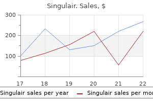
Singulair
Buy generic singulair 5 mg online
Cataplexy is sometimes mistaken not caused by disturbed nocturnal sleep or misaligned 3 for seizure activity asthma uncommon symptoms purchase singulair with mastercard, and is characterized by a bilateral loss circadian rhythms. Cataplexy is seen in approximately 70% narke, meaning numbness or stupor, and lepsis, mean- ing attack. Doing so can help relieve some of the embarrass- experiences occurring at sleep onset or upon awakening. Although the Examples of sleep hygiene practices include retiring exact cause of narcolepsy is not known, there appears to and awakening at consistent times from day to day; be a strong genetic component. Additional studies of the sion while in bed; and avoiding greasy, fatty foods and brains of narcoleptic patients have found increased levels snacks. Consistently practicing proper sleep hygiene of norepinephrine, dopamine, and epinephrine. In some techniques can greatly improve the quality of sleep cases, severe head injuries and brain tumors have been and the quality of life for both narcoleptics and normal known to cause narcolepsy. Amphetamine-like stimulants resulting symptoms similar to those of narcolepsy, such as methylphenidate and methamphetamine are diagnosing the disorder may be di? Diagnostic criteria for narcolepsy type I also ing these naps can be indicators of narcolepsy. Although these Circadian rhythm sleep?wake disorders are charac- symptoms may be common secondary symptoms in other terized by a disturbance or disruption to the normal disorders, the diagnostic criteria for this disorder require circadian rhythm, which causes the patient to experi- the absence of other sleep disorders causing them. When the sleep schedule is not a consistent part of the Kleine-Levin Syndrome circadian rhythm, it can greatly disturb the ability to Also referred to as recurrent hypersomnia or periodic initiate or maintain sleep, or the ability to achieve restful, hypersomnolence, Kleine-Levin syndrome occurs when restorative sleep. Patients may sleep 16?18 hours a Delayed Sleep?Wake Phase Disorder day during these periods, and have associated symptoms Delayed sleep?wake phase disorder is characterized by including hallucinations and confusion. A patient with hypersomnia may last as long as four weeks, and recur at this disorder is unable to fall asleep at the desired time least once a year. A typical episode lasts approximately or at a time that is considered normal, but is able to at 10 days, with some rare cases lasting several weeks. Patients with this diagnosis have the symptoms of hypersomnia, but the Advanced Sleep?Wake Phase Disorder daytime sleepiness occurs as a result of a medical disorder. Advanced sleep?wake phase disorder is character- ized by an earlier sleep time than expected or desired. Hypersomnia Associated with a Psychiatric Irregular Sleep?Wake Rhythm Disorder Disorder Irregular sleep?wake rhythm disorder is character- Patients with this disorder meet the diagnostic criteria ized by abnormal sleep and wake times. Although the for hypersomnolence, but the daytime sleepiness is asso- total sleep time during the 24-hour cycle is comparable ciated with a psychiatric disorder. Formerly called free-running disorder, non-24-hour Common alternate names include chronic sleep depriva- sleep?wake rhythm sleep disorder tion and sleep restriction. Diagnostic criteria for insuf- is characterized by a circadian rhythm not consistent? Many patients with this Sleepwalking is a disorder characterized by certain disorder are blind. Shift Work Disorder Sleepwalking can range from common behaviors such as walking calmly through the bedroom or house to violent, Patients with shift work disorder are assigned to work unusual, or dangerous behaviors such as jumping out of a shift that occurs during the late night or early morn- a second story window. Sleepwalking is common in have poor work performance, impaired judgment, and children and is considered normal in most prepubescent reduced wakefulness while at work. Sleep Terrors Sleep terrors, also known as night terrors, are character- Jet Lag Disorder ized by awakenings from slow wave sleep with feelings of intense fear. Upon going back to sleep symptoms such as gastrointestinal disturbances or poor after a sleep terror, the patient will usually return directly performance often occur. Examples of medical conditions chooses junk foods that are not typically eaten during associated with this condition include disturbances due the day. Patients with this disorder will often gain weight as Parasomnias a result of the high volume of junk foods eaten during A parasomnia is an unwanted physical movement or the night. Upon awaken- Upon awakening, a person with a confusional arousal ing, the patient is likely to remember the dream he or may be confused about who they are, where they are, and she acted out. Periods of sleep paralysis may last a few seconds sleep enuresis require the patient to be at least? Sleep paralysis is often caused by periods of the enuresis as a result of a medical condition such as sleep deprivation or shifting sleep times or habits. Nightmare Disorder Parasomnias Due to Medical Disorder A nightmare is a common occurrence in which a per- ese disorders are secondary to a medical condition son has an intense, frightening dream that causes an that leads to the parasomnia. Often upon awakening, the person is still ders such as Parkinson?s disease or dementia can lead to frightened because of the intensity of the nightmare. Nightmares are very common in children and are con- sidered normal for this age group. As a person grows into Parasomnias Due to Medication or Substance adolescence, nightmares typically reduce in frequency ese disorders are secondary to drug or medication use and intensity. Sleep Talking Other Parasomnias Talking during sleep can occur at any age, during any Exploding Head Syndrome stage of sleep, and in people who are otherwise normal and healthy. Sleep talking is often considered benign Exploding head syndrome is a sleep disorder character- unless it disturbs the sleep of the talker or the bed part- ized by an imagined loud noise or sense of explosion in ner, or is associated with other behaviors in sleep. Occasionally, people talk in their sleep without knowing it until they the patient may believe that he or she sees a? No other physical complaints occur with this disor- der, and there are no malignant physical e? Sleep-Related Movement Disorders Sleep-related movement disorders are a class of sleep Sleep-Related Hallucinations disorders characterized by simple, often repetitive move- Like sleep paralysis, sleep-related hallucinations are ments during sleep or wake that can disrupt the sleep of common features of narcolepsy. Diagnostic criteria for sleep- cally described as creeping, crawling, itchy, burning, related hallucinations require the absence of other sleep or tingling feelings. Periodic limb movements occur begin experiencing symptoms by young adulthood and within 5 to 90 seconds of each other, and at least four of continue to experience these symptoms throughout their these movements occur in a series. Certain e sample shown in Figure 2? 14 shows a series of medications such as antihistamines, antidepressants, six periodic limb movements on a 300-second epoch. Occasional sleep-related leg cramps are very common ese movements are repetitive and occur in periodic in the elderly, and have been reported only occasionally episodes, and are seen mostly in stage N2. When a patient e arrows in the sample shown in Figure 2? 18 again clenches the jaw or grinds the teeth, many muscles in point to episodes of bruxism, or grinding of the teeth or the face, head, jaw, and neck will tighten, causing disrup- clenching of the jaw. Again, the muscle activity is shown throughout body moving back and forth, whereas head banging all the leads on the head. Myoclonus, or limb jerks or movements during sleep, can occur at any age; however, it is very rarely seen in infants. Other factors in the environment can cause these disruptions, such as Medical Disorder poor room temperature or lighting, music, or leaving the is disorder is classi? Certain medical conditions such as Parkinson?s disease can cause involuntary muscle movements during sleep, disrupting the sleep period. Chapter Summary Man has known about the existence of sleep disorders for Sleep-Related Movement Disorder Due to centuries, but until the past few decades has not exten- Medication or Substance sively researched and categorized them. Seven main classes of sleep disorders have been is category is reserved for movement disorders in identi? Insomnia is the inability to initiate or maintain sleep Excessive Fragmentary Myoclonus or restful, restorative sleep. Insomnia can be caused by a Excessive fragmentary myoclonus is characterized by wide variety of factors. Poor sleep hygiene can be a common contributor to insomnia, but is easily corrected. Symptoms ries because either they overlap categories or they are of narcolepsy can include excessive daytime sleepi- relatively new or proposed disorders that need more ness, hypnagogic hallucinations, sleep paralysis, and research. Environmental acterized by disruptions to the normal 24-hour sleep? sleep disorder can consist of many di? One of the most common circadian rhythm sleep Chapter 2 Questions disorders is jet lag disorder. Why are Shift work is another common cause of circadian rhythm they grouped the way they are? What are some important features of sleep their normal sleep schedule are more likely to experience hygiene? What social impacts might the individual?s sleep and can put the patient and those narcolepsy have on an individual? Review Article Indian J Med Res 131, February 2010, pp 126-140 Overview of sleep & sleep disorders S. Circadian rhythm of sleep-wakefulness is controlled by the master clock located in the suprachiasmatic nuclei of the hypothalamus.
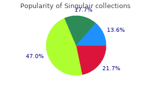
Order 10mg singulair free shipping
Screen for complications that may afect surgical risk: Nephropathy asthma definition subsequent effective 10mg singulair, cardiac disease, proliferative retinopathy, neuropathy. Resume usual therapy just afer frst meal, Check daily for dilutional hyponatreamia. Other considerations include stroke, seizures, trauma, drug overdose, infection, and ethanol intoxication. Infections, diuretic treatment, and drinking glucose-rich beverages, myocardial or cerebral ischemia may all be precipitating factors. The condition usually afects middle- aged or elderly patients and carries a high mortality. Hyper osmolar hyperglycemic state has a high mortality and immediate referral to secondary or tertiary facility is required. Heparin antithrombotic agents should be given in the absence of contraindications. People living with diabetes must change many habits, such as what they eat, when they exercise and how frequently they see your medical providers. They may need to take daily medications or insulin to keep their blood sugar levels in check. But these changes don?t mean one won?t be able to succeed at work or enjoy a healthy and fulflling life. People with diabetes have equal rights with those without the condition and should be protected from all forms of discrimination. Health-care providers should be aware of these problems so that they can give appropriate advice. Such prejudice is usually because of ignorance and the belief that all people with dia- betes have poor work performance and have regular interruptions as a result of hypoglycaemia. This must be dis- couraged as concealment may result in grave consequences in case of attacks of hypoglycaemia. Tere should be unbiased access to insurance policies (life or sickness) at a rea- sonable cost. Hypoglycaemia may even occur some hours afer exercise, possibly because the liver and muscles are still replenishing glycogen stores. Detailed advice from a health provider should be sought to reduce the risk of hypoglycaemia. Foot-Screening Assessment Sheet For Clinical Examination Patient Name? Hospital Number: ?. World Health Organization, Defnition, Diagnosis and Classifcation of? Diabetes Mellitus and its Complications. Report of a World Health Organization and International Diabetes Federation meeting. Type 2 diabetes mellitus afer gestational diabetes: a systematic review and meta- analysis 5. Canadian Diabetes Association 2003 Clinical Practice Guidelines for the Prevention and Management of Diabetes in Canada. Canadian Diabetes Association 2003 Clinical Practice Guidelines for the Prevention and Management of Diabetes in Canada. Canadian Diabetes Association 2003 Clinical Practice Guidelines for the Prevention and Management of Diabetes in Canada. The relationship of glycaemic exposure (HbA), to the risk of development and progression of retinopathy in the Diabetes Control and Complications Trial. Gaman Recommendations for Management of Diabetes During Ramadan Diabetes Care 28: 2305-2311 57 Division of Non-communicable Diseases Afya House, Cathedral Road P. Describe a neuropathy classification system (7 total) and list the prototypical condition for each 2. Myelinopathies, in which the primary site of involvement is limited to the myelin sheath surrounding the axon; 2. Axonopathies, in which the primary site of involvement is the axon, with or without secondary demyelination 3. Neuronopathies, in which the cell body of the neuron itself is the primary site of involvement, ultimately affecting the entire peripheral nerve. Note: ?Although overlap occurs, each of these prototypes has a distinctive clinical presentation, electrophysiologic profile, and microscopic appearance. Multiple lesions = multiple mononeuropathies = mononeuropathy multiplex Nonfocal = Polyneuropathies th Refer to figure 97. Up to 20% of patients remain disabled from this disease process, and about 5% will die despite therapy. Tongue weakness is associated with the development of respiratory compromise and the need for mechanical ventilation 3. Compared with adults, children have neuropathic pain more often but require mechanical ventilation less often 4. At about the level of the antecubital fossa, it bifurcates into the posterior interosseous (pure motor) rd and superficial radial (pure sensory) nerves. All motor function = extrinsic muscles of hand (unlike median and ulnar) CrackCast Show Notes Foreign Bodies January 2017 Anatomy: C7 to T1 roots - passes through the brachial plexus to descend medially, without branching, to the ulnar (medial) condylar groove at the elbow. Then goes from cubital canal, it branches to the ulnar wrist flexor and the deep flexors of the fourth and fifth digits. At the wrist it enters Guyon?s Canal between the pisiform and hook of the hamate, after which it bifurcates into the superficial terminal sensory branch and the deep motor branch. In addition to prior probability heavily favoring the elbow, the presence of sensory abnormalities in an ulnar distribution in the hand and fingers. The ulnar cutaneous innervation to the hand branches off from the main trunk proximal to the nerve entering Guyon?s Canal. Thus a lesion at the wrist should not produce sensory abnormalities, whereas one at the elbow would be expected to do so. Anatomy: C5 to T1 spinal nerve roots and exits the brachial plexus through the lower trunk. The best way to examine patients for sensory findings = touch the distal palmar tips very lightly, asking the patient whether the sensation feels ?abnormal. Last thing to note: the 7th type of peripheral neuropathy (Sensory Neuronopathy, aka Ganglionopathy) can be characterized by a selective/predominant involvement of the dorsal root ganglion. Although hundreds of apps for diabetes self-management are commercially available, we only identified health outcomes studies on 11 apps. More rigorous and longer-term research studies could determine whether apps help people manage their diabetes and reduce complications. None of the investigators have any affiliations or financial involvement that conflicts with the material presented in this report. The information in this report is intended to help health care decisionmakers?patients and clinicians, health system leaders, and policymakers, among others?make well-informed decisions and thereby improve the quality of health care services. This report is not intended to be a substitute for the application of clinical judgment. Anyone who makes decisions concerning the provision of clinical care should consider this report in the same way as any medical reference and in conjunction with all other pertinent information, i. This report is made available to the public under the terms of a licensing agreement between the author and the Agency for Healthcare Research and Quality. This report may be used and reprinted without permission except those copyrighted materials that are clearly noted in the report. Further reproduction of those copyrighted materials is prohibited without the express permission of copyright holders. Department of Health and Human Services endorsement of any derivative products that may be developed from this report, such as clinical practice guidelines, other quality enhancement tools, or reimbursement or coverage policies may not be stated or implied. Persons using assistive technology may not be able to fully access information in this report. Suggested citation: Veazie S, Winchell K, Gilbert J, Paynter R, Ivlev I, Eden K, Nussbaum K, Weiskopf N, Guise J-M, Helfand M.
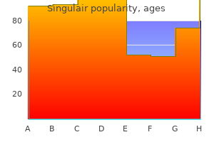
Order singulair 10mg overnight delivery
References of the identified articles were searched for additional cases and trials asthma symptoms in young child generic singulair 4mg free shipping. This fact sheet includes abstracts in the summary of published reports and considers them in determining the recommendation grade and category. Heterozygotes exhibit cholesterol of 250-550 mg/dL, xanthomata by age 20 years, and atherosclerosis by age 30. Last resort therapies include distal ileal bypass, portacaval shunting, and liver transplantation. Short-term effects include improved myocardial and peripheral blood flow as well as endothelial function. Long-term outcome studies have demonstrated significant reductions in coronary events. The columns function as a surface for plasma kallikrein generation which, in turn, converts bradykininogen to bradykinin. References of the identified articles were searched for additional cases and trials. However, the presence of such a permeability factor has not been con- firmed although some of its characteristics have been described. Unfortunately, 20-30% of transplanted patients will experi- ence a recurrence in the renal allograft, especially children. Technical notes Vascular access may be obtained through arteriovenous fistulas or grafts used for dialysis. Tapering should be decided on a case by case basis and is guided by the degree of proteinuria. Timing of clinical response is quite variable and complete abolishment of proteinuria may take several weeks to months. References of the identified articles were searched for additional cases and trials. The roughly 50% of patients who do not completely respond will suffer steroid side effects, infections and progressive end-organ complications. Maximal responses often require 2 to 6 months of treatment and most are partial rather than complete. An alternative two step process method is commonly used in Europe and for smaller body weight patients. References of the identified articles were searched for additional cases and trials. Description of the disease this inherited disorder results in iron deposition in the liver, heart, pancreas and other organs. Other mutations, in genes coding for hemojuvelin, hepcidin, transferrin receptors or ferroportin, have been described in fami- lies with syndromes of hereditary hemochromatosis. Iron accumulation in organs slowly results in liver failure (cirrhosis, hepatocellular carcinoma), diabetes, hypogonadism, hypopituitarism, arthropathy, cardiomyopathy and skin pigmentation. At diagnosis, the saturation of serum transferrin or iron binding capacity will be elevated (! Current management/treatment Because hereditary hemochromatosis is a disease of iron loading, iron removal by therapeutic phlebotomy is the mainstay of treatment. Phlebotomy therapy should be started in all patients whose serum ferritin level is elevated despite older age or the absence of symptoms. Typically, 1 unit of whole blood is removed weekly until the serum ferritin is <50 ng/mL without resultant anemia. Thereafter 2-4 phlebotomies per year are needed to maintain the ferritin 50 ng/mL. Malaise, weakness, fatigability and liver transaminase elevations often improve during the first several weeks of treatment, but joint symptoms may ini- tially worsen before eventually improving (if at all). Cardiomyopathy and cardiac arrhythmias may resolve with phlebotomy, but insulin-dependent diabetes generally will not. The risk of hepatocellular carcinoma will persist if cirrhosis was present prior to the onset of phlebotomy therapy. Rationale for therapeutic apheresis Patients typically present with upward of 20 grams of excess iron thus, with 250 mg of iron removed per phlebotomy, two years may be needed to achieve therapeutic iron depletion. Each erythrocytapheresis removes two to three times that amount of red blood cells and iron while maintaining isovolemia. For example, in a prospective series of 13 patients the goal of each procedure was to remove a maximum of 800 ml of red cells and reduce the patient?s hematocrit to 30%. A prospective, randomized trial, under way in the Netherlands, compares erythrocyta- pheresis of 300-800 ml of erythrocytes every 2-3 weeks to a target hematocrit of! Primary outcome measures are the duration and number of treatments to reach ferritin 50 ng/mL. Secondary outcome measures are decline in hemoglobin during treatment, improvement in liver function, patient discom- fort and cost. Data from the first 26 study subjects have been published, and, not surprisingly, each erythrocytapheresis procedure removes more that twice the volume of erythrocytes of a phlebotomy procedure and 2. Whether erythrocytapheresis shortens the total treatment inter- val or is cost-effective versus phlebotomy remains to be determined. In a previous pilot study, published by the same group, 6 patients achieved iron depletion with erythrocytapheresis in (mean [range]) 9. Technical notes While reported methods vary, the Dutch trial employs a schedule of erythrocytapheresis of 300-800 ml of erythrocytes every 2-3 weeks. Duration and discontinuation/number of procedures: Erythrocytapheresis every 2-3 weeks, or as tolerated, until serum ferritin <50 ng/mL. Maintenance treatment can follow with infrequent therapeu- tic phlebotomy or erythrocytapheresis. References of the identified articles were searched for additional cases and trials. Infection, pregnancy or drugs may trigger clinical disease in the presence of these mutations. All candidates for renal transplan- tation must have genetic testing, as transplantation outcome may be related to mutation type. However, 30-100% of transplant patients, depending on the type of muta- tion, have recurrence in the graft, causing graft failure. The alternative therapies may include use of purified complement factors or comple- ment inhibitors, i. These guidelines address neither continued treatment after initial therapy failure nor ongoing prophylactic treatment for patients with remission. Decisions of duration or to discontinue should be made based upon patient response and condition. References of the identified articles were searched for additional cases and trials. Myeloid blasts are larger and more rigid than lymphoid blasts, and their cytokine products may upregulate endothelial cell adhesion molecule expression and activate inflammation. These processes can lead to microvascular leukoag- gregates, hyperviscosity, tissue ischemia, infarction and hemorrhage. Clinical manifestations are not reliably predicted by the degree of hyperleukocytosis alone. The frequency and severity of leukostasis complications, particularly pulmonary, are greater with the monoblastic/monocytic subtypes. Pulmonary complications include dyspnea, hypoxemia, diffuse alveolar hemorrhage, respiratory failure and radiographic findings of interstitial and/or alveolar infiltrates. Plasma, cryoprecipitate and/or platelets are given, as indicated, for bleeding or coagulopathy. Red cell transfusions should be avoided in patients with symptomatic leukostasis prior to cytoreduction because of the risk of augmenting hyperviscosity. Adjunctive radiation therapy may be considered in cases with parenchymal brain lesions; prophylactic cranial irradiation is not indicated. A second cohort study found no decrease in early mortality and raised concerns that leukocytapheresis may delay the start of chemotherapy. Prophylactic leukocytapheresis should, therefore, be consid- ered in those patients. Severe end-organ injury or hemorrhage may not improve, however, particularly if extensive pre-existing tissue damage exists. Leukocytapheresis should be repeated in persistently symptomatic patients until clinical manifestations resolve or a maximum benefit is achieved. Chemotherapy should not be postponed and is required to prevent rapid reaccumulation of circulating blasts.
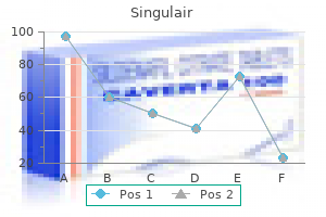
Buy cheap singulair 4mg on line
Finding 22 matches increases the chances that the encrypted files contain child pornography but such assumptions are difficult to verify without decrypting the files asthma zinc purchase singulair with american express. The memory of this process can be xi dumped to a file using a program like pmdump and searched for unencrypted data as shown here: Even if such access were available, the examiner would have to operate the computer, altering its state and potentially erasing valuable information. In practicality, memory dumps are most useful when they occur accidentally as described in the next section. Investigators may be able to obtain the decryption passphrase by searching the area surrounding a system for slips of paper containing the xii passphrase, interviewing the suspect, or surreptitiously monitoring the suspect?s computer use. Also, if it is possible to obtain passwords that the suspect uses to protect other personal data such as e-mail and personal digital assistants, these passwords should be tried since people often use the same password for multiple purposes. As noted in the previous section, accidental memory dumps may disclose information relating to encryption. In this way, if the user purposefully or unintentionally stored their passphrase on disk or an application wrote the passphrase to disk, it will be available in the keyword list. Privacy concerns related to overly broad searches are a potential problem when scouring a disk for keywords to be used in passphrase guessing. Overly broad searches of computers have been used to exclude evidence (Carey, 2000). However, generating an exhaustive list of keywords from all available media has a specific purpose and is distinct from an exploratory search of media for incriminating evidence. In one case, the suspect claimed that, in a fit of paranoia, he had changed his passphrase to something he could not recall just prior to being apprehended. To verify this claim, the forensic examiner checked the modification time of the file containing the private key and found that the passphrase had never been changed. When the prosecuting attorney presented this proof to the suspect and explained that any further lack of cooperation would make matters worse, the suspect provided the correct passphrase. Although refusing to disclose an encryption passphrase does not necessarily imply guilt, it does reflect badly on the suspect in court and can shift the burden of proof onto the defense. Faced with such a risk, offenders can be persuaded to decrypt data in exchange th for leniency in sentencing or plea bargain. In this case, however, the key was not sought for the purpose of acquiring evidence for conviction, but rather to determine whether the The prosecution argued that the contents of the file had already been uttered and, therefore, were not protected under the 5th Amendment. As long as prosecutors did not try to tie the defendant to the file by virtue of his knowing the passphrase, no incrimination was implied by disclosing the passphrase. Commercial software programs like Spector Pro and free programs such as SubSeven and Back Orifice enable key logging, screen captures, and remote file access, enabling investigators to obtain encrypted files remotely. However, these devices are not designed for Macintosh or Sun systems and do not work on laptops or personal digital assistants since the keyboard is integrated. Also, physical access to the machine is required to install and retrieve hardware devices and they are visible to the alert user. Although full details of the monitoring system are protected under the Classified Information Procedures Act, court records indicate that the system only captured keystrokes while the computer was not connected to the Internet via the modem. This explanation satisfied the court during an in camera, ex parte hearing but most key loggers do not function in this manner and this technique is of limited effect when a computer is continuously connected to the Internet or when the suspect writes e-mail offline and only connects to the Internet to send the messages. The court addressed this concern by comparing key logging to searching a closet or file cabinet. This does not, as Scarfo argues, convert the limited search for the passphrase into a general exploratory search. During many lawful searches, police officers may not know the exact nature of the incriminating evidence sought until they stumble upon it. Just like searches for incriminating documents in a closet or file cabinet, it is true that during a search for a passphrase ?some innocuous [items] will be at For instance, when a small number of passphrases are likely, it may be feasible to type in all permutations. If this approach is taken, it is important to document the process both to avoid entering a passphrase more than once and to show attorneys, jurists, and other examiners which passphrases were attempted. When manual passphrase guessing fails, an automated approach may be used with a list of common passphrases, then a dictionary in the language(s) of the suspect, and then more sophisticated permutation techniques. Therefore, examiners should attempt to access all backup copies of private keys in case some have weaker passphrases than others. Therefore, a backup copy of the private key file may be found on a floppy disk or other external media. In Windows 2000, the built-in Administrator account is the default recovery agent (an organization can override the default by assigning a domain-wide recovery agent provided the system is part of the organization?s Windows 2000 domain). Such password guessing programs cannot be relied on as the only method of recovering encrypted data since they can take a significant amount of time and often have limitations that are not immediately evident. Although it may be possible decrypt some forms of Internet, wireless, and mobile phone communications by exploiting weaknesses in the encryption protocols (Biryukov, A. Instead of attempting brute force attacks, unencrypted data can be sought at end points of a communication channel. When confronted with encryption, many computer intruders attack the end points in favor of more sophisticated attacks. Computer intruders have adapted to this challenge by attacking the end points: the client and server. On the client side, intruders are gaining unauthorized access to individuals? personal computers and capturing passwords using programs like SubSeven and Back Orifice mentioned in Subsection 4. For instance, when encrypted e-mail services like Hushmail are used, the communication is most exposed at the sender?s and recipient?s computers when they compose and read the message, respectively. Ideally, encryption detection tools would find a wider variety of encrypted data on all xxi areas of a disk. Similarly, automated mechanisms for detecting encrypted network traffic are essential to help isolate encrypted evidence in large volumes of captured data. Because plaintext is often wiped after it is encrypted, it is desirable to be able to analyze the media using scanning probe or magnetic force microscopes to recover unencrypted copies of the data. The hope is that the need for this type of equipment will drive down costs and make it more widely available, making media analysis a viable solution for encryption. As this equipment becomes more generally available, analyzing media for overwritten data will become more common. Password finding and guessing tools that can access a wider range of file types and intelligently combine keywords to create complex passphrases will make this one of the most viable ways to decrypt evidence. Similarly, as powerful brute force tools become less expensive and easier to use, it will become more practical for smaller organizations to utilize this approach. For instance, photon-based quantum cryptography make it impossible to decrypt communications because the act of monitoring alters the transmission. Fortunately, as such techniques evolve, they will also improve our ability to deal with encryption. For instance, quantum computers would significantly reduce the time it takes to break existing encryption algorithms (Schneier, 1996). Conclusion Although encryption can be a formidable hurdle in a forensic examination of digital evidence, it is not insurmountable. Providing attorneys and investigators with plaintext fragments of encrypted documents gives them leverage in a case and may even be sufficient to obtain a conviction. Furthermore, with the knowledge and tools described in this paper, a forensic examiner may be able to obtain or guess encryption passphrases, enabling them to decrypt all associated evidence. However, with the growing number and sophistication of encryption and data hiding tools, it is difficult for an individual forensic examiner to keep pace. This difficulty can be reduced by improvements in tools and increased information sharing among examiners. Some devices such as the iButton will destroy the key if physical tampering is detected but it may be possible to undermine the autodestruct feature by first disabling the power source that would be used to erase the memory of the device. He investigates computer intrusions, intellectual property theft, and cyberstalking reports, and has extensive experience analyzing evidence stored on and transmitted using computer networks. He has assisted law enforcement in a wide range of criminal investigations including homicide, child exploitation, and larceny. As an Information Security Officer at Yale University and in subsequent consulting work, Mr.
Generic 5mg singulair amex
These precautions include testing of the patient and the use of rapid testing and quarantine of samples until test results are known asthma definition vintage discount singulair 4 mg with visa. Sperm cryopreservation before cancer chemotherapy helps in the emotional battle against cancer. Membranous and structural damage that occur during cryopreservation of human sperm may be time-related events. Evaluation of chromatin integrity in human sperm using acridine orange staining with different fixatives and after cryopreservation. Effect of natural antioxidants tocopherol and ascorbic acids in maintenance of sperm activity during freeze-thaw process. Motility and respiration of human spermatozoa after cooling to various low temperatures. The effects of cooling rate and warming rate on the maintenance of motility, plasma membrane integrity, and mitochondrial function. Investigation of fertilizing capacity of cryopreserved spermatozoa from patients with cancer. The effects of cryopreservation on sperm morphology, motility and mitochondrial function. Recent developments and concepts in the cryopreservation of spermatozoa and the assessment of their post-thawing function. This information is kept on file in the European Association of Urology Central Office database. This guidelines document was developed with the financial support of the European Association of Urology. Produced in collaboration with the Ethiopia Public Health Training Initiative, the Carter Center, the Ethiopia Ministry of Health, and the Ethiopia Ministry of Education. Important Guidelines for Printing and Photocopying Limited permission is granted free of charge to print or photocopy all pages of this publication for educational, not-for-profit use by health care workers, students or faculty. All copies must retain all author credits and copyright notices included in the original document. Under no circumstances is it permissible to sell or distribute on a commercial basis, or to claim authorship of, copies of material reproduced from this publication. Except as expressly provided above, no part of this publication may be reproduced or transmitted in any form or by any means, electronic or mechanical, including photocopying, recording, or by any information storage and retrieval system, without written permission of the author or authors. This material is intended for educational use only by practicing health care workers or students and faculty in a health care field. Pathophysiology is the study of the disturbance of normal mechanical, physical, and biochemical functions, either caused by a disease, or resulting from a disease or abnormal syndrome or condition that may not qualify to be called a disease. An alternate definition is "the study of the biological and physical manifestations of disease as they correlate with the underlying abnormalities and physiological disturbances. This lecture-note will provide a summarized basis for the students by expanding the student?s knowledge in the sciences how alteration in structure (Anatomy) and function (Physiology) disrupt the human body as a whole. It is written for undergraduate students in nursing and other health oriented disciplines as prerequisite course for certain courses. In this text of pathophysiology, every attempt has been made to provide the most current available information in simplified and well- explained ways. Mesele Bezabih (Jimma University) for their intensive and meticulouse review of this lecture note. Biftu Geda from Haramaya University Faculty of Health Sciences for their invaluable comments in reviewing the lecture note. Alemayohu Bayray (Mekelle University), Wezam Tesfay (Defence College of Health Sciences) and Mr. Adaptation is a normal life cycle adjustment like in growth during puberty; changes during pregnancy or aging and stressful life style produce physiologic changes that may lead to adaptation or disease. The cell constantly makes adjustments to a changing, hostile environment to keep the organism functioning in normal steady state which is necessary to ensure the survival of the 1 Pathophysiology organism. Prevention of disease by the body depends on the capacity of the affected cells to undergo self-repair and regeneration i. The common stimuli are:- a) Physical agents o Trauma, Burn, pressure, irradiation, etc b) Chemical agents o Poisons, drugs, simple compounds, etc. As a result common changes include:- - Cellular swelling 3 Pathophysiology - Lipid accumulation (Fatty change process in the cytoplasm of cells). Changes to cellular size or numbers - Changes in size and numbers of the cells are usually as a result of response to adapt to harmful agents. Types of Hyperplasia 5 Pathophysiology a) Physiologic Hyperplasia: occurs when there hormonal stimulation - Occurs in puberty and pregnancy b) Compensatory-Hyperplasia - Occurs in organs that are capable of regenerating lost tissues. Metaplasia is a reversible change in which one type of adult cell is replaced by another type. The changes caused by this type of injury are potentially reversible if the injuring stimuli are removed. Causes of cell injury:- Causes of cell injury are the same causes of cellular adaptive changes as mentioned above. Classification of cell injury:- Cellular injury can be reversible or it may progress to irreversible change (Lethal change). Reversible cell injury:- Is cell injury which can be reversed when the stimulus or the cause of injury is removed. Example -Ischemia: o Ischemia refers to a critical lack of blood supply to a localized area. Irreversible Cell injury It is cellular injury that can not be corrected (reversed) after the stimulus or cause has been removed. Necrosis:- 10 Pathophysiology o the term necrosis refers to cell or tissue death characterized by structural evidence of this death. It is common in tuberculosis and is characterized by central area of necrosis which is soft, friable and surrounded by an area with a cheesy, crumbly appearance. Colliquative- Necrosis (liquefactive- Necrosis) 11 Pathophysiology - It frequently occurs in brain tissues and results from break down of neurons by released lysosomal enzymes resulting in formation of pockets of liquid, debris and cyst like structures in the brain tissue. Benign and Malignant Neoplasia - the capacity of undergoing mitosis is inherent in all cells. Every time a normal cell passes through a cycle of division, the opportunity exists for it to become Neoplastic. For women the most common cancers are those of the breast, lung, and colorectal respectively. Classifications of Neoplasms - Neoplasms are classified according to their cells of origin and their behavior of growth as benign or malignant. Nomenclature of Neoplasms - Naming of Neoplasia based on two main important features of the tumor. These are:- A) Based on its Behavior of growth:- i) Benign: - Add ?oma? at the end for connective tissue origin tumors. B) Based on cells of origin:- - Neoplasms are named at their prefix by their cells of origin and their suffixes are added at the end to show whether they are benign or malignant. Example: - Behavior of growth Cells of origin Benign Malignant - Fatty cells - Lipoma - Liposarcoma - Bone cells - Osteoma - Osteosarcoma - Blood vessels - Hemangioma - Hemangiosarcoma - Fibrous tissues - Fibroma - Fibrosarcoma C) Exceptional Naming (Malignant Misnomers) 16 Pathophysiology - There are some neoplasms that are named exceptionally to the above rules. It is also called Hepato-cellular carcinoma - Hodgkin?s disease: - Malignant tumors of the lymphoid tissues. Mechanisms of carcinogenesis - There are large numbers of research done in the world to know the etiology of cancer but none of the theories that attempt to explain the peculiarities of the cancer cells have been completely successful. Genetic Instability:- - the theory of somatic cell mutation supports the concept that mutational carcinogenic agents and heredity susceptibility can induce genetic abnormalities. Carcinogens - Carcinogens are those substances that are capable of inducing neoplastic growth. Some substances induce neoplastic growth at higher doses and exposure rates while others can be carcinogenic at lower doses and exposure rate. Chemical carcinogens - Many chemical agents are capable of causing Neoplasms in either humans or animals. Physical carcinogenic agents Ionizing radiation is a recognized cause of cellular mutations. A long latent period often exists between exposure and development of clinical disease. Viral carcinogens (oncogenic viruses) Viruses are thought to cause some human and Animals malignant neoplasms.
Myroxylon balsamum var. balsamum (Tolu Balsam). Singulair.
- How does Tolu Balsam work?
- Dosing considerations for Tolu Balsam.
- What is Tolu Balsam?
- Are there safety concerns?
- Bedsores, bronchitis, cancer, cough, cracked nipples, lips, reducing lung swelling (inflammation), and minor skin cuts.
Source: http://www.rxlist.com/script/main/art.asp?articlekey=96373
Generic 4 mg singulair free shipping
The work cannot be changed in any way or used commercially text of this article on the journal?s Web site ( If a donor All recommendation statements are not graded unless speci- candidate?s postdonation risk is below the transplant fied otherwise asthma symptoms in toddlers best 4mg singulair. The decision to approve donor candidates with that resolves (eg, a treated infection) may be acceptable hypertension should be individualized based on demo- for donation. These measures should be initiated be- nephrectomy (eg, computed tomography angiogram) fore donation and maintained lifelong. Transplant programs should support autonomy based clinical practice guideline on the evaluation and care through a fully informed consent process. The guideline consists of recom- mendation statements and supporting rationale, including Policies for Donor Candidate Identification summaries of systematically generated evidence on relevant 18. Outcomes staff to review the available evidence, formulate recommen- were selected and ranked by assessing patient-centeredness. Long-term outcomes were based on a mean also held regular calls until completion of the evidence review follow-up of at least 5 years. Key Question 4: What is the incidence of long-term health outcomes for living kidney donors compared with healthy nondonors? Two investigators independently screened postnephrectomy and long-term outcomes is noted in Table 2. A third ?Intermediate outcomes? are defined as events on the path- investigator resolved discrepancies. Screening criteria were Cochrane Library to identify previous systematic reviews, liberal. Bibliographic database searches sizes over 100 and mean follow-up time of at least 5 years. Citations deemed eligible by either investigator underwent outcomes were required to include a nondonor comparison S16 Transplantation August 2017 Volume 101 Number 8S These perspectives provide a framework for assessment of donor outcomes, inter- pretation of observations, patient communication, and future research design. Understanding and communicating medical risks for living kidney donors: a matter of perspective. Studies comparing living kidney donors to the for observational studies using an instrument developed general population were not eligible. Evidence profiles mographic, and outcomes data from studies eligible for full consisting of tables examining all relevant outcomes, extraction. Data fields extracted included author, year of including a summary of the results and judgments about publication, setting, donor and comparison populations, in- the certainty and quality of the evidence, were used to clusion and exclusion criteria, donor and comparison charac- facilitate this process. Evidence quality assessment criteria Study design Quality of evidence Lower if Higher if Randomized trial High Risk of Bias Large effect? Additionally, evidence quality is downgraded when estimates are imprecise and Results of the Systematic Review publication bias is likely. Evidence supplemental citation searching, yielding a total of 484 ref- quality is also increased when an effect is demonstrated after erences for full text review (Figure 2). However, the strength of a recommendation is deter- dates for living kidney donation needed to be restructured mined not only by the quality of the evidence, but also by to include a comprehensive determination of risk to the do- other, often complex judgments regarding the size of the net nor, based on simultaneous consideration of a composite medical benefit (potential risks vs benefit), values, and prefer- profile of risk factors. Formal decision analyses including for this approach to the evaluation of living donor candi- cost analysis were not conducted. The development and application of interest, taking into account explicit judgments about the rel- this clinical prediction tool are described in chapter 1 (Frame- ative importance of each outcome. The resulting 4 final cate- 7 gories for the quality of overall evidence were: ?A,?B,? work) of this guideline and in a separate publication. The development and application current guideline is notable in that many clinically important of this clinical tool are described in chapter 5 (Kidney Func- topics in living donation are not ethically or practically amena- 8 tion) of this guideline and in a separate publication. Each patient needs help substantial debate and involvement of of action, but many would not to arrive at a management decision consistent stakeholders before policy can with her or his values and preferences be determined aThe additional category ?Not Graded? is used, typically, to provide guidance based on common sense or where the topic does not allow adequate application of evidence. The most common examples include recommendations regarding monitoring intervals, counseling, and referral to other clinical specialists. Ungraded recommendations are generally written as simple declarative statements, but are not meant to be interpreted as being stronger recommendations than Level 1 or 2 recommendations. Determinants of strength of a recommendation Factors Comments Balance between desirable and undesirable effects the larger the difference between the desirable and undesirable effects, the more likely a strong recommendation is warranted. The narrower the gradient, the more likely a weak recommendation is warranted Quality of the evidence the higher the quality of evidence, the more likely a strong recommendation is warranted Values and preferences the more variability in values and preferences, or the more uncertainty in values and preferences, the more likely a weak recommendation is warranted. Review of Guideline Development Process Developing the Recommendations Several tools and checklists have been developed to assess the quality of the methodological process for systematic re- Draft recommendation statements were developed by the view and guideline development. Public Comment and Revision Format for Recommendations A draft of the guideline was distributed for open public re- Each chapter contains one or more specific recommenda- view in November 2015. Final grade for overall quality of evidence Grade Quality of evidence Meaning A High We are confident that the true effect lies close to that of the estimate of the effect B Moderate the true effect is likely to be close to the estimate of the effect, but there is a possibility that it is substantially different C Low the true effect may be substantially different from the estimate of the effect D Very low the estimate of effect is very uncertain, and often will be far from the truth S20 Transplantation August 2017 Volume 101 Number 8S Such recommendations may be deemed necessary because the work group considers such ?good practice statements? essential especially when net benefit is great and unequivocal. Goals and Principles of Evaluation candidate?s postdonation risk is below the transplant program?s acceptable risk threshold, the candidate makes 1. Donation must be voluntary (autonomous), and the scribing medical criteria that are acceptable for donation, motivation for donation must be altruistic to satisfy a addressing when possible, numeric thresholds for short- well-considered desire to help another person. There must term and long-term postdonation risks above which the be protection from undue pressure or coercion at every step transplant program will not proceed with donation. Risks in the evaluation and donation process, including the option should be expressed as absolute rather than relative risks. In addition to these ethical principles, protection estimates of short-term and long-term risks from dona- of patient privacy must be ensured. However, information re- tion, including recognition of associated uncertainty, in garding donor lifestyle, exposures or medical history that in- a manner that is easily understood by donor candidates. If a donor candidate?s postdonation risk is plantation to proceed; donor candidates should be given above the transplant program?s acceptable risk thresh- the opportunity to withdraw if they do not consent to sharing old, the risk is not acceptable for donation. The search was updated through September 2014 and supplemented by articles identified by work group members through January 2017. Public review comments were compiled and fed back to the work group, which considered the comments in its revision of the guideline (12) Update plan State whether or not there is a plan to update the guideline the requirement for an update will be assessed on an ongoing and, if applicable, an expiration date for this version basis from the publication date for potential important new of the guideline evidence that may lead to changes to the recommendations (13) Definitions Define unfamiliar terms and those critical to correct application Abbreviations and Acronyms of the guideline that might be subject to misinterpretation (14) Recommendations State the recommended action precisely and the specific this guideline contains recommendations for evaluation of and rationale circumstances under which to perform it kidney donor candidates and postdonation follow-up care. Justify each recommendation by describing the linkage Each recommendation builds on a supporting rationale with between the recommendation and its supporting evidence evidence tables if available. The strength of the recommendation Indicate the quality of evidence and the recommendation and the quality of evidence are provided in parenthesis within strength, based on the criteria described in Topic 9 each recommendation, where applicable Continued next page S22 Transplantation August 2017 Volume 101 Number 8S The estimated balance between potential benefits and harm was considered when formulating the recommendations (16) Patient preferences Describe the role of patient preferences when a the inclusion of patient values and preferences is clearly recommendation involves a substantial element of articulated where appropriate personal choice or values (17) Algorithm Provide (when appropriate) a graphical description of the No overall algorithm stages and decisions in clinical care described by the guideline (18) Implementation Describe anticipated barriers to application of the Review criteria were not suggested because implementation with considerations recommendations prioritization and development of review criteria must proceed Provide reference to any auxiliary documents for providers locally. Preservation of donor candidate autonomy and minimiza- decision making between donor candidates and their trans- 16,19-21 tion of short-term and long-term risks are high priorities in plant professionals. The transplant program has An important advance is quantification of the combined im- the responsibility to disclose anticipated risks and benefits pact of all of a donor candidate?s predonation demographic to the donor candidate and intended recipient, tailored when (eg, age, sex, and race) and health characteristics at the time 18 possible for the characteristics of each donor candidate. The transplant program must offer support for comes can be surgical, medical or psychosocial, and may oc- decision-making through education and the informed consent cur during the perinephrectomy period, in a fixed period of process, and has a responsibility to confirm that the donor long-term follow-up (eg, 15 years after donation), or for the candidate understands the likely risks and benefits of dona- remaining lifespan of the donor. The transplant program makes the final determination As described within this overall framework, a transplant of acceptance of the donor candidate, based on the program?s program can use various methods to establish its threshold policies. The transplant program must have a mechanism for for acceptable outcomes after kidney donation. For example, resolving disagreement among team members regarding ac- if a transplant program decides a lifetime postdonation risk ceptability of donor candidates that avoids conflicts of interest. Donor Decision Making candidate autonomy does not overrule medical judgment There will always be risks to living kidney donation. A and transplant professionals are ethically justified to decline central objective of donor candidate evaluation and selection a donor candidate when they believe the risk of poor 22 is to minimize risks of short-term and long-term adverse postdonation outcomes is too high. A poor outcome can outcomes after donation, and to ensure the risks are ac- have a very negative impact on the donor, on their recipient, ceptable. Consistent, transparent and defensible decision- and on public opinions about living donation. Thresholds should be both evidence-based and postdonation risk in relation to single predonation charac- consensus-based, and there are various sources of evidence teristics assessed in isolation, and generally agree on the sin- and processes by which consensus can be achieved.
Syndromes
- Weakness
- Increased blood levels of the thyroid hormones T3 and T4
- The surgeon will join together your rebuilt esophagus and stomach in your neck.
- Headache
- Do you usually have regular periods?
- Popsicles or gelatin (Jell-O) are good choices, especially if the child is vomiting.
- CT scan
- Unexplained fever with back pain
Buy singulair american express
Test Principle (Cryofibrinogen): Cryofibrinogens are complexes of fibrinogen asthmatic bronchitis in infants cheap singulair 10 mg line, fibrin split products, and plasma globulins, that are precipitable in the cold and can be associated with purpura thrombosis, and/or hemorrhage. The plasma and blood must be separated at 37oC using heated lab equipment and rapid processing techniques. One half of the plasma is maintained in a 37oC heating block for 24 hours and the other half is placed on ice in the refrigerator for 24 hours. At the end of 24 hours, if the iced sample has gel fibrin-like strands and the 37oC does not, the sample is considered positive for cryofibrinogen. The results may be further confirmed by placing the iced sample in a 37oC incubator for 30 minutes and the gel strands should disappear, confirming the presence of a cryofibrinogen. The ansence of this cold precipitable substance in serum provides substantiation that the protein is cyrofibrinogen rather than cyroglobulin. Possible results and interpretation (Cryofibrinogen): Patient samples are scored as positive or negative. Factors affecting test results (false positives and negatives) (Cryofibrinogen): Sample must be maintained at room temperature until plasma and red blood cells are separated or the cryofibrinogen may deposit on red blood cells and give a false negative result. The activated partial thromboplastin time is the coagulation test that is used to measure the functional levels of the coagulation factors in the intrinsic pathway. Analysis is performed by making serial dilutions of the reference plasma in buffer (with assay values for the factor that needs analysis). Each dilution is then mixed with an equal volume of ?substrate? plasma that is known to contain normal levels of all factors, except the factor that is being assayed. The results are plotted on a log/log graph (polynomial curve) with percent factor on the abscissa and the time in seconds on the ordinate. The test results of the above dilutions will form a straight line that forms the basis for a standard curve at the connected points which can then be used to determine the activity of that coagulation factor in patient samples. To analyze a factor activity level on an unknown sample, 1:5 (100% activity) and 1:10 (50% activity) dilutions are prepared and mixed with ?substrate? plasma. The percent activity is read from the abscissa of the graph by finding where the clotting time obtained for the unknown sample intercepts the standard curve. All of this now is in fact programed into our analyzers, and graph paper can no longer be found in our laboratory. Isolated deficiency of Factor V activity may be inherited (incidence 1/1,000,000) or rarely acquired in patients suffering from amyloidosis. Cases of congenital factor V deficiency associated with the production of a dysfunctional molecule have also been reported. Inhibitors directed against factor V can be seen in patients exposed to fibrin glue prepared with bovine thrombin. Note: Factor V deficiency is not synonymous with Factor V Leiden (see hypercoagulable state discussion). Acquired factor X deficiency is most commonly seen in conjunction with vitamin K deficiency or therapy with vitamin K antagonists. Factors affecting test results (false positives and negatives) (Factor Assays Studies): Factor activity results can be influenced by the type of coagulometer or the reagents used to perform the test. Dilution errors can adversely affect the obtained factor activity results as well. The presence of the Lupus Anticoagulant has been associated with falsely decreased results. Diagnosis of deficiency states or inhibitors affecting factors in the intrinsic pathway of the coagulation cascade. Analysis is performed by making serial dilutions (1:5 to 1:320 dilutions of calibration reference plasma or factor assay reference plasma in buffer (both with known assay values for the factor of interest). Each dilution is mixed with an equal volume of ?substrate? plasma that is known to contain normal levels of all factors except the factor of interest. The results are plotted on a log/log graph (polynomial curve) with the percent dilution on the abscissa and the time in seconds on the ordinate. The connected points from the above dilutions form a straight line that is the basis for a standard curve. This standard curve is then used to measure the factor activity of patient samples with unknown factor content. To analyze the factor activity level on an unknown sample, 1:5 (100% activity) and 1:10 (50% activity) dilutions are prepared and mixed with immunoabsorbed, factor deficient (<1%) plasma. The percent factor activity of the patient sample is read from the abscissa of the standard curve graph by finding where the time obtained on the unknown intercepts the standard curve. Hemophilia A is inherited in an X-linked recessive manner and occurs in approximately 1 in every 10,000 live male births. These autoantibodies are associated with autoimmune disorders, malignancies, medications and the peripartum period. Like hemophilia A, hemophilia B is inherited in an X-linked recessive fashion but its incidence is about 10-fold less common occurring in 1 out of every 50-100,000 male births. These antibodies are associated with anaphylactic reactions among hemophilia B patients when receiving factor replacement. The presence of the Lupus Anticoagulant has been associated with falsely decreased results. The urea stability test is sensitive to levels of 1-5%, while the acetic acid method is sensitive to below 10%. There are other assays available but only the Berichrom is approved in the United States. Of note there are reports of the thromboelastogram being abnormal, with reduced maximum amplitude and strength and increased clot cyis at 30 minutes. By comparing the difference in factor activity of the patient incubation mixture and a control mixture, the amount of inhibitor present is calculated in Bethesda units. A modification of the Bethesda assay, the Nijmegen modification, dilutes patient plasma in buffered plasma to eliminate the decrease in factor activity that occurs with pH shifts during incubation. The imprecision introduced with unbuffered plasma is relevant particularly for lower titer inhibitors. Inhibitors to specific coagulation factors can be classified as being either autoantibodies or alloantibodies. Immunosuppressive therapy is effective in eradication of these antibodies in 60-70% of patients. In contrast to autoantibodies, alloantibodies develop in patients who lack a particular coagulation factor and generate an alloantibody upon exposure to this factor during treatment with coagulation factor concentrates. The prevalence of inhibitors is much lower among patients with hemophilia B where their occurrence has been estimated to be 2%. Patients with low titer inhibitors can be effectively treated with factor concentrates by doubling or tripling the dose. The advantage of this approach is that factor activity levels can be followed to objectively assess the adequacy of therapy. The ability to measure factor levels can be very useful in situations in which objective confirmation of the adequacy of replacement therapy is important such as during major surgical procedures or treatment for life-threatening bleeds. This assay cannot distinguish between specific factor inhibitors and fibrin degradation products, heparin, or heparin like inhibitors that may also lead to a positive mixing test result. Four major type 2 variants are recognized: type 2A, type 2B, type 2M and type 2N von Willebrand disease. It is an autosomal recessive form in which one abnormal gene is inherited from both parents. Ristocetin Cofactor Assay the Ristocetin Cofactor Assay Measures the Functional Level of von Willebrand Factor in Plasma Indications (Ristocetin Cofactor Assay): 1. The Ristocetin Cofactor Assay is performed by agglutinating a standardized suspension of platelets in the presence of von Willebrand Factor (provided by the patient plasma) using the antibiotic, Ristocetin. Although the platelets play a passive role in such agglutination, there is an absolute requirement that the Ristocetin-dependent receptor be intact. Levels of Ristocetin Cofactor activity are determined by the ability of the test plasma and Ristocetin to induce aggregation in a standardized platelet suspension. Following reconstitution, lyophilized platelets are treated with Ristocetin in the presence of dilutions of normal standardized human plasma with a known amount of Ristocetin Cofactor Activity. A standard curve is prepared, after which patient plasma is then used as a source of Ristocetin Cofactor Activity.
Cheap singulair 5mg fast delivery
Comment: An incision in the infero-lateral abdominal wall flap extending superiorly relaxes the flap and opens the abdominal cavity fully asthma symptoms 10 month old buy cheap singulair 4mg. At this point, it will be useful to have a preliminary inspection of the peritoneal cavity. Now is the time to take photographs, swabs, or to collect samples of ascites or pus, if indicated. Comment: In addition, if indicated, look for perforations or sources of haemorrhage. Palpate and inspect for any pathology that should be identified before continuing with the evisceration. Divide the jejunum between the ties preparatory to removing the small and large bowel. Comment: the removal of the small and large bowel serves to debulk the subsequent en-bloc removal, and enables better examination of the bowel. About 1 cm from the bowel wall, divide the mesentery from the jejunum and then the ileum down to the ileo-caecal junction. Dividing the mesentery closer to its root in the posterior peritoneum is quicker but leaves the small bowel in loops which makes subsequent opening of the bowel more difficult and time consuming. With experience, this can be done by holding the scalpel (or scissors) so that the mesentery is gently pulled across the cutting edge by the other hand, drawing the bowel towards the place where it will rest awaiting later opening and inspection. The ileo-caecal junction is then freed along with the ascending, transverse and descending colon, dividing the greater omentum as required (which can usually be done manually) at the same time. Comment: Care needs to be taken in dividing the mesocolon in the region of the hepatic flexure and the spleen. Some divide the mesocolon of the descending colon leaving the splenic flexure for last, as it is easy to perforate the flexure and/or damage the spleen if one is not careful. Comment: the neatest way to do this is again to make two ties and divide the rectum between them. Following this, the remainder of the separation of the diaphragm can be undertaken manually by pulling on the free diaphragm with one hand and using the other hand to assist with dislodging the diaphragm from its more posterior attachments. Collect urine via a needle and syringe through the dome of the bladder immediately behind the symphysis pubis. Comment: If there is not much urine present, it may be necessary to open the bladder to collect the urine under direct inspection. If done sufficiently well, it should then be possible with relatively little extra 63 Forensic AutopsyForensic Autopsy dissection to remove the neck, chest and abdominal organs in continuity. Hold the aorta and posterior mediastinal structures with one hand, and ease the remaining organs and tissues out using the other hand to remove them from the posterior abdominal wall by traction. The fingers of both hands are used to free the bladder from the symphysis pubis and in the same tissue plane the pelvic organs are separated from the pelvic wall, with the rectum being manually drawn anteriorly from the sacrum, often requiring both hands being used together. Comment: this is a manual process, thus care needs to be taken if there is any possibility of pelvic fractures being present as bony splinters can cause injury with the attendant risks of transmission of viral infections or sepsis. The pelvic organs can then be divided using a knife at the floor of the pelvis through the membranous urethra (then the vagina in females) and lower rectum. Comment: the pelvic organs should be freed to a point distal to the prostate, which can be palpated. Returning now to the chest, using a scalpel, divide the intercostal muscles along the line you will use to divide the costal cartilages or ribs. Some people at this stage use a scalpel or short knife to divide the sterno- clavicular joint. This is followed by using bone cutters or a saw to divide the ribs, starting at the lower end of the rib cage. Others divide the ribs, and with the saw or bone cutters, divide the clavicle at about its mid-point. Comment: There is no particular merit in one or other approach, except that dividing the sterno-clavicular joint requires less effort and is less destructive. While pulling anteriorly on the divided central segment of the rib cage, the diaphragm is divided from the back of the segment inferiorly. Any adherent mediastinal tissues are also divided and the central segment is completely removed; additional tethering around the sterno-clavicular joints may need to be divided. Comment: Following this, the pleural cavities are inspected, as are the mediastinal contents. Measuring the volume of effusions or haemorrhage in the pleural cavities, and in the pericardial sac is a mandatory requirement. Reflecting the skin and musculature of the trunk 65 Forensic AutopsyForensic Autopsy Figure 11. There should be a low barrier to undertaking in situ dissection, as removal before dissection introduces artefact which may be indistinguishable in some cases from ante-mortem injury. In situ dissection of the neck (See Figure 12) the head is opened first and the brain removed. Comment: As mentioned above, this helps drain the neck of blood and reduces (but does not completely remove) the risk of artefactual haemorrhage occurring during the dissection. The body should be resting on a block between the scapulae so that the neck is extended. A high ?Y? incision (see Figure 12) best facilitates this dissection, although lower incisions are possible (but less desirable from a technical point of view) if there is technical support to manually hold the upper chest flap while dissection is undertaken beneath it. Comment: the high ?Y? incision means the best possible neck dissection can be undertaken by the pathologist alone without assistance. Consider also dividing the neck structures after their descent through the thoracic inlet. Comment: this reduces to an absolute minimum the risk of artefact by further draining the neck of blood. A disadvantage of this manoeuvre is that the neck structures lose their anchorage and, with it, some of the ease of their dissection. Dissect the remaining subcutaneous fat from the front of the neck to expose the underlying strap muscles. Identify the sternal and clavicular heads of the sternocleidomastoid muscle; divide them and reflect the muscle to the edge of the field or up to the mastoid process. Identify omo-hyoid muscle and dissect to the lateral edge of the field from the hyoid and the underlying neck structures. Identify the individual strap muscles and reflect from below upwards: sterno- hyoidmuscle, sterno-thyroid muscle. Divide the thyroid gland in the mid line and dissect from the underlying trachea, leaving it attached posteriorly. Gently palpate the wings of the thyroid cartilage and its superior horns for fractures, and the hyoid bone as well. Divide the tissues of the floor of the mouth from the inner aspect of the mandible exposing the base of the tongue. With fingers, or forceps, pull the anterior tongue inferiorly through the floor of the mouth to expose the roof of the mouth and naso-pharynx. Divide the tissues in the retro-pharyngeal region on the front of the cervical spine, and dissect the pharyngeal tissues from the spine, pulling on the tongue at the same time to keep the area exposed and to keep tension/traction on the tissues to assist dissection. Comment: There is a great temptation during this dissection to grab the neck structures around the thyroid cartilage and the hyoidbone region. This must not be allowed as the possibility exists of causing fractures of these structures. Decide in advance whether or not to include the carotid arteries in this dissection, or whether to leave them attached to the retro-pharyngeal/para-oesophageal tissues. Comment: the carotid arteries are easily seen at this point and can either be left attached to the neck structures or not. If not opened for inspection, they are then available for embalming purposes if this is important. In this way the neck structures are reflected to the thoracic inlet where they can be divided from the thoracic structures (or simply removed, if the thoracic structures have been previously divided). Open the descending aorta from its distal arch down to , and open, each common iliac artery. Dissect the opened descending thoracic aorta from its posterior mediastinal attachments. Open the oesophagus from the superior posterior pharynx down about one quarter or one third of its length. Comment: this is the last opportunity the pathologist has to separate the neck structures from the chest organs if necessary for later, separate, more detailed examination.
Order singulair 10 mg amex
It was prepared by international asthma bronchitis icd 9 code buy singulair on line, Arab and local experts with an advanced level of expertise and will help the integrity and efficiency of forensic medical services. This manual outlines the importance of autopsies and the cultural and social obstacles faced in conducting them and includes the legal framework for autopsies as well as protocols to be followed regarding visiting the place of death, exhumation, and transporting a body to the morgue. It also provides information about how to operate a morgue, including safety regulations and the steps taken in storing a body. In a highly professional manner, it also deals with the smallest medical details of autopsies for different parts of the body, protocols for dealing with different liquids and tissues of the body, clarification of the medical certificate, preparing the judicial medical reports and submitting them to the competent authorities independently and impartially and in accordance with the laws and ethics of the profession. I highly appreciate all the experts? efforts who participated in preparing this guide. I also thank the sponsors of this great achievement hoping that this guide will act as a constitution in the work of forensic medicine. The manual was produced by the Project Implementation Unit (Mutasem Awad, George Bullata, Hanan Kamar, Nour Nasser, Khulod Nijem) under the supervision of Sarah Donnelly, Project Coordinator. The development of this manual would not have been possible without the contributions of a panel of forensic medicine experts, which included forensic doctors from the State of Palestine, Jordan and further afield. Chebi Kipsaina, Public Health Physician who provided the material on certification of the cause of death and fatal injury surveillance Ms. Understanding medical certification of cause of death and fatal injury surveillance. The project aims to strengthen the Palestinian criminal justice system by providing reliable and credible forensic evidence and improved capacity to manage forensic services. Amongst a number of initiatives to strengthen forensic medicine structures and to improve knowledge and competencies, the project provides for:. A panel of forensic medicine experts, which included forensic doctors from the State of Palestine, Jordan and further afield, recommended that the manuals should meet the following objectives. Chapter 2 sets out in brief a factual account of how the autopsy system works in the State of Palestine. This is not an evaluation of the strengths and weaknesses of the system, but serves as context for what follows. There is necessarily some repetition of existing published material, but we believe this is justified on the grounds of the format of this manual, its forensic emphasis, and to render the work useful by not referring the reader to other texts, many of which are old and difficult to obtain. Abrasion Superficial injury involving the epidermis resulting from a scratch, scrape, compression or friction. Also used to refer to such injuries extending to involve the dermis (and thus can bleed). Accountability A system or process designed to assure the proper discharge of responsibility by a person or institution. Accreditation A formal audit by independent external auditors of institutional processes against agreed industry wide standards. Anatomical pathology A sub-discipline of pathology comprising morbid anatomy (the macroscopic or naked-eye evaluation or diagnosis of diseased tissue and organs) and histopathology, which is the microscopic evaluation or diagnosis of diseased tissue. Anatomical planes Planes of the body used to help describe the location and course of body structures. Anterior (or ventral) the front of the body or limbs; term used to help describe the relationship of one thing to another in or on the body. In relation to the palm of the hand, the term palmar is used, while plantar is used in descriptions of the sole of the foot. In relation to autopsy, it is a sign (for example resulting from resuscitation or post-mortem damage) imitating pathology, disease, or injury occurring in life. Autolysis A form of post-mortem decomposition mediated by enzymes and chemicals already present in organs and tissues Autonomy Literally, self-rule. Autopsy (necropsy) A post-mortem examination of a body involving its external and internal examination and incorporating the results of special tests. In a full autopsy, the internal examination involves, but is not limited to , examining the contents of the cranium, chest, and abdomen. Autopsy room Part of the mortuary; the place in the mortuary where the external and internal examination of the body occurs. Blanket (mattress) stitch A stitch used in reconstructing the body following autopsy. Bruise(contusion, An injury characterized by extravasation of blood into the surrounding ecchymosis) tissues. Cause of death and these four terms have been included together because they are related terms important, are related to each other and are used inconsistently. Cause of death the underlying cause (the disease, condition or circumstance initiating the chain of events resulting in death), possibly proceeding through intermediate and immediate (or proximate) causes, ends the logically linked statements which constitute the cause of death. Thus, the cause of death of a young man who was driving a car that crashed into a tree was: I(a) Haemorrhagic shock(due to) I(b)Multiple fractures and a ruptured liver (due to) I(c) Being the driver of a car which crashed into a tree. Mechanism of death the phrase ?mechanism of death? is confusingly used in two different ways. First, it is used to describe the physical or other means by which the manner of death (in the example above: accident) or underlying cause of death (in the example above: being the driver of a car which crashed into a tree) led to the immediate and intermediate cause of death (for example: Haemorrhagic shock due to multiple fractures and a ruptured liver). The second way the phrase is used is to represent the pathophysiological or biochemical means by which the underlying cause of death leads to death. Used in this way, the mechanism of death in the above example is haemorrhagic shock. Used in this way, the mechanism of death is the same as the mode of death see below. Manner of death the manner of death is the summary of the circumstances of the death; thus: homicide, suicide, accident or natural. Mode of death the mode of death is the word or phrase that best describes the pathophysiological events preceding death in the example above, haemorrhage or haemorrhagic shock. Chain of custody (of A process enabling the complete history of the custody of an exhibit to exhibits) be tracked and recreated; that is, who has had care and control of the exhibit from the time it was first secured to the present. Clinical forensic medicine the medical discipline at the interface of clinical medicine and the law. Clinical pathology Hospital- or laboratory-based pathology consisting mainly of anatomical (surgical) pathology including cytopathology, haematology, microbiology, chemical pathology (biochemistry), immunology, cytogenetics, molecular biology, and others. Consent (informed An agreement, freely given, to something; the agreement is not consent) induced by fraud, and is based on the required information that has been provided. Contamination the location on a person or object of material, whether obvious or not, from another source. Such contamination can be useful forensically (see Locard?s Principle) or confusing and damaging to justice (e. Contextual bias An unjustified preference for a particular professional opinion induced by circumstantial or other contextual information that is not relevant for forming that opinion. A vertical plane of the body, at right angles to the sagittal or median plane, which passes through or parallel to the central part of the coronal suture of the skull. Narrowing of the arteries of the heart (the coronary arteries) by fatty material (atheroma, comprised of cholesterol and related material). When the narrowing of the arteries is severe (greater than 75%) it is regarded as capable of causing sudden death. In Ontario, Canada, coroners are doctors (including, in some cases, forensic pathologists). This system of death investigation is often contrasted with the medical examiner system. Death certification Better described as the ?cause of death? certification system. The vast system bulk of deaths are dealt with by doctors who certify the natural cause from which the patient died. Death investigation the system that deals with those deaths not certified as natural causes system deaths by doctors. Death in custody Deaths that occur in police or prison custody; in some jurisdictions this extends to deaths in other forms of state supervised custody. Decomposition (post the process after death of decomposition comprises one or more of: mortem) autolysis, putrefaction, mummification, adipocere formation, skeletalization. Distal Term used in preference to superior/inferior in describing the location of things on or in the limbs. Document control the process of creating, and keeping up to date, documents setting out the institution?s policies and procedures. Effectiveness A measure of whether the output achieved matched what was expected or required.