
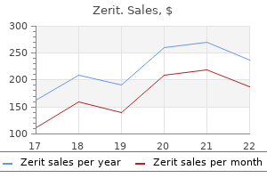
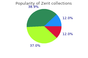
Zerit
Order zerit 40mg free shipping
Strength of Evidence Recommended medications side effects buy generic zerit 40mg online, Evidence (C) Rationale for Recommendations There is one moderate-quality trial suggesting needling or bursoscopy is superior to a non-interventional control. Another moderate-quality trial suggested adding needling is effective when used as an adjunct with shockwave therapy. Nevertheless, there is insufficient evidence to support a recommendation of needling compared to arthroscopic surgery. Additional quality trials appear necessary prior to recommending its widespread use. Bursal arthroscopic removal/excision is more invasive, but is selective in its ability to remove tissue, has evidence of efficacy, and thus is recommended. The indications for emergent surgery for red-flag conditions including unstable fractures, abscess, or hematoma, etc. Early recognition of red-flag conditions that require expedited referral to a surgeon qualified to deal with shoulder emergencies is recommended (see Red Flags). This section of this guideline addresses surgical indications including rotator cuff tears and surgery for impingement syndrome. Many patients function normally with rotator cuff tears, while others have incapacitating problems that may require physical theapy (Moosmayer 10, 14; Ainsworth 07) and/or attempts at surgical repair or debridement. There also are reports of improved overall health status after rotator cuff surgery. Repairs of larger tears have increased rate of healing failure which correlates with outcomes. There are many purported and documented risk factors for poorer surgical outcomes. These most common risk factors include low-volume surgical practice (physician performs less than 6 rotator cuff repairs per year), (Sherman 08) age (older patients), (Ogilvie-Harris 90; Boehm 05; Sherman 08; Watson85) female gender, (Boehm 05; Lindh 93) larger rotator cuff tears, (Milano 07; Wilson 02; Warner 01Habernek 99; Bartolozzi 94; Rokito 96; Iannotti 06) retraction, (Milano 07) concomitant subscapularis tears, (Milano 07) fatty tendon degeneration, (Milano 07; Costouros 07) diabetes, smoking, (Mallon 04) overweight or obesity, weakness of shoulder (strength of abduction and external rotation), pre-operative activity level, (Iannotti 96; Ellman 86) preoperative stiffness, (Namdari 10) abnormal mental status, involvement in litigation or workers’ compensation (Ogilvie Harris 90; Spangehl 02; Kempf 99; Misamore 95) or sick-leave, (Brox 99) regular “pain medication use,” (Brox 99) excessive post-operative hyperalgesic crises, (Kempf 99) non-compliance with rehabilitation programs, and otherwise unhealthy individuals. If surgery is a consideration, counseling regarding likely outcomes, risks, and benefits, and especially expectations, is important. Ideally, this education begins with the referring physician who may note that post-operative physical or occupational therapy exercises are essential in comparison to non-operative treatment for good clinical results. These exercises might be difficult to comply with for some rotator cuff repair patients. The decision as to which type of rotator cuff repair procedure to perform – arthroscopic, open, or mini-open repair – should be left to the surgeon and patient until quality evidence demonstrating procedural superiority becomes available to provide evidence-based guidance. Achievement of a plateau in improvement and assessment for final results after surgical repair of a rotator cuff tear has been found to require 1 year. Recommendation: Rotator Cuff Repair for Small, Medium, or Large Tears © Copyright 2016 Reed Group, Ltd. Patient must agree to participate fully in post operative active rehabilitation and understand there is a long recovery time. Pre-operative physical therapy is an option (but not a pre-operative requirement) as many pataients sufficiently recover without surgery. Recommendation: Addition of Claviculectomy or Subacromial Decompression to a Rotator Cuff Repair for Isolated Supraspinatus Tears Adding claviculectomy or subacromial decompression to a rotator cuff repair is moderately not recommended for treatment of isolated supraspinatus tears. Strength of Evidence – Moderately Not Recommended, Evidence (B) Rationale for Recommendations While surgery tends to produce modestly superior outcomes over 1 to 5 years (Moosmayer 10,14), non operative treatment is often successful. Surgical cuff repair is believed to be a superior option among patients for whom occupational shoulder exposures and demands are greater, although quality data that address this issue are not available. Many quality studies necessitated non-operative treatment prior to surgery (see evidence table). Rotator cuff repair has evolved from open to mini-open to all arthroscopic techniques. Currently, arthroscopic techniques are evolving with the advent of new technology and instrumentation. There is high-quality evidence there are no long-term differences associated with arthroscopic repair and mini-open compared to open repair, (Mohtadi 08; vi Spangehl 02) although evidence suggests a modest short-term advantage of arthroscopic mini-open repair versus open repair of rotator cuff tears. The re-tear rate for a single row arthroscopic repair has been estimated at 40%, but varies considerably depending on the size of original tear. Isolated side-to-side repair or margin convergence means that there is an incomplete repair as is usually present in cases of chronic massive tears. Tendon to bone repair has been suggested to be modestly better than side-to side repair in one moderate-quality study. Patients who are candidates for surgery generally have pain and impaired function. There are no quality studies suggesting better or worse results for earlier or delayed surgery (see evidence table), and current evidence does not support a need to rush surgical decisions. Until quality evidence becomes available to provide evidence-based guidance, the decision as to which surgical procedure to perform should be left to the surgeon and patient as there appear to be only modest short-term improvements for arthroscopic rotator cuff repair over open rotator cuff repairs (Mohtadi 08) or for impingement syndrome including trends towards shorter sick leave in one study (mean 10 versus 5. Surgery is invasive, involves prolonged recovery (many months), has adverse effects, and is costly. However, benefits appear to outweigh risks for most patients and surgery is recommended. Recommendation: Rotator Cuff Repair for Acute Massive Tears Rotator cuff repair is recommended for treatment of acute massive tears (>5cm). Recommendation: Rotator Cuff Repair for Chronic Massive Tears Rotator cuff repair is not generally recommended for treatment of chronic massive tears (>5cm). Strength of Evidence – Not Recommended, Evidence (C) © Copyright 2016 Reed Group, Ltd. Recommendation: Rotator Cuff Repair for Massive Tears Using Porcine Xenograft Material Porcine small intestine submucosa graft for surgical repair is not recommended for treatment of large or massive tears that are otherwise unrepairable. Recommendation: Rotator Cuff Repair for Massive Tears Using Tissue Augmentation There is no recommendation for or against tissue augmentation to surgically repair large or massive tears that are otherwise unrepairable. Strength of Evidence – No Recommendation, Insufficient Evidence (I) Rationale for Recommendations Repair of massive rotator cuff tears is technically more difficult and has a worse prognosis. Some chronic massive tears can be repaired and some can also undergo successful partial repair, although this does not apply for most patients. A study of 27 shoulders found primary rotator cuff repair was often infeasible when the length was greater than 4cm, the width was greater than 4cm, the supraspinatus muscle was thin at the superior glenoid margin, and the signal intensity was high. Cases of margin convergence may be amenable to a primary closure, if the tendon edges can be approximated without undue tension on the patient’s remaining rotator cuff. A few of these repairs were included in the available quality literature (see evidence table), but did not present stratified analyses specific to massive rotator cuff tears. Even so, there is some limited evidence suggesting repair is superior to debridement with considerably better results in the surgical repaired group (Melillo 97) and thus, there is limited evidence to recommend attempted repair of massive rotator cuff tears. Additional materials interposed include porcine dermal xenograft (Badhe 08) and porcine small intestinal submucosa. Hemiarthroplasty has also been used to treat select patients with massive tears (see Arthroplasty), but there are no quality studies of hemiarthroplasty for treatment of massive rotator cuff tears. It also is used to treat selected patients with unrepairable massive rotator cuff tears. It has been suggested that the outcomes for patients with larger tears are inferior to smaller tears. Infections are generally rare and are most commonly associated with mini-open repair. Surgical repair of massive rotator cuff tears is invasive, has adverse effects, and is costly. Rehabilitation is often considerably longer and more complicated than for smaller rotator cuff tears. However, particularly in younger patients with massive rotator cuff tears, benefits appear to outweigh risks for most patients and surgery is generally recommended. In Cochrane Library, we found and reviewed 17 articles, and considered 1 for inclusion. Of the 23 articles considered for inclusion, 13 randomized trials and 10 systematic studies met the inclusion criteria. Author / Scor Sample Size Comparison Group Results Conclusion Comments Title Study e (0 Type 11) Rotator Cuff Tear: Open vs Mini-Open Arthroscopic Repair Mohtadi 8. Data treatment of with a general from baseline not undergoing the suggest slight at least 3 anesthetic and were significant at 6 months. After 6 months, Constant-Murley score improved in both groups compared to baseline; however, there were no significant differences between groups. There were no significant differences in mean range of motion between groups © Copyright 2016 Reed Group, Ltd. However, there and quality-of-life meaningful clinical and acromioplasty were no significant scores between the differences imaging intervention (n = differences between groups up to two years between groups.
Purchase discount zerit online
The future challenge in this field of research will be to address a number of fundamental questions relevant to human health and disease including: 1) Is there a microbiome associated with human health—the “healthy gut microbiome”; 2) Are there causal relationships between the gut microbiome and human disease? The answers to these symptoms for pneumonia order zerit us, and other fundamental questions in the field of gut microbial ecology await further studies in human subjects in whom clinical metadata is carefully collected together with continued investigation in animal models. Ultimately, the answers to these questions could lead to a fundamental shift in the way that we treat many common diseases. The availability of the therapeutic substrate (feces), together with the ease of administration, has advanced the practice in the field of gut microbiota modulation much more rapidly than our scientific understanding in the field. Indeed, two of the four goals of the registry are: 1) To assess short-term and long-term safety and 2) To promote scientific investigation. Patients who experience one recurrence have a 40% risk of an additional recurrence and those with 2 or more episodes face a 60% risk of another episode21,29. While the first recurrence is generally treated with a second course of metronidazole or vancomycin, current guidelines30 recommend a tapering course of oral vancomycin. Interventions that have been used, but with little or no data to support efficacy, include intravenous immunoglobulin, rifaximin, nitazoxanide, and probiotics15. Fecal transplantation from the lean donors improved peripheral insulin sensitivity during the short period in which this parameter was followed19. The gut microbiota is a complex consortium with many components that have never been characterized. A priori knowledge is not available regarding the impact of transferring these complex communities from one individual to another, although many studies in mice indicate that the composition of the gut microbiota can affect host susceptibility to diseases. Recall the unexpected consequence of the hepatitis C virus epidemic from the transfusion of contaminated blood before its presence was recognized. Protocols need to be developed about donor sample preparation, characterization, archiving (so that follow-up analyses can be performed), and host preparation/administration/dosing. Critical methodologic data such as donor/recipient screening, fecal preparation, modality of delivery, and patient consent practices are lacking. Based on this information void, national societies have attempted to provide guidance to practitioners through published guidelines and editorials48 50 but the degree of adherence to these recommendations is unknown. For example, can transfer of fecal microbiota lead to chronic diseases such as diabetes, obesity, or cardiovascular disease? Animal models have suggested such a possibility, as transfer of specific phenotypes. Participation in the registry does involve the potential risk of breach of confidentiality of medical information and associated privacy of the participants. The human-to-human transfer of feces may be associated with long-term health risks to the recipient because the gut microbiota is composed of many components that have not been characterized and can change over time in ways that cannot be currently predicted. In addition to infections, the possibility that gut microbiota associated with a disease phenotype. A prospective registry in a large sample of patients with long-term follow-up is the only practical method to achieve this aim at present. The registry database will be available to investigators, with study proposals submitted for review by a Data Access and Publications Committee. Together with the clinical metadata collected within the registry, information from analysis of specimens in the biobank will enhance short and long-term safety surveillance as well as be a rich source of data to investigate modulation of the human gut microbiome. In addition, gut-related-microbiota products, from processed stool to defined microbiota consortia, require submission for regulatory approval. The Registry may function as an important vehicle for sponsors of gut-related microbiota products to satisfy pre approval and post-approval assessment of product safety and efficacy. Follow-up information collected will be designed to assess potential short-term and long-term safety and effectiveness. Sites must demonstrate proper data protection procedures prior to participant enrollment. To enhance representation of a geographically diverse population in the registry, centers will be related to their location in population-based regions. Participants will have as much time as they would like to review the documents and ask questions regarding participation. Registry mailing address and electronic address will be provided to all participants. If the participant informs the study staff that they wish to withdraw consent, no further data will be sought from them or their healthcare provider. Data that was previously collected when the patient was actively enrolled will be retained in the registry. No costs will be incurred by participants related to participation in the Registry. For sites that employ a donor questionnaire, Supplement 2 provides potential questions used on a questionnaire, based on those used for blood transfusion. Finally, Supplements 5 and 6 detail data elements for short-term and long-term safety outcomes. However, participants have the option to enroll in a Biorepository Sub-Study and provide stool specimens to be stored in a biorepository that will be linked to their registry data. The biorepository will be maintained by the American Gut Project at University of California at San Diego. This variance in practice also raises potential concerns about risks for both short-term and long-term risks. In addition, patients will be contacted directly by email and text at least annually for up to 10 years to provide follow-up information via a dedicated portal maintained by the Icahn School of Medicine at Mt. All participants are given a toll-free number to contact the Investigators directly if they have any safety issues or concerns. Laine and Kelly, will serve as the Chair and Co-Chair of the Steering Committee and will be responsible for the daily management of the registry and its associated activities. All investigators are required to conduct themselves at all times in accordance with Good Clinical Practices. Investigators are expected to commit themselves to ethical and professional conduct of clinical research. Commitment to preserving the physician’s, medical community, and public’s trust is ensured by conducting research at the highest scientific and ethical standards. The planned sample size will also provide high confidence in ruling out rare adverse events. For more common outcomes, the statistical power for specific relative risk estimate is greater. For example, with 2,000 patients in each group there would be 90% power to detect a difference between two approaches for which the incidence rates of events were 3% and 5%. There will be several major advantages of the registry design as opposed to a system of spontaneous reporting. In contrast, systems that rely on spontaneous reporting have incomplete numerator data and lack denominator data. The ability to calculate incidence rates greatly facilitates both descriptive epidemiology and signal detection. Thus, descriptive statistics with 95% confidence intervals will be determined for the safety and effectiveness outcomes to provide precise estimates of incidences. To assure the feasibility of this approach, we have conducted a preliminary analysis of these data to identify the number of patients who will be included in the control cohort. The sample size should be sufficient to address questions on relatively rare outcomes. For example, at age 68 years (the median age in our control cohort), the probability of death in the next 12 months is 1. Comparative effectiveness studies rely on the ability to adequately account for differences between the treatment groups. Such data will support standard statistical methods including logistic, Cox, and other regression methods, and the development of propensity scores and risk scores for risk adjustment. There is a theoretical risk of breach of confidentiality or a data security loss although exhaustive efforts will be made to minimize this inherent risk. All sites must demonstrate proper physical and electronic security measures in order to participate in the registry. Securing in a separate location and limiting access to information linking codes assigned to the registry information with direct participant identifiers 3. All computers will be used to collect and send data during implementation of the study or to receive or store data at the central location will be password protected. A password will be required to open Windows and a second, different password will be required to open the electronic data capture system, Viedoc™. Electronic forms will be stored on a secure dedicated server with appropriate firewalls.
Buy zerit 40 mg mastercard
Always wearing a mask when caring for patients Correct answer: B Performing hand hygiene in compliance with the World Health Organization or Centers for Disease Control and Prevention guidelines is the most effective in reducing the risk of health care–associated infections medications hyponatremia buy zerit canada. Keeping employee health records up-to-date (Option A), providing annual influenza vaccinations (Option C), and always wearing a mask when caring for patients (Option D) aren’t the most effective ways to reduce the risk of health care–associated infections. Which action should the nurse take when receiving a telephone order from a physician? Correct answer: C When receiving a telephone or other verbal order, the nurse should write down the order and then read back the complete order to the physician to verify its accuracy. Options A, B, and D aren’t appropriate actions for the nurse to take when receiving a telephone order from a physician. A secondary latency phase that occurs in some diseases that is commonly followed by another acute phase is referred to as: A. Correct answer: A A secondary latency phase that occurs in some diseases that is commonly followed by another acute phase is referred to as remission. The acute phase (Option C) refers to the disease at its full intensity, possibly with complica tions. The subclinical acute phase (Option D) occurs when the patient is in the acute phase but still func tions as if the disease weren’t present. Which term in qualita tive research describes the researcher laying aside what is known about the experience being studied? Theoretical sampling Correct answer: A Bracketing requires the researcher to lay aside what’s known about the experience being studied and be open to new insights. Saturation (Option B) describes the point at which data collec tion is ended because continuing would result in acquiring more of the same information or data. Intuit ing (Option C) refers to the focused awareness on the phenomena being studied. Theoretical sampling (Option D) is the selecting of subjects on the basis of concepts that have theoretical relevance to an evolv ing theory. Confidentiality If a patient receives appropriate information and refuses care or treatment against medical advice, the physician or Confidential information is any information that the health care provider is responsible for documenting that the patient communicates to the nurse or practitioner for informed consent conversation took place, the general facts diagnosis or treatment with the expectation that it discussed, and the patient’s decision. Nurses have an ethical and legal duty to avoid disclosing such confidential information to Exceptions to informed consent unauthorized people who aren’t involved in the patient’s Informed consent isn’t required if a delay to obtain consent care and treatment. The nurse can the patient waives the right to consent and asks not to be be liable for invasion of privacy, defamation, intentional or informed, or if compulsory treatment is mandated by law or negligent infliction of emotional distress, or breach of an a court order. Some states provide a statu tory penalty against health care providers who violate the Right to refuse treatment patient’s right to confidentiality. However, confidential information can be disclosed Adults are presumed to be legally capable of refusing treat under certain circumstances. In fact, mentally competent patients with terminal disclose such information when authorized by a patient (but conditions can refuse life-sustaining treatment without cre only to the extent authorized), when a patient is a danger ating legal liability for their health care providers. In some persons to express their wishes about life-sustaining treat cases, the court may order disclosure of confidential patient ment should they become legally incapacitated. Under this the law presumes that adults are legally capable of con act, the health care facility must provide patients with writ senting to treatment. To provide a valid consent, a patient ten materials explaining their rights under their state laws must be mentally capable of understanding the nature and to make decisions concerning medical care, including the consequences of treatment. Expressed consent is obtained right to accept or refuse treatment and the right to execute orally or in writing; implied consent is obtained by the an advance directive. A living will expresses a patient’s patient’s voluntarily submitting to treatment. Medical treat wishes about withholding or withdrawing life-sustaining ment performed without a patient’s expressed or implied treatment. A durable power of attorney designates a person consent may result in legal claims of battery or negligence. Informed consent requires the prohibits covered health care facilities from conditioning health care provider (physician, nurse practitioner, physician the provision of care or discriminating against an individual assistant, nurse midwife) to provide appropriate informa based on whether the individual has executed an advance tion. Did the patient receive authorize a surrogate decision maker to accept or refuse sufficient information such that a reasonable person in the medical treatment for the patient based on the patient’s same circumstances could make an informed decision? Employers and supervisors ◆ An employer is automatically liable for its employees’ actions within the scope of their employment; the employer is thereby encouraged to hire competent employees ◆ A nurse in a management or supervisory position may be liable for the actions of a negligent nurse under her supervision; liability can result if the supervisor didn’t adequately assess the nurse’s Possible civil actions ❍ 25 competence or the assignment’s requirements, didn’t adequately supervise the nurse’s performance, or knew the nurse’s limitations and didn’t provide adequate training or staffing Independent contractors ◆ An independent contractor is one who contracts with another to do a specific job; in nursing, the pri vate duty nurse who is employed by an agency but hired on a per diem basis by a health care facility is the most common example of an independent contractor ◆ the independent contractor’s actions aren’t directly controlled by the employer; the contractor has independent discretion in performing the job ◆ the employer may not be liable for the negligent actions of an independent contractor unless the employer knew or should have known of the independent contractor’s incompetence Corporations ◆ A health care facility is obligated to carefully monitor the credentials and competence of employees and independent contractors ◆ A health care facility that doesn’t ensure its workers’ competence may be liable for injuries caused by the workers’ negligence ❖ Possible civil actions Torts ◆ A tort is a civil action for damages for injury to a person, property, or reputation ◆ Torts are classified as unintentional or intentional Unintentional torts ◆ Professional negligence and professional malpractice are the most common legal claims against nurses ◗ Negligence is the failure to exercise the degree of care that a person of ordinary prudence would exercise under the same circumstances; for example, if a nurse notices water on the floor of a room and doesn’t wipe it up, and the water causes a patient to fall and injure himself, this constitutes negligence ◗ the plaintiff in a negligence suit must prove that the nurse’s actions caused harm ◗ Professional malpractice or professional negligence requires a plaintiff to introduce proof of duty, breach of duty, proximate cause, and damages or harm; proof of the nurse’s standard of care is criti cal to establishing the first three elements ◗ Unlike negligence cases, professional negligence cases require expert testimony as to the duty of care, its breach, and its causal relationship to the injury ◆ A nurse also can be sued for negligent infliction of emotional distress ◗ In many states, a plaintiff can be awarded damages for severe emotional distress resulting from a nurse’s negligent actions ◗ Some states require that the plaintiff have physical manifestations of the emotional distress, such as palpitations, gastric discomfort, or insomnia Intentional torts ◆ Assault is an act that places a patient in fear of harmful or offensive touching ◆ Battery is touching a patient without justification or permission ◆ Defamation results when a nurse communicates false information verbally (slander) or in writing (libel) about a patient that damages the patient’s reputation or causes the patient to be shunned or avoided by the community ◆ Invasion of privacy occurs when a nurse gives unauthorized access to the patient or information about the patient; taking photographs of a patient without permission is an invasion of privacy ◆ Fraud and misrepresentation are false or misleading statements by the nurse that the patient relies on to his detriment ◆ False imprisonment is unjustifiable restriction of patient movement ◆ Intentional infliction of emotional distress results when a nurse’s actions produce distress so severe that no reasonable person could be expected to endure it 26 ❍ Legal and ethical aspects of nursing Contract actions ◆ Contract actions are determined by whether the parties performed obligations agreed to in a contract ◆ A breach of contract results when one party fails to perform as required by the contract ◆ Nurses are most commonly involved in employment contracts and malpractice insurance contracts ❖ Possible defenses to a health care negligence suit Comparative and contributory negligence ◆ With comparative negligence (the more common defense), the jury compares the degree of negli gence of the parties or of various defendants and apportions damages (that is, compensation or indem nification) accordingly ◆ With contributory negligence, because the plaintiff’s conduct contributes to the cause of his injury and falls below the standard by which individuals are expected to conform, he can’t recover damages, even though the defendant violated a duty of care to the plaintiff and would be liable Statutes of limitation ◆ A statute of limitation sets a time limit within which the legal action must be brought; a nurse can’t be sued for negligence if the claim is made after the time limit expires ◆ Statutes of limitation for health care negligence are established by the state legislature ◆ these statutes may not apply in some cases, such as those involving fraudulent concealment of negligence or later discovery of negligence Good Samaritan laws ◆ All states have Good Samaritan laws to protect people who render assistance at the scene of an emergency ◆ Some statutes protect all citizens; others cover only specified health care providers ◆ Those protected under Good Samaritan laws are liable only for grossly negligent acts Statutory defenses ◆ Many states have statutes that prescribe special procedures for health care negligence cases ◆ A suit may be dismissed if the statutory requirements aren’t met ❖ Professional liability insurance for nurses Types of policies ◆ A claims-made policy provides coverage for claims made during the policy period ◆ An occurrence policy provides coverage for negligence that occurs during the policy period Obligations of the insurer and the insured ◆ Insurers must provide and pay for legal counsel to defend the nurse in the lawsuit and must pay damages (within coverage limits) for which the nurse is judged liable ◆ the nurse must notify the insurer that a claim has been made and must assist as needed in preparing the defense (see Reducing nursing liability, page 28) ❖ Patient’s bill of rights Self-determination ◆ Self-determination is often used synonymously with autonomy ◆ It means having a form of personal liberty to choose and implement one’s own decisions, free from deceit, duress, constraint, or coercion ◆ It involves the right of patients to decide what will or will not happen to their bodies ◆ Basic elements of self-determination include the patient’s ability to decide, the power to act upon his own decision, and respect for the individual autonomy of others ◆ Although self-determination is often addressed in relation to death and dying, it concerns all aspects of consent and its refusal ◆ To practice this right, the individual must be competent to make his own decisions about accepting or refusing treatment ◆ Some states uphold the patient’s in orally expressed desires, provided they’re documented, including the patient’s awareness of consequences of his actions Ethical aspects of nursing ❍ 27 ◆ the Patient Self-Determination Act has three basic premises ◗ Patients who are informed of their rights are more likely to take advantage of them ◗ If patients are more actively involved in decisions about their medical care, then that care will more closely respond to their needs ◗ Patients may choose care that is less costly Informed consent ◆ Informed consent is the voluntary authorization by a patient to a care provider to do something to the patient ◆ Some states accept oral forms of consent as being equally as valid as written consent ◆ Contents of an informed consent include the procedural information, associated risks and benefits, alternatives to the procedure, and the name of the person who will perform the procedure ◆ Generally, physicians have the responsibility to obtain informed consents ◆ Nurses can witness and have the responsibility to advocate for the patient to ensure that all criteria for autonomous decision making are met ◆ Four exceptions to consents are emergency situations, therapeutic privilege, patient waiver, and prior patient knowledge Living wills ◆ Living wills, or health care directives, are directives from competent individuals to medical person nel regarding treatment they wish to receive ◆ A living will takes effect when the previously competent person becomes sick and can no longer make decisions for himself ◆ It typically contains information about the conservator or health care agent, the patient’s code status, and the patient’s desire for organ donation; it may also contain other information ◆ Because living wills aren’t typically legally enforced, medical practitioners may choose to abide by them or to ignore them as they see fit ◆ There’s no protection for the practitioner against criminal or civil liabilities for proceeding under a living will’s directions Durable power of attorney ◆ Durable (or medical) power of attorney for health care allows competent patients to appoint a surro gate to make decisions for them in the event that they lose competence ◆ the power includes the right to ask questions, select and remove physicians from the patient’s care, assess risks and complications, and select treatments ❖ Ethical aspects of nursing General information ◆ Ethics is a branch of philosophy that examines values, actions, and choices to determine right and wrong ◗ Nursing ethics is part of normative ethics, a type of ethics that’s based on the criteria by which people make moral judgments ◗ As the basis for professional codes of ethics, ethical theories attempt to provide a system of prin ciples and rules for resolving ethical dilemmas ◗ the American Nurses Association’s “Code of Ethics for Nurses” provides guidance for carrying out nursing responsibilities consistent with the ethical obligations of the profession ◆ Morality involves rules of conduct about right and wrong ◗ It’s based on norms of conduct determined by society ◗ Society’s moral codes guide what people ought to do; professional codes, such as the code of ethics for nurses, communicate the goals and ideals of a profession ◆ Although ethics and morals are theoretically distinct terms, they’re used interchangeably to describe right and wrong actions Professional code of ethics for nurses ◆ Nurses have a contract with society to behave in accordance with rules dictated by society and the nursing profession 28 ❍ Legal and ethical aspects of nursing Reducing nursing liability the nurse can take measures to reduce liability in several areas. Liability area Prevention measures Competent practice; Know your practice area, and stay current in the field. Access to medical records; Follow Health Insurance Portability and Accountability Act guidelines as developed by the health care facility. Common sources of injuries; Know the facility’s fall-risk policies and procedures. Medications; Know the facility’s protocols and procedures regarding specific drugs, such as insulin and anticoagulants. Inadequate patient; Assess the patients cognitive ability and willingness to learn, and document education accordingly. Ethical aspects of nursing ❍ 29 Reducing nursing liability (continued) Liability area Prevention measures Abandonment; Don’t leave a patient without arranging for continuing care. Malfunctioning equipment; Check equipment according to the manufacturer’s recommendation and the facility’s protocol. The nurse participates in establishing, maintaining, and compassion and respect for the inherent dignity, worth, and improving health care environments and conditions of uniqueness of every individual, unrestricted by consider employment conducive to the provision of quality health ations of social or economic status, personal attributes, or care and consistent with the values of the profession the nature of health problems. The nurse participates in the advancement of the pro whether an individual, family, group, or community. The nurse promotes, advocates for, and strives to project istration, and knowledge development. The nurse is responsible and accountable for individual and the public in promoting community, national, and inter nursing practice and determines the appropriate delegation national efforts to meet health needs. The nurse owes the same duties to self as to others, ing values, for maintaining the integrity of the profession including the responsibility to preserve integrity and safety, and its practice, and for shaping social policy. The nurse leaves a patient who is elderly and confused to find someone to assist with transferring the patient to bed. The better courses of action are to turn on the call bell or elicit help on the way to the patient’s room. Options A and C are incorrect because neither excuses the nurse from her responsibility for ensuring the patient’s safety. Option B is incorrect because restraints are only to be used as a last resort, when all other alternatives for ensuring patient safety have been tried and have failed; moreover, restraints won’t ensure the patient’s safety. The nurse is caring for a patient admitted to the emergency department after a motor vehicle accident. Under the law, the nurse must obtain informed consent before treatment unless the patient: A. Correct answer: C the law doesn’t require informed consent in an emergency situation when the patient can’t give consent and no next of kin is available. Option A is incorrect because even though a patient who is declared mentally incompetent can’t give informed consent, mental illness doesn’t by itself indicate that the patient is incompetent to give such consent. Option B is incorrect because a mentally competent patient may refuse or revoke consent at any time. Option D is incorrect because although the nurse may act as a patient advocate, the nurse can never give substituted consent. Abandonment Correct answer: A Battery, touching a patient without justification or permission, is an intentional tort. Option B is incorrect because although a nurse who breaches a patient’s confidentiality can be subject to a lawsuit or disciplinary action, the act isn’t an intentional tort. Option C is incorrect because negligence, the failure to exercise the degree of care that a person of ordinary prudence would exercise under the same circumstances, is an unintentional tort. Option D is incorrect because although abandonment is a liability for nurses, the act isn’t an intentional tort. Options A and B are incorrect because it’s the responsibility of workers’ compensation to compensate workers for injuries occurring in the workplace and to provide rehabilitative services. Option D is incor rect because it’s the employer’s responsibility to improve the safety and health of employees.
Purchase generic zerit line
Skeletal malous muscles about the volar aspect of the wrist and Radiol 23:127–131 forearm symptoms stomach cancer purchase generic zerit pills. A closer look at the nerve sheaths demonstrates an References 133 external sheath – the outer epineurium – which sur rounds the nerve fascicles. Each fascicle is invested in turn by a proper connective sheath – the perineu rium – which encloses a variable number of nerve M. Then, the individual nerve fibers are invested “Giannina Gaslini”, Largo Gaslini 5, 16148 Genova, Italy by the endoneurium. Individual fascicles are invested by a thin sheath – the perineurium (arrowheads) – and are separated from each other by a loose connective tissue envelope – the epineurium (green) – containing small intraneural vessels (white arrows). In complex motor and sensory nerves (3), fascicles (asterisks) are of different size and may be grouped in function-related areas within the nerve. This drawing (3) recalls the structure of the sciatic nerve, in which the nerves fibers for the tibial nerve (light gray) and for the peroneal nerve (dark gray) remain grouped tightly throughout the course of the nerve, even proximally epineurium (internal epineurium), as opposed to the impulse transmission and axonal transport. The outer epineurium which surrounds the entire nerve vascular supply is form ed by an interconnected trunk. Generally speaking, the amount of connective system of perineural vessels that course longitudi tissue of the epineurium is more abundant in large nally in the external epineurium and branch among multifascicular nerves and in regions in which the the fascicles (endoneural vessels). This thickening of the connective tissue seems to provide more cushioning for the nerve and, therefore, more 4. The improved performance of these traction on its blood supply during joint motion transducers has made it possible to recognize (George and Smith 1996). In a, the nerve fascicles (white arrow) are depicted as well-circumscribed individual structures of different size separated by echogenic epineurium. In this segment, 11 fascicles are distinguished in the cross-sectional area of the me dian nerve. In b, the nerve fascicles appear as elongated hypoechoic bands (white arrows) that run parallel to each other. The internal epineurium (white arrowheads) separates them more clearly, while the external epineurium (open arrowheads) helps to define the outer boundaries of the nerve color and power Doppler systems are, for the most for following the nerves contiguously throughout part, unable to recognize the weak and small blood the limbs (Martinoli et al. Long-axis scans flow signals from the perineural plexus and the are less effective for this purpose because the elon intraneural branches. Generally speaking, nerves gated fascicles may be easily confused with echoes are compressible and alter their shape depending from muscles and tendons coursing along the same on the volume of the anatomic spaces within which plane. Even proximally and distally, shifting the transducer up with slight pressure applied with the probe, they or down according to the nerve’s course. With this may be seen sliding over the surface of an artery technique – which we can call the “lift technique” or a muscle. As a general rule, each individual fas – the examiner is able to explore long segments of cicle in a nerve runs independently of the others. If intrinsic or extrinsic nerve anatomic passageways – the osteofibrous tunnels abnormalities are encountered during scanning, the – that redirect their course. Although all main nerves – that prevent dislocation and traumatic damage of can be readily displayed in the extremities due to the structures contained in the tunnel during joint their superficial position and absence of intervening activity (Martinoli et al. In fact, most cra osteofibrous tunnels, subtle echotextural changes nial nerves – except for the vagus – and the spinal can be seen, with a more homogeneous hypoe accessory nerve (Giovagnorio and Martinoli, choic appearance caused by tighter packing of the 2001; Bodner et al. In addi anatomic relationships with surrounding structures tion, the perineural structures greatly influence is essential for recognizing peripheral nerves with nerve detection in the limbs and extremities. Unlike other structures of the musculoskeletal nerves course deeply, as in obese patients, their system, nerves do not show anisotropic properties. As a general rule, nerves Therefore, appropriate probe orientation during of the lower extremity run deeper than those of the scanning is not needed to image them; however, sys upper extremity and are more difficult to visual tematic scanning in the short-axis plane is preferred ize. Nerves coursing among hypoechoic muscles are Nerve and Blood Vessels 101 a b c Fig. Due to the flexibility of the epineurial sheath, the nerve flattens, whereas the fascicles – which are noncompressible structures – redistribute according to the nerve shape changes. Then, the transducer is swept upward (dashed arrow) along the course of the nerve in the forearm. This technique, which we can call the “lift technique,” allow a simple and reliable evalu ation of long nerve segments in a single sweep, excluding possible intrinsic and extrinsic abnormalities along the nerve path. Among these, the proxi condition – which is also referred to as neural fibrol mal bifurcation of the median nerve at wrist has ipoma, perineural lipoma, fatty infiltration of the been extensively reported in the literature (see nerve, lipofibroma, or neural lipoma – has a definite Chapter 10) (Propeck et al. Similarly, some inher with lower extremity involvement (plantar nerve, ited and developmental anomalies of the peripheral sciatic nerve) reported as being rare (Marom and nervous system, such as the fusiform enlargement Helms 1999; Wong et al. Fibrolipomatous of the median nerve by fibrofatty tissue (so-called hamartoma may be associated with local gigantism fibrolipomatous hamartoma), the hypertro of an extremity, usually the hand or foot, related to phy of nerves in Charcot-Marie-Tooth syndrome bony overgrowth, fat proliferation in the soft tis (Martinoli et al. Note the atrophic changes in the flexor hallucis longus muscle (asterisk) and the adjacent posterior tibial artery (a) and veins (v). Note the equivalent size of the flexor carpi radialis (arrowheads) and palmaris longus (white arrow) tendons in the two images. The magnification scale is indicated on the right constantly changing (because not all the causative diameter of the fascicles and the resulting nerve area genes have yet been described), the most common are more than twice those seen in healthy subjects forms include the autosomal dominant types 1A and and in type 2 and the X-linked type (Fig. There is no correlation chromosome 17 which codes for a peripheral myelin between the maximum fascicular size of the nerve protein, and the X-linked type that is related to a and electrophysiologic features, such as distal laten mutation in the gene which codes for connexin 32, cies, velocities, and amplitude (Martinoli et al. The degree of electrophysiologic used to help the neurologist identify unrecognized alterations varies widely among patients with differ disease in patients with nonspecific symptoms, to ent forms of the disease, especially in the type 1A, differentiate the 1A genetic subtype, and to provide a as a result of phenotypic differences and the action useful screening tool for a first selection of the indi of stochastic factors or environmental modulation viduals in an affected kindred who are to undergo of disease severity (Schenone and Mancardi genetic assessments. Nerves appear larger than normal but retain a normal fascicular echotexture (Heinemeyer and Reimers 1999; Martinoli et al. Zamorani an autosomal dominant inherited disorder charac cation of the ulnar nerve over the medial epicondyle terized by a tendency to develop focal neuropathies if the retinaculum is loose or absent (Jacobson et al. Histopathologically, a sausage-shaped drome, the ulnar nerve dislocation is secondary to myelin sheath swelling, the so-called tomacula, is the snapping triceps and dynamic scanning demon responsible for multifocal nerve enlargement. Elec strates the medial head of the triceps and the ulnar trophysiologic studies demonstrate one or more nerve remaining in close continuity as they dislocate entrapment neuropathies on a background of motor over the medial epicondyle (see Chapter 8). The more frequently involved nerves are: the peroneal nerve at the fibu lar tunnel, the ulnar nerve at the cubital tunnel, the 4. Short periods of constriction result in course of nerves throughout the limbs (Fig. It is slowing and failure of conduction across the con conceivable that the “sausage-shaped” myelin swell striction point, whereas the nerve portion distal to ings (tomacula) found at teased nerve fiber studies the region that was compressed retains a normal in patients with this disorder are responsible for function. The conduction abnormalities, which are nerve enlargement (Beekman and Visser 2002). If local compression is prolonged, ischemia with or without snapping triceps syndrome. This induced by direct severe compression, mechani finding typically occurs in the cubital tunnel, an cal distortion of the nerve architecture, may cause osteofibrous tunnel formed by a groove between the more significant damage in the myelin sheath and olecranon and the medial epicondyle and bridged by axonal degeneration (Wallerian degeneration) of the Osborn retinaculum. As described in Chapter the nerve fibers and persistent nerve deficit due to 8, dynamic scanning during full elbow flexion can disruption of the axoplasm after the compression allow continual depiction of the intermittent dislo has been relieved (Delfiner 1996). Hereditary neuropathy with liabil ity to pressure palsies in a 42-year-old man with mild median and ulnar neuropathy. Zamorani close proximity to the compression level, where the pression site and proximal to it (Fig. Although nerve flattening should be regarded of the individual fascicles and decreased echogenic as the main sign of nerve compression, quantitative ity of the epineurium. Depiction of such changes sistent criterion for the diagnosis at various entrap may increase confidence in the diagnosis and in ment sites (Chiou et al. As an ancillary finding, of entrapment by scar tissue, diagnostic difficulties dynamic scanning may show a reduced mobility of may arise in distinguishing echotextural changes the nerve over the mass or beneath the retinaculum, related to the compressed nerve from the scar itself, but this latter sign is too subjective and hard to quan because of a similar hypoechoic appearance. At an enhanced depiction of intraneural blood flow sig least at the carpal tunnel level, the cross-sectional nals can be appreciated with color and power Doppler area of the median nerve has also been regarded as techniques as a sign of local disturbances in the nerve an index for selecting patients with severe disease for microvasculature that occur in a compressive context which surgical decompression is indicated (Lee et al. It is conceivable that loss of axons may be asso is more clearly appreciated in swollen hypoechoic ciated with nerve enlargement as an expression of an nerves of patients with chronic, longstanding dis increased amount of endoneural edema (Beekman ease. In entrapment neuropathies, the nerve vessel pedicles that enter the nerve from the superfi echotexture may become uniformly hypoechoic with cial epineurium to run perpendicular to the fascicles loss of the fascicular pattern at the level of the com (Fig. As the nerve (arrows) approaches the site of compression, increasing hypoechoic changes are detected due to crowding of edematous fascicles and reduced echogenicity of the epineurium. These vessels give off intraneural branches that pierce the outer epineurium (2) and distribute longitudinally (3) among the fascicles. A characteristic injury is the avulsion of the nerve for the sciatic nerve (see Chapter 12) (Graif et al. Another typical site 2000b); the tarsal tunnel for the tibial nerve (see of nerve traction is the popliteal fossa, where the Chapter 16) (Martinoli et al.

Cheapest generic zerit uk
For treatment of progressive disseminated histoplasmosis in a nonimmunocompro mised infant or child symptoms 7 days before period 40mg zerit with amex, amphotericin B is the drug of choice and is given for 4 to 6 weeks. An alternative regimen uses induction with amphotericin B therapy for 2 to 4 weeks and, when there has been substantial clinical improvement and a decline in the serum concen tration of histoplasmosis antigen, oral itraconazole is administered for 12 weeks. Longer periods of therapy can be required for patients with severe disease, primary immunode fciency syndromes, acquired immunodefciency that cannot be reversed, or patients who experience relapse despite appropriate therapy. Stable, low concentra tions of urine antigen that are not accompanied by signs of active infection may not nec essarily require prolongation or resumption of treatment. Exposure to soil and dust from areas with signifcant accumulations of bird and bat droppings should be avoided, especially by immunocompromised people. If exposure is unavoidable, it should be minimized through use of appropriate respiratory protec tion (eg, N95 respirator), gloves, and disposable clothing. Old structures likely to have been contaminated with bird or bat droppings should be moistened thoroughly before demolition. Guidelines for preventing histoplasmosis have been designed for health and safety professionals, environmental consultants, and people supervising workers involved in activities in which contaminated materials are disturbed. Chronic hookworm infection in children may lead to physical growth delay, defcits in cognition, and developmental delay. Pneumonitis associated with migrating larvae is uncommon and usually mild, except in heavy infections. Colicky abdominal pain, nausea, and/or diarrhea and marked eosinophilia can develop 4 to 6 weeks after exposure. Blood loss secondary to hookworm infection develops 10 to 12 weeks after initial infection and symptoms related to serious iron-defciency anemia can develop in long-standing moder ate or heavy hookworm infections. After oral ingestion of infectious Ancylostoma duodenale larvae, disease can manifest with pharyngeal itching, hoarseness, nausea, and vomiting shortly after ingestion. Hookworms are prominent in rural, tropical, and subtropical areas where soil contamination with human feces is common. Although the prevalence of both hookworm species is equal in many areas, A duodenale is the predominant species in the Mediterranean region, northern Asia, and selected foci of South America. N americanus is predominant in the Western hemisphere, sub-Saharan Africa, Southeast Asia, and a number of Pacifc islands. Larvae and eggs survive in loose, sandy, moist, shady, well-aerated, warm soil (optimal temperature 23°C–33°C [73°F–91°F]). These larvae develop into infective flariform larvae in soil within 5 to 7 days and can persist for weeks to months. A duodenale transmission can occur by oral ingestion and possibly through human milk. Approximately 5 to 8 weeks are required after infection for eggs to appear in feces. A direct stool smear with saline solution or potas sium iodide saturated with iodine is adequate for diagnosis of heavy hookworm infection; light infections require concentration techniques. Quantifcation techniques (eg, Kato Katz, Beaver direct smear, or Stoll egg-counting techniques) to determine the clinical signifcance of infection and the response to treatment may be available from state or reference laboratories. Although data suggest that these drugs are safe in children younger than 2 years of age, the risks and benefts of therapy should be con sidered before administration. In 1-year-old children, the World Health Organization recommends reducing the albendazole dose to half of that given to older children and adults. Reexamination of stool specimens 2 weeks after therapy to deter mine whether worms have been eliminated is helpful for assessing response to therapy. Nutritional supplementation, including iron, is important when severe anemia is present. Treatment of all known infected people and screening of high-risk groups (ie, children and agricultural workers) in areas with endemic infection can help decrease environmental contamination. Wearing shoes may not be fully protective, because cutaneous exposure to hookworm larvae over the entire body surface of children could result in infection. Despite relatively rapid reinfection, periodic deworming treatments targeting preschool-aged and school-aged children have been advocated to prevent morbidity associated with heavy intestinal helminth infections. Three distinct genotypes have been described, although there are no data regarding antigenic variation or distinct serotypes. In temperate climates, seasonal clustering in the spring associated with increased transmission of other respiratory tract viruses has been reported. However, prolonged shedding of virus in respira tory tract secretions and in stool may occur after resolution of symptoms, particularly in immune-compromised hosts. Appropriate hand hygiene, particularly when handling respiratory tract secretions or diapers of ill children, is recommended. Roseola is distin guished by the erythematous maculopapular rash, which appears once fever resolves and can last hours to days. Other neurologic manifestations that may accom pany primary infection include a bulging fontanelle and encephalopathy or encephalitis. Some initial infections can present as typical roseola and may account for second or recurrent cases of roseola. The clinical circumstances and manifestations of reactivation in healthy people are unclear. Essentially all postnatally acquired primary infections in children are caused by variant B strains, except infections in some parts of Africa. Virus-specifc maternal antibody, which is present uniformly in the sera of infants at birth, provides transient partial protection. As the concentration of maternal antibody decreases during the frst year of life, the rate of infection increases rapidly, peaking between 6 and 24 months of age. A fourfold increase in serum antibody concentration alone does not necessarily indicate new infection. An increase in titer also may occur with reactivation and in association with other infections, especially other beta-herpesvirus infections. However, seroconversion from negative to positive in paired sera is good evi dence of recent primary infection. In regions with endemic disease, a primary infection syndrome in immu nocompetent children has been described, which consists of fever and a maculopapular rash, often accompanied by upper respiratory tract signs. In areas of Africa, the Amazon basin, Mediterranean, and Middle East with endemic disease, seroprevalence ranges from approximately 30% to 60%. Low rates of seroprevalence, generally less than 5%, have been reported in the United States, Northern and Central Europe, and most areas of Asia. Sexual transmission appears to be the major route of infection among men who have sex with men. Studies from areas with endemic infection have suggested transmission may occur by blood transfusion, but in the United States, such evidence is lacking. These serologic assays can detect both latent and lytic infection but are of limited use in the diagnosis and manage ment of acute clinical disease. In the 1 For a complete listing of current policy statements from the American Academy of Pediatrics regarding human immunodefciency virus and acquired immunodefciency syndrome, see aappolicy. Local symptoms develop secondary to an infammatory response as cell-mediated immunity is restored. Group M viruses are the most prevalent worldwide and comprise 8 genetic subtypes, or clades, known as A through H. Three principal genes (gag, pol, and env) encode the major structural and enzymatic proteins, and 6 acces sory genes regulate gene expression and aid in assembly and release of infectious viri ons. Although B-lymphocyte counts remain normal or somewhat increased, humoral immune dysfunction may precede or accompany cellular dysfunction. Increased serum immunoglobulin (Ig) concentrations of all isotypes, particularly IgG and IgA, are manifes tations of the humoral immune dysfunction, but they are not directed necessarily at spe cifc pathogens of childhood. Specifc humoral responses to antigens to which the patient previously has not been exposed usually are abnormal; later in disease, recall antibody responses, including responses to vaccine-associated antigens, are slow and diminish in magnitude. A small proportion (less than 10%) of patients will develop panhypogamma globulinemia. Latent virus persists in peripheral blood mononuclear cells and in cells of the brain, bone mar row, and genital tract even when plasma viral load is undetectable.

Purchase cheap zerit line
Multifactorial disorders are those disorders shows improvement with every passing week at this stage medications to treat bipolar order 40 mg zerit free shipping. In infancy, the major health problems are related to and environmental influences. Some common examples of congenital anomalies, infections of lungs and bowel, and such disorders in which environmental influences mask the sudden infant death syndrome (often during sleep). Cleft lip and cleft palate of sustaining injuries, and manifest certain congenital 2. Congenital heart disease injuries from accidents and have other problems related to 6. Specific tumours peculiar to infants and affecting infancy and childhood are genetic or developmental children are discussed along with discussion in related in origin. Here, other diseases affecting the period from birth chapters of Systemic Pathology. However, a short note on to puberty are discussed under the heading of paediatric general aspects of this subject is given below. Benign tumours Late childhood: 5-14 years are more common than malignant neoplasms but they are Each of these four stages has distinct anatomic, generally of little immediate consequence. Another aspect physiologic and immunologic development compared to requiring consideration here is the difficulty in differentiating adults and, therefore, has different groups of diseases unique benign tumours from tumour-like lesions. Histogenetic evolution of tumours at these stages can be made: different age groups takes place as under: 264 Some tumours have probably evolved in utero and are 1. Hamartomas are focal accumulations of apparent at birth or in immediate postnatal period. Such cells normally present in that tissue but are arranged in an tumours are termed developmental tumours. Choristoma or heterotopia is In embryonic tumours, proliferation of embryonic cells collection of normal cells and tissues at aberrant locations occurs which have not reached the differentiation stage essential. Tumours of infancy and childhood have some features of Malignant Tumours normal embryonic or foetal cells in them which proliferate under growth promoting influence of oncogenes and suffer from Cancers of infancy and childhood differ from those in adults mutations which make them appear morphologically in the following respects: malignant. Cancers of this age group more commonly pertain Under appropriate conditions, these malignant embryo to haematopoietic system, neural tissue and soft tissues nal cells may cease to proliferate and transform into non compared to malignant tumours in adults at sites such as proliferating mature differentiated cells. Many of paediatric malignant tumours ganglioneuroma; tissues in foetal sacrococcygeal teratoma have underlying genetic abnormalities. These tumours have unique histo development in embryonal tumours represent two opposite logic features in having primitive or embryonal appearance ends of ontogenesis, with capability of some such tumours to rather than pleomorphic-anaplastic histologic appearance. Many of paediatric malignant tumours are curable by chemotherapy and/or radiotherapy but may Benign Tumours and Tumour-like Conditions develop second malignancy. Many of the benign tumours seen in infancy and childhood A few generalisations can be drawn about paediatric are actually growth of displaced cells and masses of tissues cancers: and their proliferation takes place along with the growth of In infants and children under 4 years of age: the most the child. Some of these tumours undergo a phase of common malignant tumours are various types of blastomas. Blastomas Neuroblastoma Neuroblastoma Hepatocellular Hepatoblastoma Hepatocellular carcinoma carcinoma Retinoblastoma Nephroblastoma (Wilms’ tumour) 3. Others Teratoma — Thyroid cancer Based on these broad guidelines, classification of common presented in Table 10. These have been discussed in related paediatric malignant tumours at different age groups is chapters later. As mentioned in the beginning of this book, surgical iv) Cytodiagnosis has a major role in the detection and diag pathology developed as a prospective diagnostic branch nosis of clinically silent early cancer. As a result, cytopathologic for response to chemotherapy in carcinoma of the urinary diagnosis was initially introduced purely as Exfoliative bladder. This application is evolved over the next three decades mainly in Scandinavian derived from its ability to distinguish between benign and countries in Europe and later spread to the rest of the world malignant neoplasms. In this role, cyto interpretation of cells from the human body that either diagnosis complements histopathologic diagnosis. Nuclear size : Usually larger than benign nuclei; variation in size (anisonucleosis) more Role of Diagnostic Cytology significant. Among the numerous applications of cytodiagnostic (N:C) ratio techniques, the following are more important: 3. Nuclear membrane : Irregular thickening, angulation and oncology, establishing a ‘tissue diagnosis’. Nuclear chromatin : Hyperchromatic (less significant), an essential pre-requisite for proper management of a cancer uneven distribution, coarse irregular patient. Number of nuclei : Multinucleation unreliable; nuclear ii) Cytologic techniques also provide a preliminary diagnosis character more important. Mitoses Increased mitoses unreliable; abnormal detection of ovarian cancer cells in ascitic fluid. Cell samples Exfoliated from epithelial surfaces Obtained by intervention/aspiration 2. Smears Require screening to locate Abundance of cells for study in most suitable cells for study smears 3. Diagnostic basis Individual cell morphology Cell patterns and morphology of groups of cells 4. Morphologic criteria of Nuclear characteristics most Nuclear characteristics important; cytoplasmic diagnostic significance important character and background equally significant do not routinely require surgical intervention. Cytology of female genital smears accurately reflect changes in female sex hormonal tract is discussed in detail below while brief mention is made levels. For example, identifi cation of spermatogenic elements in aspirates from the testes I. For cytomorphological recognition of cancer, nuclear charac Smears from the female genital tract have traditionally been teristics are used to determine the presence or absence of known as ‘Pap smears’. Gastrointestinal tract — Endoscopic lavage/brushing Exfoliative cytology is facilitated by the fact that the rate of 4. Urinary tract — Urinary sediment exfoliation is enhanced in disease-states thereby yielding a — Bladder washings larger number of cells for study. In addition, cells for study — Retrograde catheterisation may also be obtained by scraping, brushing, or washing — Prostatic massage (secretions) various mucosal surfaces (abrasive cytology). They offer the advantages of both and are recommended for routine population screening as they allow detection of up to 97% of cervical cancers and about 90% of endometrial cancers when properly prepared. Lactation cells are parabasal cells with strongly acidophilic vi) Endocervical and endometrial smears may also be prepared cytoplasm. Nuclei of endocervical cells are combined smear contains two types of cells: epithelial and vesicular, with fine granular chromatin and contain 1-2 others (Fig. A few variants of morphological forms and appear as tight rounded clusters of overlapping cells with other epithelial cells are as under: moderately dark oval nuclei and scanty basophilic, Navicular cells are boat-shaped intermediate cells with vacuolated cytoplasm. These cells appear in latter half of the Trophoblastic cells are seen following abortion or after menstrual cycle, during pregnancy and menopause. Cell Type Size Nuclei Cytoplasm Morphology Superficial 30-60 μm < 6 μm Polyhedral, thin, broad, dark, pyknotic acidophilic or cyanophilic with keratohyaline granules. Intermediate 20-40 μm 6-9 μm Polyhedral or elongated, vesicular thin, cyanophilic with folded edges. Parabasal 15-25 μm 6-11 μm Round to oval, thick, vesicular well-defined, basophilic with occasional small vacuoles. Basal 13-20 μm Large, (> one-half of cell Round to oval, volume), hyperchromatic, deeply basophilic. Leucocytes in cervical smear include polymorphonuclear Stage Maturation Index* (%) Comment neutrophils (in large numbers normally), lymphocytes Neonatal 0/90/10 As in pregnancy (isolated and entrapped in mucus normally), plasma cells (in chronic cervicitis), macrophages (normally in first 10 days Infancy 90/10/0 With infections shows midzone of the menstrual cycle) and multinucleate cells (in specific shift inflammation). Preovulatory 0/40/60 Shift-to-right Döderlein bacilli (Bacillus vaginalis/Lactobacillus acidophilis), Post-ovulatory 0/70/30 Midzone shift which belong to the group of lactobacilli, are the predominant Pregnancy 0/95/5 Midzone shift organisms of the normal vaginal flora. It is a slender, gram positive, rod-like organism staining pale blue with the Postpartum 90/10/0 Shift-to-left Papanicolaou technique. These organisms utilise the Menopausal 0/80/10 Estatrophy (early) glycogen contained in the cytoplasm of intermediate and parabasal cells resulting in their disintegration (cytolysis). Menopausal 95/5/0 Teleatrophy (late) They are most numerous in the luteal phase and during pregnancy.
Diseases
- Odontotrichomelic hypohidrotic dysplasia
- Hypophosphatasia, infantile
- Lurie Kletsky syndrome
- Ataxia telangiectasia
- Fetal methyl mercury syndrome
- Pulmonary venous return anomaly
- Erdheim disease
- Lymphedema distichiasis syndrome
Purchase 40 mg zerit amex
Because they are formed by anechoic fluid surrounding the fragments greatly hyperechoic cortical bone covered by a thin layer of enhances the diagnostic confidence (Fig symptoms 0f brain tumor buy zerit once a day. This can be achieved by asking the examining the trochlea during forced flexion of the patient to contract the quadriceps or simply by push knee allow a correct diagnosis. Note the severe narrowing of the medial joint space (arrow) due to cartilage thinning and the presence of marginal osteophytes (white arrowheads). The medial meniscus (M) is extruded and stretches the overlying superficial layer of the medial collateral ligament (black arrowheads) Knee 737 a b c Fig. In a, note some unaffected fascicles (arrowhead) displaced at the periphery of the tumor. It can be an isolated condition or may help to distinguish lipoma arborescens from can be found in association with chronic arthropa synovitis. Differentiation other cases it is painful and can be associated with of this condition from the normal superolateral fat a decreased range of knee motion and joint effu pad is based on the typical location, the smaller size sion. Note the characteristic internal structure of the muscle made up of hypoechoic bundles and hyperechoic fibroadipose septa. Br J Radiol patient with secondary hyperparathyroidism: implication 65: 485–490 for diagnosis and therapy. Clin Orthop 195: Bianchi S, Martinoli C (1999) Detection of loose bodies in 185–190 joints. J Clin Ultrasound lar tendon and adjacent structures in pediatric and adult 14: 355–360 patients. Skeletal Radiol Bone Joint Surg Am 84: 2274–2276 33: 429–431 Clark K (1961) Ganglion of the lateral popliteal nerve. Eur J Vasc Endovasc Surg 16: Coari G, Iagnocco A, Zoppini A (1995) Chondrocalcinosis: sono 218–222 graphic study of the knee. Bianchi Frediani B, Falsetti P, Storri L et al (2002) Ultrasound and Kartus J, Rostgard-Christensen L, Movin T et al (2000) Evalua clinical evaluation of quadricipital tendon enthesitis in tion of harvested and normal patellar tendons: a reliability patients with psoriatic arthritis and rheumatoid arthritis. Knee Surg Sports Traumatol Arthrosc 8: 275–280 Friedman L, Finlay K, Jurriaans E (2001) Ultrasound of the Kellner H, Zoller W, Herzer P (1990) Ultrasound findings in knee. Victorian Institute of Sport Gebhard F, Authenrieth M, Strecker W et al (1999) Ultrasound Tendon Study Group. Clin Orthop 111: 163–171 Klebuc M, Burrow S, Organek A et al (2001) Osteochondroma Grassi W, Lamanna G, Farina A et al (1999) Sonographic imag as a causal agent in popliteal artery pseudoaneurysms: ing of normal and osteoarthritic cartilage. Radiol Clin North Am between symptoms of jumper’s knee and the ultrasound 35: 45–76 characteristics of the patellar tendon among high level Jarvela T, Paakkala T, Kannus P et al (2004) Ultrasonographic male volleyball players. Scand J Med Sci Sports 6: 291–296 and power Doppler evaluation of the patellar tendon ten Lindgren G, Rauschning W (1979) Clinical and arthrographic years after harvesting its central third for reconstruction studies on the valve mechanism in communicating pop of the anterior cruciate ligament: comparison of patients liteal cysts. Anatomy and Jozwiak M, Pietrzak S (1998) Evaluation of patella position histology. Skeletal Radiol 27: 325–329 ances of medial retinacular complex injury in transient Martino F, De Serio A, Macarini L et al (1998) Ultrasonography patellar dislocation. Clin Radiol 58: 636–641 versus computed tomography in evaluation of the femo Parkes A (1961) Intraneural ganglion of the lateral popliteal ral trochlear groove morphology: a pilot study on healthy, nerve. J Martinoli C, Bianchi S, Spadola L et al (2000b) Multimodality Ultrasound Med 9: 333–337 imaging assessment of meniscal ossicle. Skeletal Radiol 29: Peiro A, Ferrandis R, Garcia L et al (1975) Simultaneous and 481–484 spontaneous bilateral rupture of the patellar tendon in Masciocchi C, Innacoli M, Cisternino S et al (1992) Myxoid rheumatoid arthritis: a case report. Eur J Radiol 14: temic lupus erythematosus: Case report and review of the 52–55 literature. Ann Rheum Dis 43: 347–349 Mathieu P, Wybier M, Busson J et al (1997) the medial collat Ptasznik R, Feller J, Bartlett J et al (1995) the value of sonography eral ligament of the knee. Radiol Clin North Am 37: 653–668 muscle in the region of the popliteal fossa: case report. RadioGraphics 24: 467– Stener B (1969) Unusual ganglion cysts in the neighborhood 469 of the knee joint. Acta Orthop Scand 40: 392–401 Yamazaki H, Saitoh S, Seki H et al (1999) Peroneal nerve palsy Sureda D, Quiroga S, Arnal C et al (1994) Juvenile rheumatoid caused by intraneural ganglion. Am J Orthop 25: nosis of pathology of the anterior and posterior cruciate 702–704 ligaments of the knee joint. Pathology of nerves and vessels of the involved in dorsal flexion, pronation and supination lower leg is less common but should be recognized of the ankle by the examiner nevertheless due to important clini the anterior muscles of the lower leg lie in a more cal implications and possible association with other medial position. Martinoli Dynamic scanning during passive extension–flexion movements of either the greater or the lesser toes in a group may be helpful to distinguish them. Longitudi nal planes may show the circumpennate appearance of the tibialis anterior muscle. These muscles are separated from the anterior leg muscles by the anterior crural intermuscular septum. The peroneus brevis arises more distally from the lower half of the lateral aspect of the fibula. It descends more distally than the peroneus longus and contin ues in a flat tendon located anteromedially to that of a the peroneus longus (Fig. More caudal images demonstrate the medial aspect of a tendon located posteriorly and then inferiorly to the tendon of the peroneus brevis. Note that the flat tendon (arrowheads) of the peroneus longus arises from the super FibulaFibula ficial aspect of the muscle and, more caudally, overlies the belly of the peroneus brevis. Martinoli cular septum, which is also referred to as the deep strong posterior tendon which reflects in an osteo transverse fascia of the leg. The the deep muscles are: the flexor digitorum longus, flexor digitorum and flexor hallucis longus are flex the tibialis posterior and the flexor hallucis longus ors of the toes. The flexor digitorum longus takes Superficial to these muscles, the triceps surae, its origin from the middle third of the posterior face the largest and most powerful muscle of the leg, is of the tibia, just distal to the soleal line and medial composed of the soleus and the gastrocnemius. The soleus is a broad and flat muscle which lies deep and tibialis posterior muscle, the most deeply seated, bulges on each side of the gastrocnemius extending lies between the flexor digitorum longus and the more caudally than it (Fig. It arises from from the middle third of the medial tibial surface, the the middle third of the posterior aspect of the tibia, proximal third of the posterior aspect of the fibular the posterior surface of the interosseous membrane shaft including a strong fibrous arcade which joins and the upper two thirds of the medial aspect of the the tibial and fibular insertions. The tibialis posterior muscle has a bipen ented obliquely from anterior to posterior and attach nate appearance consisting of a central aponeuro down into the deep face of a broad aponeurosis. Just sis which continues downward in a strong tendon superficial and posterior to the soleus, the gastroc reflecting over the medial malleolus. It acts as an nemius consists of two heads – medial and lateral extensor of the foot and also plays a role as a foot – which join together in the midline (Fig. It arises from the posterior the gastrocnemius take their origin from the poste aspect of the distal two thirds of the fibula, the pos rior aspect of the medial and lateral epicondyles. In terior surface of the interosseous membrane and the each head of the muscle, the proximal tendon forms posterior crural septum which separates it from the a flat “superficial aponeurosis. Schematic drawings of a coronal view of the posterior leg showing the (a,b) deep and (c,d) superficial muscles of the posteromedial compartment. Note that its long, central tendon (in black) arises at the proximal third of the muscle. The small plantaris muscle (Pl) and its long thin tendon (black arrow) lies on the dorsal aspect of the soleus. Between the medial head and the soleus, posterior muscles, extended field-of-view images are the thin plantaris tendon can be appreciated as a well suited to display the relationships among the very small flattened hyperechoic structure. The gastrocnemius hyperechoic intermuscular septum is usually visible overlies the flat soleus and is separated from it by at the point where the two heads join together in the two well-defined hyperechoic layers reflecting the midline. In the in obese patients with thick legs, a careful adjust distal gastrocnemius, intramuscular fibroadipose ment of image setting parameters, including pen septa appear as a series of parallel hyperechoic lines etration and focus positioning, may be required for arranged obliquely to reach their insertion into the this purpose. On the other hand, the fibers of scanning during flexion and extension movements the soleus are less evident because of the deep loca of the greater and lesser toes can help to distinguish tion of this muscle and its less organized internal the flexor muscles from one other. Note the deeper position of the tibialis posterior muscle, which lies between the tibia (T) and the fibula (F), relative to the other muscles. Note the relationship of this muscle with the other flexors, the soleus (So) and the gastrocnemius (G). The posterior tibial artery (white arrowhead) and the tibial nerve (black arrowhead) intervene between the superficial and deep layers of these muscles. The photograph at the right of the figure indicates probe positioning Leg 753 the most common accessory muscle in the lower muscles. In the medial head of gastrocnemius, there leg is the accessory soleus, which is located between are two or three pairs of intramuscular veins (each the Achilles tendon and the soleus muscle (Bianchi pair enclosing a small artery) characterized by a et al. In most cases, this muscle takes its origin larger diameter than the other veins in the lateral from the posterior aspect of the tibia and the anterior head and in the soleus (Fig.
40 mg zerit with mastercard
In addition treatment knee pain buy 40 mg zerit with mastercard, skeletal metastases from tumors of prostate, lung, thyroid, kidney, rectum, and uterine cervix are quite common. Postherpetic neuralgia is pain that persists for longer than 1 month after the rash of acute herpes zoster (reactivated chickenpox virus) resolves. The pain can be lancinating or manifest as a steady 484 the Spine burning or ache along a thoracic dermatomal pattern. Postherpetic neuralgia can mimic thoracic radiculopathy or referred pain of thoracic spine origin. Frequently it is referred pain from thoracic or rib dysfunction, probably in the corresponding vertebral level. If the patient demonstrates inhibition or difficulty in activating the lower trapezius muscle, what should the therapist consider? Segmental mobilization or manipulation to improve extension may result in immediate improvement of lower trapezius muscle activation. The mechanism is unclear; it could be secondary to localized pain that inhibits maximal muscle firing. If the patient demonstrates inhibition of the serratus anterior muscle or has difficulty in stabilizing the scapula during arm movements, what should the therapist consider? In the absence of long thoracic neuropathy, the therapist should screen the T3-T7 vertebral segments for flexion restrictions. Segmental mobilization or manipulation to improve flexion often results in immediate improvement of serratus anterior muscle activation. The mechanism is unclear; it may be secondary to localized pain that inhibits maximal muscle firing. The cervical zygapophyseal joints, especially those at the C5-C6 and C6-C7 spinal levels, and the cervical intervertebral disks and nerve roots, especially at the C5-C6 and C6-C7 spinal levels, commonly refer pain into the middle region of the back. Assessment and treatment of the thoracic spine should be performed in patients presenting with this syndrome. Thoracic spine manipulation has been used in this population with subsequent reduction in pain and dystrophic symptoms. Can treatment of the thoracic spine and rib cage aid in the management of shoulder dysfunction? Bang and Deyle have demonstrated that manual therapy procedures targeted at impairments of the cervical and thoracic spine result in decreased pain and improved function in patients with shoulder impingement syndrome. In addition, in a small case series Boyle reported that apparent shoulder impingement syndrome was relieved by mobilization of the second rib. Boyle J: Is the pain and dysfunction of shoulder impingement lesion really second rib syndrome in disguise? Thoracic Spine and Rib Cage Dysfunction 485 Browder D, Erhard R, Piva S: Intermittent cervical traction and thoracic manipulation for management of mild cervical compressive myelopathy attributed to cervical herniated disc: a case series, J Orthop Sports Phys Ther 34:701-712, 2004. Cleland J et al: Immediate effects of thoracic manipulation in patients with neck pain: a randomized clinical trial, Manual Ther 10:127-135, 2005. Fruergaard P et al: the diagnoses of patients admitted with acute chest pain but without myocardial infarction, Eur Heart J 17:1028-1034, 1996. Greigel-Morris P et al: Incidence of common postural abnormalities in the cervical, shoulder, and thoracic regions and their association with pain in two age groups of healthy subjects, Phys Ther 72:425-431, 1992. Hamberg J, Lindahl O: Angina pectoris symptoms caused by thoracic spine disorders: clinical examination and treatment, Acta Med Scand Suppl 644:84-86, 1981. Kikta D, Breder A, Wilbourn A: Thoracic root pain in diabetes: the spectrum of clinical and electromyographical findings, Ann Neurol 11:80-85, 1982. Lee D: Biomechanics of the thorax: a clinical model of in vivo function, J Manual Manipulative Ther 1:13-21, 1993. In Flynn T, editor: the thoracic spine and ribcage: musculoskeletal evaluation and treatment, Newton, Mass, 1996, pp 107-120, Butterworth-Heinemann. Lindgren K-A: Conservative treatment of thoracic outlet syndrome: a 2-year follow-up, Arch Phys Med Rehabil 78:373-378, 1997. Lindgren K-A, Leino E: Subluxation of the first rib: a possible thoracic outlet syndrome mechanism, Arch Phys Med Rehabil 68:692-695, 1988. Lindgren K-A, Leino E, Manninen H: Cervical rotation lateral flexion test in brachialgia, Arch Phys Med Rehabil 73:735-737, 1989. Lindgren K-A et al: Cervical spine rotation and lateral flexion combined motion in the examination of the thoracic outlet, Arch Phys Med Rehabil 71:343-344, 1990. In Grieve G, editor: Modern manual therapy of the vertebral column, New York, 1986, pp 370-376, Churchill Livingstone. Menck J, Requejo S, Kulig K: Thoracic spine dysfunction in upper extremity complex regional pain syndrome type I, J Orthop Phys Ther 30:401-409, 2000. There are over 1 million spine injuries per year in the United States alone; 50,000 of these injuries include fractures to the bony spinal column. The improvement in automobile restraint systems has increased survival rates from major spinal column injury. An estimated 16,000 people sustain spinal cord injuries each year, with 11,000 of the injured surviving to reach the hospital. Overall, 10% to 25% of spinal column injuries are associated with at least some neurologic changes. These changes are more common with injuries at the cervical level (40%) than at the lumbar level (20%). In children falls account for only 9% of significant spine injuries, whereas in older patients they account for 60%. Organized football accounts for 42 cervical fractures and 5 cases of quadriplegia per year. This statistic has decreased from 110 and 34, respectively, in 1976 (before the spear tackling rules were enacted). Injuries are most commonly missed in patients with a decreased level of consciousness, intoxication, head trauma, or polytrauma. The presence of one obvious spinal injury increases the chance of missing another, more subtle injury. Red flags to alert the practitioner to subtle spine injury are facial trauma, calcaneus fracture, hypotension, and localized tenderness or spasm. Significant injury is also more likely in patients with osteopenia or neuromuscular disease. Incomplete cord syndromes reflect injuries in which only part of the cord matter is damaged. For children older than 8 to 10 years, the spine behaves biomechanically like an adult. Younger children have more elastic soft tissues that make multiple, contiguous fractures much more common than in adults. The large size of the child’s head relative to the body places the fulcrum for spinal flexion at C2-C3 in children. Younger children are therefore far more likely to have upper cervical spine injuries (occiput to C3). The marked elasticity of the pediatric spinal column is greater than the elastic limit of the cord. More than half of these children will have delayed onset of neurologic symptoms, and therefore close and repeated exams are needed. Because there is little ligamentous injury associated with civilian weapons, most can be treated closed with external immobilization. In trauma patients, the spine is assumed to be unstable until a secondary survey and radiographs have been performed. Directly examine the back by log-rolling the patient while maintaining in line traction on the neck. Ecchymosis, lacerations, or abrasions on the skull, spine, thorax, and abdomen suggest that force was imparted to underlying spinal elements. Deformity, localized tenderness, step-off, or interspinous widening warrants further evaluation. After radiographs and a secondary survey have excluded major instability, transfer the patient to a regular bed. Maintain a hard cervical collar until the cervical spine has been formally cleared. Until definitive stabilization can be undertaken, patients with significant thoracolumbar injury should be transferred to a rotating frame or other protective bed.
Discount zerit 40mg
Capacitive impedance medicine x 2016 zerit 40 mg sale, 79 Cell membrane depolarization, 77 Capitate, 415 Cell membrane potential, 75 Capitellar fracture, 398 Cellulitis, 207 Capitolunate angle, 311, 312 Central cervical stenosis, 465 Capsaicin Central cord syndrome, 487 for chronic pain, 253 Central nervous system for complex regional pain syndromes, 62 effects of immobility on, 295 Capsular pattern, 109 physiologic changes with aging, 189 Capsular shift, 346 role in chronic pain, 247 Carbamazepine, 262 Central neuropathic pain, 65 Carbohydrates, daily-recommended percentages during Ceramic-on-ceramic hip prosthesis, 542 heavy training, 277 Cerebral palsy, 227 Cardiac output, 38 Cervical headache, 255-258 Cardiovascular disorders, 198-202 Cervical lymph node resection, 369 nutrition and, 275-277 Cervical myelopathy, 465 older adult and, 293 Cervical nerve root compression injury, 164 pain in, 199-201 Cervical quadriplegia, 187 signs and symptoms of, 201 Cervical radiculopathy, 115-116, 160, 161 systemic involvement in, 197 Cervical ribs, sacralization and, 509 Cardiovascular medications Cervical rotation lateral flexion test, 480 exercise and, 138, 298 Cervical spinal curve, 445 increased risk of falling and, 294 Cervical spine Cardiovascular system basilar invagination of, 309 of athlete versus sedentary individual, 38 disk herniation of, 456 effects of immobility on, 294-295 dural movement with flexion and extension, 450 endurance training and, 40 facet joints of, 447 exercise in hot environment and, 46 fractures and dislocations of, 486-495 physiologic changes during pregnancy, 231 burst fracture in, 493 physiologic changes with aging, 188, 298-299 compression injuries and, 488, 490, 493 Carisoprodol, 135 distractive flexion injuries and, 490 Carpal arcs, 313-314 Ferguson-Allen classification of, 489 Carpal bones, 415 flexion teardrop fracture in, 490 anatomic mnemonic for, 269-270 hangman’s fracture in, 491 articular relationships of, 313-314 Jefferson fracture in, 491 642 Index Cervical spine—cont’d Child—cont’d fractures and dislocations of—cont’d spondylolisthesis in, 472 odontoid fracture in, 490-491 Sprengel’s deformity in, 228 pediatric spine and, 487 standardized tests for, 224 radiography in, 487-488 torticollis in, 225 seat-belt injury and, 493 wheelchair and, 223-224 surgical management of, 494-495 Chin halter, 116 whiplash and, 491-492 Cholecystitis, 482 functional anatomy of, 445-451 Chondroitin supplements, 136, 191 ligaments of, 446 Chondromalacia, 553 loose-packed versus close-packed position of, 109 Chondroplasty, 28-29 radiologic evaluation of, 307, 308 Chopart’s joint, 602 range of motion of, 445 Chronic burner syndrome, 380 ratio of disk height to vertebral body height of, 449 Chronic compartment syndrome, 191 referred pain in thoracic region and, 484 Chronic emphysema, 202 spinal nerve roots and, 448 Chronic injury, cold application for, 71 stenosis of, 465-466 Chronic obstructive pulmonary disease, 44 Cervical spondylosis, 455 Chronic pain, 247-254 Cervical traction, 115-116 antidepressants for, 132, 253 Cervicogenic headache, 255-257 central nervous system role in, 247 cervical traction for, 116 diskogenic nonradicular, 252 manual therapy for, 106 exercise programs for, 252 Cesarean section, exercise after, 233 inflammatory cascade and, 249 Chamberlain’s line, 309 local anesthetics for, 136, 251, 253 Chance fracture, 493 low back, 250 Charcot joint, 242, 622 medications for, 253 Charleston bending brace, 476 nerve blocks for, 250 Chauffeur’s fracture, 432 neuropathic, 248 Cheiralgia paresthesia, 405 trigger points and, 248-249, 250-251 Chemotaxis, 6, 26 in whiplash, 491-492 Chest pain Chronic renal failure, 210 musculoskeletal versus ischemic, 298-299 Chronic tension headache, 113 in pulmonary disorders, 204 Chronic venous insufficiency, 57 Chest wall pain, 299 Chvostek’s sign, 144 Chi-square, 173-174 Ciprofloxacin, 134 Child, 223-230 Classic migraine, 260 brachial plexus palsy in, 226-227 Claudication, neurogenic, 462 cerebral palsy in, 227 Clavicle, 326 clubfoot in, 226 acromioclavicular joint injuries and, 359-363 complex regional pain syndromes in, 61 fracture of, 371 deformational plagiocephaly in, 225 Mumford procedure and, 333 developmental dysplasia of hip in, 225-226 sternoclavicular injuries and, 363-365 developmental milestones of, 223 Claw toes, 615 fracture in, 34 Cleland’s ligament, 423 condylar, 397 Climara. Colon disorders, 208 Coracoacromial arch, 331 Colorectal cancer, 276 Coracoacromial ligament, 328 Colton classification of olecranon fractures, 399 coracoacromial arch and, 331 Combination-lasting athletic shoes, 632 subacromial decompression and, 332 Comminuted fracture, 31 Coracobrachialis muscle, 322 Common fibular neuropathy, 591 Coracohumeral ligament, 323 Common iliac artery, 509 adhesive capsulitis and, 350 Common migraine, 260 Coronary ligament, 548 Compartment syndrome, 247-248 Coronoid process fracture, 399-400, 401 chronic, 191 Correlation coefficient, 173 foot and ankle fractures and, 623-624 Corset Complete blood count, 144 for lumbar spinal stenosis, 465 Complete cord syndrome, 487 lumbosacral, 449 Completely randomized design, 170 Cortef. Completely randomized factorial design, 170 Corticosteroids Complex regional pain syndromes, 60-65, 248 delay in healing and, 249 thoracic spine dysfunction and, 484 for lateral epicondylitis, 391-392 Compound fracture, 31 for migraine, 262 Compression-flexion injury, 490 physiologic effects on collagen, 22 644 Index Corticosteroids—cont’d D for trochanteric bursitis, 527 D-dimer assay, 58 Cortisone, 133 Dakin’s solution, 241 Cortone acetate. Costoclavicular syndrome, 379 Dantrolene, 135 Costovertebral angle, 210 Darifenacin, 236 Cotrel-Dubousset system, 477 Darrach procedure, 51 Cotylbutazone. Creep equilibrium, 20 de Quervain’s disease, 233, 424-425, 436 Crohn disease, 514 Dead arm syndrome, 380 Crossover cut maneuver, 569 Debridement, 240-241 Cross-over hop test, 188 Decadron. Deep venous thrombosis, 55-59, 207 Cubital tunnel syndrome, 403, 406 after total hip arthroplasty, 540 Cuboid subluxation, 613 after total knee arthroplasty, 577-578 Cuff test, 404 pharmacologic prevention of, 137 Cuprimine. Cyproheptadine, 262 Deltoid ligament of foot, 602 Cyriax end-feel classification, 108 Deltoid muscle, 322, 329 Cyriax transverse friction massage, 113-114 Dementia, exercise and, 301 Cyst Demerol. Dihydropyridine receptor, 8 Developmental dysplasia of hip, 225-226, 314-315 Dilantin. Deweighted treadmill ambulation in lumbar spinal Diphenylhydantoin, 262 stenosis, 464 Direct current, 77-80, 90 Dexamethasone, 133 Direct manual therapy techniques, 104 Dexone. Disability, manual therapy and, 107 Dextran, 58 Discrete variable, 169 Diabetes insipidus, 215 Disease-associated muscle atrophy, 11 Diabetes mellitus Disease-modifying antirheumatic drugs, 136 aging and, 300 Disk herniation, 454-457 Charcot deformity and, 242 classification of, 455 cold applications and, 71 effects on proprioception and postural control, 459 effects of exercise on, 44 lumbar spinal stenosis versus, 462 foot ulcer in, 242, 631 thoracic, 479 hyperglycemia and, 146 at various spinal levels, 456 metabolic syndrome and, 300 Diskectomy, exercise after, 458-459 wound healing and, 242 Diskogenic back pain, 452-460 Diagnosis, 194 Diskogenic nonradicular chronic pain, 252 Diagnostic rules of thumb, 182 Diskoid meniscus, 567-568 Diagnostic ultrasound, 303-304 Dislocation Diastasis recti abdominis, 232 elbow, 401 Diazepam, 135 foot and ankle, 617-624 for migraine, 262 avascular necrosis and, 621 for spasticity, 227 calcaneal, 619, 620-621 Diclofenac, 128 Charcot neuroarthropathy and, 622 Didronel. Distal radius, 416 Dynamic exercise, cardiovascular responses to , 298 fracture of, 293-294, 432 Dynamic receptors, 454 Distal realignment of patella, 562-563 Dysesthesia, 61, 164 Distal tibiofibular ligament rupture, 618 Dysfunction syndrome, 280 Distal transverse arch, 601 Dyskinesia, scapular, 366-368 Distention arthrography, 351 Dysphagia, 208 Distractive flexion injuries, 490 Dysplastic spondylolisthesis, 467-473 Disuse, physiologic effects on collagen, 22 Dystrophin, 3 Ditropan. Edema Dorsal intercalated segment instability, 433 in ankle sprain, 612 Dorsal interossei muscles, 417, 605 cold treatment for, 70-71 Dorsal interossei tendons, 420 in complex regional pain syndromes, 60 Dorsal proximal interphalangeal joint dislocation, 431 electrotherapeutic control of, 88 Dressings, 242-244 in integumentary disorders, 205-206 Drop-arm test, 335, 336 in posterior tibialis tendon dysfunction, 609 Drop finger, 430 in prepatellar bursitis, 557-558 Drop sign, 336 Eden-Lange procedure, 376 Drug therapy, 126-139 Effective radiating area of transducer, 92 acetaminophen and, 131-132 Effexor. Electrocardiography in pulmonary embolism, 57 Erb-Duchenne palsy, 226 Electrode Ergotamine tartrate, 262 in electrotherapy, 82-83 Error iontophoretic, 91 in clinical reasoning, 221-222 Electromotive force, 77 statistical, 169 Electromyography, 151-159 Erythrocyte disorders, 213 in carpal tunnel syndrome, 438-439, 441, 442 Erythrocyte sedimentation rate, 145, 150 classifications of nerve injuries and, 153-154 Eskalith. Fibrillations, 152 Fluid film lubrication, 23 Fibrin clot, 566 Fluocinonide gel, 95 Fibroblast, 26 Fluoride, 238 Fibromyalgia Fluoroquinolones, 134 antidepressants for, 132 Flurbiprofen, 128 manual therapy for, 106 Flushing in Horner syndrome, 165 signs and symptoms of, 219 Foam dressing, 244 trigger points and, 248-249 Focal demyelinating process, 151 Fibula, 601 Focal dystonia, 426 Fibular nerve, 507 Folic acid supplementation, 275, 276 entrapment of, 591, 593 Fondaparinux, 58, 59 Fick’s angle, 601 Foot, 597-634 Film dressing, 244 Achilles tendon rupture and, 607-608 Finger Achilles tendonitis and tendonosis and, 606-607 extensor mechanism of, 414-415 ankle sprain and, 611-613 fracture of, 430 claw toes and, 615 infection in, 426 forefoot disorders of, 615 motions of, 416 fractures and dislocations of, 617-624 Finger to nose test, 166 avascular necrosis and, 621 Finkelstein’s test, 425, 436 calcaneal, 619, 620-621 First-degree sprain, 20 Charcot neuroarthropathy and, 622 First metatarsophalangeal joint classification of, 617-618 first ray cut-out in orthosis and, 628 compartment syndrome and, 623-624 hypomobile, 614 Jones, 622 turf toe and, 191 Lisfranc joint injury and, 621-622 Fissured fracture, 31 pilon, 620 Fitzgerald’s acetabular labral test, 531 radiography of, 618 Fixation plate, 24 stress, 306, 620 Flatfoot surgical management of, 618 acquired, 608-609 talar, 619, 622-623 congenital, 601 functional anatomy of, 599-605 Flexeril. Implant Insall-Salvati ratio, 306 joint biomechanics and, 23 Instability, 18 magnetic resonance imaging and, 304 elbow, 386 ultrasound and, 93 patellar, 549-550, 558 Impulse, 15, 16 pubic symphysis, 515 Imuran. Hill-Sachs lesion and, 344 Indirect manual therapy techniques, 104 multidirectional, 342, 346 Indocin. Lauge-Hansen classification of ankle fractures, 617-618 Lipase, 142 Laugier fracture, 398-399 Lipid-lowering medications, 138 Le Fort-Wagstaffe fracture, 618 Lipitor. Lee test, 531 Lisfranc joint, 602 Leflunomide, 49 injury of, 621-622 Left lower quadrant, 209 Lisfranc ligament, 602 Left upper quadrant, 209 Lithium, 263 Leg Little league elbow, 229, 390 alignment in child, 226 Little leaguer’s shoulder, 229 chronic compartment syndrome and, 191 Liver distal femoral fracture and, 584-586 alkaline phosphatase and, 141 exercise-induced changes in physiology of, 39 aminotransferases and, 141-142 innervation of, 507 Load-bearing role of facet joint, 449 Legg-Calvé-Perthes disease and, 228 Load-shift test, 343, 347 lumbar spinal stenosis and, 461-467 Local anesthetics for chronic pain, 136, 251, 253 malalignment of, 186 Locked knee, 565 manual therapy of, 106 Lodine. Malgaigne fracture, 395, 536 Lumbar disk prolapse, 455 Malignant melanoma, 207 Lumbar disk replacement surgery, 449 Mallet finger, 422, 430 Lumbar diskectomy, 450 Malocclusion, 500 Lumbar radiculopathy, 117, 160 Malunion of ankle fracture, 618 Lumbar spinal curve, 445 Mandibular muscles, 497 Lumbar spine Manipulation therapy, 103 disk herniation of, 456 for adhesive capsulitis, 351, 352 facet joints of, 447 contraindications for, 108-109 ligaments of, 446 for disk herniation, 457 loose-packed versus close-packed position of, 110 drug contraindications in cervical manipulation, 137 lumbar spinal stenosis and, 461-467 effects on healing, 249 McKenzie’s classification of disorders of, 280 spinal, 105-106, 107 muscles in flexion and extension of, 449 Manual muscle testing muscular stabilization of, 458 in hamstring length assessment, 525 nerve root movement during straight leg test, 450 isokinetic testing and exercise versus, 286-287 range of motion of, 445 Manual therapy, 102-111 ratio of disk height to vertebral body height of, 449 for cervicogenic headache, 106, 259, 260 spinal nerve roots and, 448 for chronic pain, 251-252 spondylolysis and, 307 for conditions of extremities, 106 Lumbar support, 448 contraindications for, 108-109 Lumbar traction, 117 for disk herniation, 457 Lumbosacral corset, 449 end-feel and, 108 Lumbrical muscles, 417, 605 following total knee arthroplasty, 577 Lumbrical plus finger, 426 grading systems for joint mobilization in, Lunate, 415 104-105 Kienböck’s disease and, 423, 433 healing process and, 249 Lunotriquetral dissociation, 434 indications for, 102 Lupus arthritis, 51 joint play and, 102-103 Luschka’s tubercle, 369 loose-packed and close-packed positions in, 109-111 Lyme disease, 219 for migraine, 260 Lymphatic drainage, massage and, 112 physiologic and anatomic barriers in, 103 Lymphedema, 236 popping of joint in, 104 Lymphocyte, 145, 150 for range of motion, 107 side effects of, 107 M for spinal conditions, 105-106 M line, 3, 4 for thoracic outlet syndrome, 480 M line protein, 3 types of, 103 Macrophage-secreted myogenic factors, 29 Marcaine. Magnetic resonance arthrogram, 304 Marinacci communication, 403 Magnetic resonance imaging, 304-305 Martin-Gruber anastomosis, 403, 419 in adhesive capsulitis, 350 Massage, 112-114 of anterior cruciate ligament, 317 for cervicogenic headache, 260 Index 659 Massage—cont’d Medical laboratory tests—cont’d for groin pull, 524 calcium and, 143-144 for muscle strain, 526 complete blood count and, 144 Masseter muscle, 497, 500 creatinine phosphokinase/creatine kinase and, Master knot of Henry, 604 148-149 Masticatory musculature, 500-501 differential white blood cell count and, 145 Maximal oxygen uptake, 37-38, 42 erythrocyte sedimentation rate and, 145 McBurney point, 209 human leukocyte antigen and, 147-148 McGowan’s classification of ulnar nerve compressions, 402 hyperglycemia and, 146 McGregor’s line, 309 hypoglycemia and, 145-146 McKenzie’s extension philosophy, 280 international normalized ratio and, 144 McMurray test, 565 normal values in, 150 McRae’s line, 309 pancreatic amylase and lipase and, 142 Mean, statistical, 170 partial thromboplastin time and, 144 Mean corpuscular hemoglobin, 150 potassium and, 146-147 Mean corpuscular hemoglobin concentration, 150 prothrombin time and, 144 Mean corpuscular volume, 150 rheumatoid factor and, 147 Measurement reliability, 169 sensitivity and specificity in, 140 Measurement validity, 170 sodium and, 149 Mechanical back pain, 52, 452-460 thrombocytopenia and, 146 Mechanical debridement, 240 Medical meniscus injury, 565 Mechanical neck pain, 481 Medications. Mechanical power, 17 Mediopatellar plicae, 547 Meclofenamate, 128 Medium-frequency stimulation, 79 Meclomen. Medial collateral ligament, 570 Membrane potential, 75 injury of, 574 Meniscal cyst, 567 of rear foot, 602 Meniscal injuries, 564-568 Medial cord lesion, 378 Meniscal transplant, 567 Medial epicondyle apophysis, 229 Meniscus, 551 Medial epicondyle fracture, 401 Menopause Medial epicondylitis, 393 heart attack and stroke after, 238-239 Medial hamstring reflex, 161 osteoporosis and, 237 Medial ligamentous complex, 386 Menostar. Medial longitudinal arch, 601 Menstruation, delayed menarche and, 236 Medial meniscus, 551 Meperidine, 127 Medial pectoral nerve, 328-329 Meralgia paresthetica, 532, 591, 592 Medial plica injury, 557 Mercer-Merchant patellar view, 589 Medial pterygoid muscle, 497, 501 Meta-analysis, 180 Medial scapular winging, 327 Metabolic acidosis, 216 Median, statistical, 170 Metabolic alkalosis, 216-217 Median antebrachial cutaneous nerve, 388 Metabolic disorders, 197, 215-217 Median nerve, 411, 420 Metabolic syndrome, 300 carpal tunnel syndrome and, 403, 405, 406, 436-442 Metabolism diagnostic tests for, 439-441 effect of ice application on, 69 laser therapy for, 95 effects of immobility on, 295 during pregnancy, 233 exercise and, 40 therapeutic ultrasound for, 93 physiologic changes with aging, 189 elbow and, 388 Metacarpophalangeal joint, 419 palmar triangle numbness and, 425 loose-packed versus close-packed position of, 110 pronator teres syndrome and, 404-405 scar contracture and, 428 splinting for nerve injury of, 422 splinting of, 422 Median nerve compression test, 440 Metal, bone and, 24 Mediators of inflammatory response, 26 Metal implant Medical laboratory tests, 140-151 magnetic resonance imaging and, 304 albumin and, 140-141 ultrasound and, 93 alkaline phosphatase and, 141 Metal-on-metal hip prosthesis, 542 aminotransferases and, 141-142 Metal-on-polyethylene hip prosthesis, 543 antinuclear antibodies and, 142 Metatarsalgia, 615 bilirubin and, 142-143 Metatarsophalangeal joint blood urea nitrogen and, 143 hallus rigidus and limitus and, 614 C-reactive protein and, 148 loose-packed versus close-packed position of, 111 660 Index Metatarsus adductus, 604 Mulder’s sign, 616 Methadone, 127 Mule, 630 Methotrexate, 49, 136 Multidirectional shoulder instability, 342, 346 Methyl salicylate, 95 Multifidus muscle, 282, 458, 459 Methylprednisolone, 133 Multipennate muscle, 7 Methysergide, 263 Multiple pain syndromes, 62 Mexate. Muscle, 3-12 Microfracture technique, 28-29 aching of, 248 Middle glenohumeral ligament, 325 actions of, 16-17 Midfoot arthrosis, 605 active insufficiency of, 7-8 Midrin. Occipital notch, 257 Ordinal scale, 173 Occipital wedge, 116 Ornish low-fat diet, 273, 274 Occipitocervical junction, 309 Orthosis Occiput anterior facet joint, 447 foot, 625-630 Occult osteochondral lesion, 570 for lateral epicondylitis, 391 Oculomotor nerve, 162 for patellofemoral pain, 562 Odds, 176 Orthostatic hypotension, 129, 294 Odds ratio, 177 Ortolani sign, 226 Odontoid fracture, 490-491 Os acromiale, 331 Ofloxacin, 134 Osgood-Schlatter disease, 228, 556 Ogden classification, 230 Ossification of elbow, 388 Ohm’s law, 77 Osteitis pubis, 530 Older adult, 293-302 Osteoarthritis, 54 cancer and, 301 drug therapy for, 135-136 cardiovascular issues and, 298-299 of hand and wrist, 425 contraindications for exercise, 297 of hip, 521-522 diabetes mellitus and, 300 of knee, 124 falls and, 293-294 lumbar spinal stenosis versus, 462 hip fracture in, 534 Osteoblast, 31 hypertension and, 296-297 Osteochondral fracture of patella, 588 lumbar spinal stenosis in, 462 Osteochondral lesion musculoskeletal effects of aging and, 295-296 anterior cruciate ligament rupture and, 570 orthostatic hypotension and, 294 of femoral condyle, 316 physiologic effects of bed rest and, 294-295 Osteochondral talar dome fracture, 623 pulmonary disease and, 299-300 Osteochondritis dissecans resistance training and, 296 of knee, 227-228, 554 Olecranon of radial head, 390 bursitis of, 393 radiography of, 316 fracture of, 399 Osteoclast, 31 Olfactory nerve, 162 Osteocyte, 31 Oligomenorrhea, 236 Osteokinematics, 12 Omega-3 fatty acids, 276 of hip joint, 520 Omohyoid muscle, 322, 501 Osteomalacia, 217 Open acromioplasty, 332, 339 Osteopenia, 61 Open carpal tunnel release, 442 Osteoporosis, 31, 217, 236-238 Open-chain exercises in complex regional pain syndromes, 61 anterior cruciate ligament and, 573 exercise machine and, 300 for patellofemoral pain, 561 female athlete triad and, 189 Open fracture, 31 medications for, 137-138, 237 Open kinetic chain isokinetic testing, 289, 290-291 radiology in, 307 Open lock, 498 of thoracic spine, 481-482 Open reduction, 33-34 vertebral compression fracture and, 495 Open reduction and internal fixation Osteotomy, proximal tibial, 579-580 of capitellar fracture, 398 Outsole of shoe, 632 of distal femoral fracture, 585 Overflow incontinence, 235 of femoral neck fracture, 534 Oxaprozin, 128 of humeral shaft fracture, 375 Oxford shoe, 630 of intercondylar fracture, 398 Oxybutynin chloride, 236 of patellar fracture, 583 Oxycodone, 127, 253 of radial head fracture, 400-401 Oxycontin. Posterior tibialis tendon dysfunction, 608-609 Piriformis muscle, 521 Posterolateral elbow dislocation, 401 Piriformis syndrome, 531-532, 591 Posterolateral rotary instability, 386, 572 Piroxicam, 128 Postherpetic neuralgia, 483-484 Index 667 Postmenopausal women Pronator teres syndrome, 404-405 calcium supplementation for, 276 Prone extension with external rotation of teres minor, 333 osteoporosis in, 237 Prone knee flexion test, 512 Postoperative hip dislocation, 539 Propoxyphene, 127 Postoperative pain Propranolol, 260, 263 cold treatment for, 70-71 Proprioception, 188 in disk herniation surgery, 459 disk herniation and, 459 Postpartum period, carpal tunnel syndrome and de gait and, 123 Quervain tenosynovitis in, 233 surgical repair of shoulder instability and, 347 Posttraumatic compartment syndrome, 247-248 Proprioceptive training, 188 Postural receptor, 454 Prostaglandins Postural syndrome, 280 inflammatory response and, 26 Posture nonsteroidal antiinflammatory drugs and, 130 cervical headache and, 258 Protein changes during pregnancy, 231 athlete need for, 190-191, 277 disk herniation and, 459 daily-recommended percentages during heavy training, lumbar pressures in, 450 277 spinal loads and, 448 Prothrombin time, 59, 144 temporomandibular joint dysfunction and, 498 Protrusion of temporomandibular joint, 497 Potassium, 146-147 Provocation elevation test, 379 Power, 15 Provocative maneuvers Predental space, 309 for chronic pain, 251-252 Prednisolone, 133 in sacroiliac joint pain, 511-512 Prednisone, 133, 262 Proximal femoral fracture, 293-294 Preemptive analgesia, 247 Proximal humeral replacement, 373, 374 Pregnancy, 230-234 Proximal humerus, 323-324 physiologic changes during, 230 average articular version of, 329 cardiovascular, 231 fracture of, 371-375 effects on collagen and, 22 Proximal interphalangeal joint, 419 effects on exercise and, 42 boutonniére deformity of, 424 respiratory, 231 lateral collateral ligament injury of, 431 Prelone. Progressive resistance exercises, 9-10, 41 Pubic symphysis Prolapse changes during pregnancy, 231 lumbar disk, 455 instability of, 515 pelvic organ, 234-235 Pubofemoral ligament, 520 Prolotherapy, 515 Pudendal nerve, 507 Pronation Pulley system of hand, 413, 428 elbow, 387, 388 Pulmonary disorders, 202-205 foot, 627 chronic obstructive pulmonary disease in, 44 rear foot, 600 exercise in older adult and, 299-300 wrist, 416 Pulmonary embolism, 57 Pronation-dorsiflexion fracture, 619 Pulsatile lavage, 244 Pronator quadratus muscle, 388 Pump, 630 Pronator teres muscle, 388 Putti-Platt procedure, 346 668 Index Q Radiography—cont’d Q-angle, 552 pelvic, 315 Q10 effect, 11 reading of radiograph, 303 Quadrangular space, 325 in rotator cuff tear, 337 Quadriceps avoidance, 122 of sacroiliac joint, 509 Quadriceps femoris reflex, 161 safety of, 302 Quadriceps muscle in spinal cord injury, 487-488 anterior cruciate ligament rehabilitation and, 88 in spondylolysis, 307 contusion of, 529 in sternoclavicular injury, 364 hip fracture and, 537 in stress fracture, 305-306 L3-4 radiculopathy and, 160 in temporomandibular joint dysfunction, 499 strain of, 525 of wrist, 310-313, 431-432 strengthening in patellofemoral disorders, 559-560, 561 Radiologic studies, 302-318 Quadriceps tendon rupture, 583 of anterior cruciate ligament, 317 Quadriga, 422 anterior humeral line and, 310 Qualitative variable, 169 of anterior shoulder dislocation, 316 Quantitative computed tomography in osteoporosis, 237 of basilar invagination, 309 Quantitative variable, 169 bone scan in, 305 in cervical headache, 258 R of cervical spine, 307, 308, 309 Radial artery, Allen test and, 422 in complex regional pain syndromes, 61 Radial collateral ligament, 386 computed tomography in, 303 Radial deviation of wrist, 416 in developmental dysplasia of hip, 314-315 Radial head diagnostic ultrasound in, 303-304 dislocation of, 310 femoral neck-shaft angle and, 315 fracture of, 34, 400, 401 of greater tuberosity fracture, 318 osteochondritis dissecans of, 390 in Hill-Sachs lesion, 344 radial nerve compression and, 406 magnetic resonance imaging in, 304-305 Radial inclination, 311, 313 of osteochondral lesion of femoral condyle, 316 Radial neck fracture, 34 in osteoporosis, 307 Radial nerve, 411 in patella alta, 306 compression of, 403-404 in patellofemoral disorders, 558-559 elbow and, 388 positron emission tomography in, 305 humeral shaft fracture and, 374, 375 predental space and, 309 injury of, 158 radiocapitellar line and, 310 Saturday night palsy and, 405-406 reading of radiograph in, 303 splinting for injury of, 422 in rotator cuff tear, 337 superficial branch of, 420 in sacroiliac joint pain, 515 Radial tunnel syndrome, 392, 403-404 safety of x-rays and, 302 Radicular disorders, 195-196 in scoliosis, 307 Radicular pain, 164, 195 in shoulder instability, 344 in spondylolisthesis, 470 in spondylolisthesis, 471 Radiculopathy, 455-456 in spondylolysis, 307 Radiocapitellar joint, 385 in stress fracture, 305-306 closed-chain upper extremity exercise and, 387 sulcus angle and, 306 Radiocapitellar line, 310 in temporomandibular joint dysfunction, 499-500 Radiocarpal joint, 419 of thoracolumbar spine, 308 Radiofrequency neurotomy, 515 ulnar variance and, 310 Radiography in whiplash, 492 in acromioclavicular injuries, 360-361 of wrist, 310-314 in adhesive capsulitis, 350 x-ray versus arthrogram in, 302 in ankle fracture, 618 Radioulnar joint, 110 in ankle sprain, 611-612 Radius in anterior shoulder dislocation, 316 distal, 416 arthrography versus, 302 radiocapitellar line and, 310 in calcaneal fracture, 619 Raloxifene, 237 in cervical headache, 258 Random error, 169 in complex regional pain syndromes, 61 Randomized controlled trial, 180 in developmental dysplasia of hip, 314-315 Rang classification, 230 in lumbar spinal stenosis, 463 Range of motion in osteoporosis, 307 active insufficiency and, 8 in patellar malalignment, 589 cervical spine, 450 Index 669 Range of motion—cont’d Rehabilitation—cont’d elbow, 387 shoulder—cont’d electrotherapy protocols and, 87-88 in anterior shoulder dislocation, 345-346 following total knee arthroplasty, 577 in long thoracic nerve palsy, 369 foot and ankle, 600 in rotator cuff tear, 334-335, 338 hip, 522, 523, 540 in total shoulder arthroplasty, 356-357 lumbar spinal stenosis and, 463 in spondylolisthesis, 471-472 manual therapy and, 107 Reiter syndrome, 52, 219, 514 of mouth opening, 496-497 Relafen. Spinal loading, 450 Snapping hip syndrome, 530 Spinal manipulation, 105-106, 107 Snapping scapula, 369-370 Spinal nerve, 448 SnNouts, 182 Spinal traction, 115-118 Sodium, 149 Spine, 443-502 Sodium hypochlorite solution, delayed healing and, 241 articular receptor distribution in, 454 Index 673 Spine—cont’d Spine—cont’d back pain and, 452-460. Trauma—cont’d Tolerance to opioid analgesics, 129 in elbow fractures and dislocations—cont’d Tolmetin, 129 distal humerus and, 394-395 Tolterodine, 236 epicondylar, 397 Tongue, resting position of, 499 intercondylar, 397-398 Topical agents, delayed healing and, 241 olecranon, 399 Topical analgesics, 132 radial head, 400-401 Topical growth factors, 245-246 supracondylar, 395-396 Toradol. Velocity, 15 Ulcer Venlafaxine, 132 plantar, 242 Venography in deep venous thrombosis, 58 venous versus arterial, 241 Venous thrombosis, 55-59 Ulcerative colitis, 514 Venous ulcer, 241 Ulnar artery, Allen test and, 422 Ventilatory threshold, 38 Ulnar deviation of wrist, 416 Vertebral fracture Ulnar nerve, 420 compression, 495 compression of, 402-403, 437 older adult and, 293-294 elbow and, 388 Vertebral landmarks, 450 injury in clavicle fracture, 371 Vertebroplasty, 495 loss of function following total elbow joint arthroplasty, Vertical compression injury, 490 406-407 Vesicare. Unipennate muscle, 7 Volar intercalated segment instability, 433 Unipolar hemiarthroplasty, 534 Volar plating of distal radius fracture, 432 Unipolar neuromuscular electrical stimulation, 83 Volar proximal interphalangeal joint dislocation, 431 Upper cervical manipulation, drug contraindications in, 137 Volar tilt of distal radius, 313 Upper extremity Voltage, nerve cell membrane depolarization and, 77 exercise-induced changes in physiology of, 39 Voltaren. Wrist flexion test, 439-440 Wilk classification of patellofemoral pain, 554-556 Writer’s cramp, 426 Williams’ flexion exercises, 279 Wilson test, 227 X Wind-up of wide dynamic range neuron, 248 X-ray. Windlass mechanism of foot, 601, 614-615 Within-subjects factorial design, 170 Y Wolff’s law, 33 Yergason’s test, 336 Women’s health issues, 230-239 Yield point, 19, 20 anterior cruciate ligament injuries in, 186-187 Young’s modulus, 19 heart attack and stroke in, 238-239 Youth strength training program, 185-186 lymphedema in, 236 oligomenorrhea and amenorrhea in, 236 Z osteoporosis in, 236-238 Z disk, 3, 4 pelvic floor dysfunction in, 234 Zanaflex. All of these structures a standardized, often sequential search for what can and may be injured by compression or stretch. There may lead to fracture in bones or neural dysfunction in is always an ultimate decision: rule in or rule out referable nerves. Joints can be further divided into weight bear rule out fracture (and its complications such as neu ing and non-weight bearing. Non-weight-bearing joints ral or vascular damage), dislocation, and gross may be transformed into weight-bearing joints through instability. One error is to think of all joints cal processes that involve seeding of infection or cancer as distinctly different because the names of structures, as well as the development of primary cancer and the im disorders, or orthopaedic tests are different for each joint. Clues to rheumatoid and seronegative arthritides erates as an independent contractor without accountability include a pattern of involvement with a specific predilec to other joints. The first error leads to an overspecial tion to a joint or groups of joints coupled with laboratory ization effort that often leaves the doctor unwilling to investigation. The second error leads the examiner to an approach tal complaint is also directed by a knowledge of common that excludes important information that may contribute conditions affecting specific structures (regardless of the to the diagnosis of a patient’s complaint. Following is a list of these structures and in extremes: the first is that too much knowledge is as the disorders or conditions most often encountered with sumed necessary; the second assumes that too little base each: line information is needed for making diagnostic and bone treatment decisions. Consider mus tendinosis cle strain, tendinitis, trigger points, or peripheral nerve entrapment. If extra Clarify the mechanism if traumatic (for extremities see articular, attempt to differentiate between bursal Table 1–1). Trauma may indicate an underlying A mnemonic approach to the patient’s complaints may be fracture requiring radiographic evaluation. A careful history will usually indicate the diagnosis or, Weakness at the very least, narrow down the possibilities to two or Weakness may be due to pain inhibition, muscle strain, or three. Physical examination and imaging studies more neurologic interruption at the myoneural junction, pe often are used as a confirmation of one’s suspicion(s). Generalizing a history approach allows the doctor to ad Weakness may be a misinterpretation by the patient when dress any complaint regardless of region. Generally speak instability or a “loose” joint is present or the patient has stiff ing, damage to structures locally is due to (1) exceeding ness that must be overcome by increased muscularactivity. Although the first two categories are acquired as a result of repetitive overstretch positioning. Suspicion of specific structures is based on a basic knowledge of what causes damage to any similar structure Restricted Movement regardless of which region or joint is involved. Ligament Restricted movement may be due to pain, muscle spasm, or capsular injury is often the result of excessive force on stretching of soft tissue contracture, or mechanical block the opposite side of the ligament/capsule. Although more dra abrasions, swelling, and a patient’s subjective sense of matically evident in an acute injury, it must be remembered numbness or paresthesias. Often Pain is nonspecific; however, the cause usually will be re when ligaments are damaged, muscle/tendon groups are vealed by combining a history of trauma, overuse, or in also involved. Muscle/tendons often act as static stabilizers sidious onset with associated complaints and significant simply because when they cross the joint they are in the examination findings. Following are some guidelines: muscles will often contract in an attempt to protect the Referred pain from scleratogenous sources: Scler joint and either incur damage or impose more damage to atogenous pain presents as a nondermatomal pat the joint. This occurs especially when a joint is in exten tern with no hard neurologic findings such as sion (such as the knee and elbow) or in neutral (such as significant decrease in myotomal strength or deep the wrist and ankle). Usually an overexertion prob broadly, here we are referring mainly to facet and lem, concentric injury often occurs when too heavy a disc-generated pain. Concentric injury occurs as the muscle is short a historical screening of patients will reveal pri ening. Eccentric injury occurs while the muscle is mary or secondary visceral complaints.
Buy line zerit
Based on this medicine xalatan discount 40mg zerit with amex, the thesis falls into two sections; Firstly, the investigation of how B. Secondly, identification of spore coat specific functions during infection, focusing particularly on the role of the BclA proteins on the C. Activation of a mucosal immune response by this potential vaccine rather than parenteral delivery of antigens has previously been shown to be successful in preventing disease (Permpoonpattana et al. The use of spores provides a treatment that is simple to produce and store, which can be delivered through simple oral doses. By use of strains with mutations in bclA genes, spores lacking these proteins can be investigated for any anomalies in infectious behaviour. Both in vitro and in vivo characteristics of the bclA spores will be investigated in order to ascertain whether these proteins contribute to significant processes associated with infection. For ClosTron mutagenesis and mutant analysis an erythromycin-sensitive derivative 630Δerm was used (provided by N. R20291 is an epidemic strain of ribotype 027 isolated from Stoke Mandeville Hospital in 2006 and was obtained from T. After o growth for seven days at 37 C spores were harvested from agar plates and purified using HistoDenz as follows. The spore pellet was recovered and washed three times with ice-cold sterile water. Toxin detection o Faecal samples were collected fresh and kept at -20 C until assay. Caecal samples were treated in the same manner as faecal samples for extraction of toxins. Toxins were extracted using a protease inhibitor buffer as described previously (Permpoonpattana et al. For detection, monoclonal antibodies (AbD Serotec) against toxin A (1/500) and toxin B (1/500) were used. The titres of toxin in samples were calculated against a serial dilution of commercial reference toxins A or B. Samples were pelleted by centrifugation at 10,000 x g for 5 minutes and ethanol was removed from the sample. Pellets of faecal/caecal matter were resuspended in 1ml sterile H O, and serially diluted. Colonies were counted and number of spores/g calculated based on the original weight of the 2 sample. A detection limit of 10 spores/g was applied to prevent over estimation of spore content. Animal studies All animal work was carried out under Home Office Project License 70/7025. Spores were harvested using ice cold sterile H O washed over agar and2 sterile loops to mix bacterial overlay into a suspension. The2 spore pellet was recovered and washed three times with ice-cold sterile water. Spore pellets were suspended in formaldehyde solutions and2 incubated at 37°C for between 2 and 24 hours in Protein LoBind tubes (Eppendorf) for preliminary assessment of inactivation. Spore suspensions were washed three times to remove traces of formaldehyde and then checked for residual levels using a titration based formaldehyde quantification kit (Hach, Düsseldorf, Germany). Inactivated spores were stored in sterile H O at 4°C over 7 days to assess longevity of2 inactivation. Spores used for immunisations were incubated with 4% formaldehyde for 4hours at 37°C. Immunisation schedule Groups of 6-8 week C57/Bl6 mice (Charles River) received oral or sublingual immunisations 10 on days 1, 15, 36, 64 and 85. Oral doses of spores were 4x10 /dose and were delivered by oral gavage with mice under light sedation. Due to volume restrictions of this dosing method, the total sublingual dose 9 was delivered over three days, with mice receiving 2 x 10 spores per day. To account for this disparity in dose regimen, control oral dosed groups were included that received the 10 oral dose over three days, with one dose of 1. Mice were weighed regularly to ensure immunisation doses or use of anaesthesia had no ill effects. Samples were gently shaken for 30 min at 4°C to disrupt solid material and then centrifuged (14,500 g 15 min). Replicate samples were used together with a negative control (pre-immune serum or faecal extraction). Dilution curves were created for each sample and end-point titres for each specific antibody were estimated at the maximum dilution of serum giving an absorbance reading of 0. Cytokine flow cytometry Immunised mice were culled 14 days post the last immunisation and splenocytes were 5 recovered. After incubation, 1ml of wash buffer was added to each reaction tube then tubes were centrifuged to pellet beads. Challenge studies After the final immunisation, mice were given oral antibiotic doses as described elsewhere (Chen et al. Doses on days 1, 2 and 3 were antibiotic cocktails (kanamycin, gentamycin, colistin, metronidazole and vancomycin). A single oral dose of clindamycin (30mg/kg) was given on day 5, followed by orogastric challenge with 4 10 C. Animals were then monitored for the appearance of symptoms including diarrhoea, lethargy, pilo-erection and weight loss. Symptoms were scored on a numeric scale, where 0 represented no symptoms (no diarrhoea, active and no weight loss), and 3 represented severe disease (diarrhoea, weight loss, reduced activity and obvious lethargy). Spores of this strain were prepared as described previously (Harwood & Cutting 1990). Pure suspensions of spores were obtained using lysozyme treatment to remove vegetative cells. Phase bright microscopy was then used to visually assess spore suspension for presence of vegetative cells or debris. If debris 48 Chapter 2 Materials and methods remained in suspension, spores were washed in sterile H O until suspension only contained2 spores as seen under the microscope. Probiotic doses were delivered via orogastric gavage as stipulated by treatment group (Table 2. Animals were monitored for appearance of symptoms, with faecal 49 Chapter 2 Materials and methods samples and weights recorded daily. Animals were culled at the clinical end point of disease, which was considered when animals lost 20% of original body weight. Hamster challenge Golden Syrian hamsters weighing approximately 100g were obtained from Charles River. A single oral dose of clindamycin (30mg/kg) was used to induce susceptibility to C. Animals were monitored for appearance of symptoms, and culled as clinical end point of infection was reached, based on severity of symptoms. Wells were washed twice with fresh2 7 growth medium and then medium containing 10 spores per ml was added to cell monolayers. Cells were incubated with spores for 24hrs before growth medium was removed for use in assays. Histology C57Bl/6 mice (Charles River) were culled three days post infection with C. Colon and caecum were removed from animals that had received probiotic treatment either pre or post infection, using non-treated animals as control for apparent tissue damage. Mice were culled 48hrs after infection and the caecum removed intact with contents. Spores were pelleted and spore coats were extracted as described elsewhere (Harwood & Cutting 1990). The membrane was then removed from the apparatus and toxins were detected using western blot. Monoclonal α-mouse primary antibody (AdB Serotec) for toxin detection was added, either α-toxin A or α-toxin B at 1/500 dilution for 1 hr. Cells were washed twice with fresh growth medium, then media 7 containing infecting agent (spores or vegetative cells of B. Macrophages were then lysed in situ and homogenised by passing cell lysate through a 20 –gauge needle five times. Rotor-Gene 6000 series software was used for analysis of real time data, and 53 Chapter 2 Materials and methods relative gene expression was calculated using relative standard curve method, with β-actin as a reference gene.